OPEN ACCESS
molecules
ISSN 1420-3049 www.mdpi.com/journal/molecules
Article
Induction of Apoptosis in Human Breast Adenocarcinoma Cells
MCF-7 by Monapurpyridine A, a New Azaphilone from
Monascus purpureus NTU 568
Li-Chuan Hsu 1,2, Ya-Wen Hsu 1,2, Yu-Han Liang 1,2, Chia-Ching Liaw 2, Yao-Haur Kuo 2,3,* and
Tzu-Ming Pan 1,*
1 Department of Biochemical Science and Technology, National Taiwan University, Taipei 10617,
Taiwan
2 Division of Herbal Drugs and Natural Products, National Research Institute of Chinese Medicine,
Taipei 11221, Taiwan
3 Graduate Institute of Integrated Medicine, China Medical University, Taichung 40402, Taiwan
* Author to whom correspondence should be addressed; E-Mail: [email protected] (Y.-H.K.); [email protected] (T.-M.P.); Tel.: +886-2-2820-1999 ext. 7061 (Y.-H.K.); +886-2-3366-4519 ext. 10 (T.-M.P.); Fax: +886-2-2823-6150 (Y.-H.K.); +886-2-3366-3838 (T.-M.P.).
Received: / Accepted: / Published:
Abstract: A new azaphilonidal derivative, monapurpyridine A (MPA), has recently been isolated from the fermented products of Monascus purpureus NTU 568. The structure of MPA was elucidated by nuclear magnetic resonance (1H NMR, 13C NMR, COSY, HMQC,
and HMBC) and other spectroscopic analyses. Biological evaluation revealed that MPA could induce cell death in human breast adenocarcinoma cells MCF-7, and it has no significant toxicity to normal mammary epithelial cells M10. The MTT assay and flow cytometric analysis were employed to investigate cell viability and cell cycle influenced by MPA. Moreover, we used Western blot and caspase activity assay to demonstrate the activation of caspase-3, -8 and -9 resulted from MPA. All evidence supported that MPA was suitable for developing into a chemotherapeutic or chemopreventive agent against breast cancer.
Keywords: Monascus purpureus NTU 568; azaphilone; monapurpyridine A; cytotoxicity; apoptosis 12 3 4 5 6 7 8 9 10 11 12 13 14 15 16 17 18 19 20 21 22 23 24 25 26 27 28 29 30 31 32 33 34 35 36
1. Introduction
Monascus species has traditionally been used as food additives in Asian nations for thousands of
years. Recently, Monascus-fermented rice, also called red mold rice (RMR), has been reported for various biological functions, such as: hypolipidemic effects [1], antifatigue activities [2], neuroprotective properties against Alzheimer’s disease [3], preventive ability for obesity [4], and prevention of carcinogenesis [5] or tumor progression [6], etc.
Some bioactive secondary metabolites from Monascus species have been identified and proved for their biological activities. For example, monacolin K was a kind of HMG-CoA reductase inhibitor [7], -amino butyric acid (GABA) could reduce hypotension [8-9], and dimerumic acid, which was an anti-oxidant [10]. In addition, yellow pigments possessed anti-tumor and anti-inflammatory effects [11-13]. Recently, a variety of new azaphilones were isolated and characterized from Monascus-fermented products. For example, monapurones A-C were isolated from the extract of RMR and showed selective cytotoxicity against human lung cancer cell line A549, while exhibiting no significant toxicity to human normal lung cells MRC-5 and WI-38 [14]. Four new pyridine derivatives, monasnicotinates A-D were isolated from Monascus pilosus BCRC 38093 and evaluated for their inhibitory effects against lipopolysaccharide (LPS)-induced nitric oxide production [15].
In our laboratory, five new azaphilone pigments, including two blue fluorescent monapurfluores have been isolated from Monascus purpureus NTU 568. These new azaphilones were reported to be cytotoxic to cancer cell lines or anti-inflammatory on LPS-stimulated Raw 264.7 cells [16-17]. We also executed large-scale preparation for monaphilone A, one of these new azaphilones, to explore the apoptosis-related and anti-inflammatory properties by some experiments about inducing death of human laryngeal carcinoma cell line HEp-2 and reducing inflammatory responses on RAW 264.7 cells [18]. Here, we reported the structural elucidation of the new isolated compound, monapurpyridine A (MPA; Figure 1), and its apoptosis-related mechanisms. For these purposes, we prepared a large scale MPA and designed some experiments to induce apoptosis in human breast cancer cell line MCF-7.
Figure 1. The structure of monapurpyridine A.
2. Results and Discussion
2.1. Structure Determination
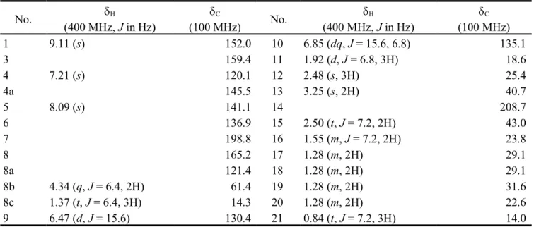
Monapurpyridine A (MPA) was obtained as a yellow oil. The HRESIMS of MPA showed a molecular ion at m/z 400.2480 ([M+H]+ , C
24H34NO4), indicating a molecular formula of C24H33NO4
4 37 38 39 40 41 42 43 44 45 46 47 48 49 50 51 52 53 54 55 56 57 58 59 60 61 62 63 64 65 66 67
bands at 1716 and 1675 cm-1, consistent with the presence of conjugated ketone and carboxylic ester
groups. The UV maximum at 261 and 308 nm inferred pyridine-chromophore system. The 1H and 13C
spectra (Table 1) of MPA disclosed the signals for the presence of ketones (δC 208.7, 14; 198.8,
C-7), ester carbonyl (δC 165.2, C-8), trisubstituted pyridine unit (δC 152.0, 159.4, 120.1, 145.5,
and 121.4; δH 9.11, s, H-1; 7.21, s, H-4), trisubstituted olefinic unit (δC 141.1, 136.9; δH 8.09, s,
H-5), trans-double bond (130.4, 135.1; δH 6.47, d, J = 15.6; 6.85, d, J = 15.6, 6.8), one ethyl group (δH
4.34, 2H, J = 6.4; 1.37, 3H, J = 6.4; δC 61.4, 14.3), seven methylene carbons (δC 40.7, 43.0, 23.8, 29.1,
29.1, 31.6, 22.6), as well as three methyls (δH 2.48, s; 1.92, d, J = 6.8; 0.84, t, J = 7.2). As shown in
Figure 2, the correlations of three partial structures (H2-8b/Me-8c, H-9/H-10/Me-11, and H2-15/H2
-16/H2-17/H2-18/H2-19/H2-20/Me-21) were confirmed by analysis of 1H-1H COSY (the bold) and
HMBC (the arrows) correlations. From the HMBC spectrum, the methylene protons at δH 3.25 (H2-13)
were correlated between trisubstituted olefinic unit and two ketone carbonyls. Addtionally, the (CH2)6CH3 group and Me-12 (δH 2.48) were determined at C-14 and C-7, respectively, due to the
correlations of H2-15/C-14 and Me-12/C-7. Furthermore, the key HMBC correlations between
H-1/C-3, C-4a, C-8a, H-4/C-H-1/C-3, C-8a, C-9, H-5/C-4, C-7, C-6, C-1H-1/C-3, H2-8b/C-8 revealed that the
fragments were located at C-3, C-4a and C-8a positions of trisubstituted pyridine unit. Thus, the planer structure of MPA was completely assigned by 2D NMR experiments, especially 1 H-1H COSY, HMQC, and HMBC. The relative stereochemistry of MPA was further determined, based
on the NOESY (Figure 2) spectrum showing the correlations between 9/Me-11, 4/H2-13, and H-5/Me-12 indicating E-forms of C-9/10 and C-5/6 double bonds. Together with the above findings, the MPA was established as ethyl 4-((E)-2-acetyl-4-oxoundec-1-enyl)-6-((E)-prop-1-enyl)nicotinate, and
has been named monapurpyridine A.
Table 1. 1H- and 13C-NMR spectroscopic data of MPA (in CDCl 3) a,b. No. (400 MHz, J in Hz) C (100 MHz) No. (400 MHz, J in Hz) C (100 MHz) 1 9.11 (s) 152.0 10 6.85 (dq, J = 15.6, 6.8) 135.1 3 159.4 11 1.92 (d, J = 6.8, 3H) 18.6 4 7.21 (s) 120.1 12 2.48 (s, 3H) 25.4 4a 145.5 13 3.25 (s, 2H) 40.7 5 8.09 (s) 141.1 14 208.7 6 136.9 15 2.50 (t, J = 7.2, 2H) 43.0 7 198.8 16 1.55 (m, J = 7.2, 2H) 23.8 8 165.2 17 1.28 (m, 2H) 29.1 8a 121.4 18 1.28 (m, 2H) 29.1 8b 4.34 (q, J = 6.4, 2H) 61.4 19 1.28 (m, 2H) 31.6 8c 1.37 (t, J = 6.4, 3H) 14.3 20 1.28 (m, 2H) 22.6 9 6.47 (d, J = 15.6) 130.4 21 0.84 (t, J = 7.2, 3H) 14.0
a Assignments were confirmed by 1H-1H COSY, HMQC, HMBC. b m: multiple signal.
7 69 70 71 72 73 74 75 76 77 78 79 80 81 82 83 84 85 86 87 88 89 90 91 92
Figure 2. Key 1H-1H COSY (▬), HMBC(H→C) and NOESY (H↔H) correlations of
MPA.
2.2. Cytotoxicity of MPA on MCF-7 and M10 Cells
We utilized MTT assay for a two-day course to study the inhibition on cell viability of MCF-7 and M10 cells treated with MPA (Figure 3). Up to the concentration of 100 M, MPA showed dose-dependent and moderated cytotoxic activity against MCF-7 cells, but no significant cytotoxicity to normal M10 cells. The results suggested that MPA was selectively cytotoxic to breast cancer cell line.
Figure 3. The effects of MPA on cell viability of MCF-7 and M10 cells. (A) Cells were treated with 25, 50 and 100 M MPA for 24 hr. (B) Cells were treated with 100 M MPA for 12 and 24 hr. Data were expressed as means ± SD (n = 3). * Significantly different (p < 0.01) versus the negative control (without any treatment).
A 10 93 94 95 96 97 98 99 100 101 102 103 104 105 106 107
2.3. Cell Death Induced by MPA on MCF-7 Cells
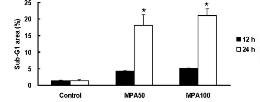
To study cell deaths of MCF-7 cells induced by MPA, we utilized flow cytometry (propidium iodide staining) to analyze the ratio of sub-G area for 12 and 24 hr (Figure 4). MPA (50 M, 24 hr)
significantly induced about 10 folds of cell deaths than control group. To make clear the cell death was resulted from apoptosis or necrosis, we designed some apoptotic approaches in the next step.
Figure 4. Flow cytometric analysis of sub-G1 area of MCF-7 cells treated with MPA.
MCF-7 cells were treated with 50 and 100 M MPA for 12 and 24 hr. Data were expressed as means ± SD (n = 3). * Significantly different (p < 0.05) versus the negative control (without any treatment).
2.4. Caspase Activation of MPA on MCF-7 Cells
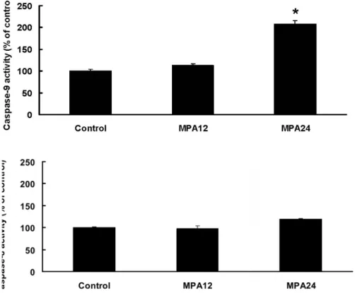
MCF-7 cells were treated with 50 and 100 M of MPA for 12 and 24 hr, and further analyzed for the cleaved caspase-3 by Western blot (Figure 5) and enzyme activity of caspase-8 and -9 by colorimetric assay kit (Figure 6). Treatment of MPA (50 and 100 M, 24 hr) exhibited increases of cleaved caspase-3, which were estimated as a down-streamed event of apoptosis. As to the up-streamed caspase-9 and caspase-8, treatment of MPA (50 M, 24 hr) exhibited significant increases of caspase-9 activity, but showed no significant increase of caspase-8. Thus, MPA was demonstrated to induce apoptosis through caspase-9 activations
Figure 5. The effects of MPA on caspase-3 activation in MCF-7 cells. Cleaved caspase-3 and -actin were detected by Western blot. MCF-7 cells were treated with 50 or 100 M of MPA for 12 or 24 hr. (A) From the left side: Lane 1, control; lane 2, MPA 50 M for 12 hr; lane 3, MPA 100 M for 12 hr; lane 4, MPA 50 M for 24 hr; lane 5, MPA 100 M for 24 hr. (B) Quantification of cleaved caspase-3 presented above.
A 13 109 110 111 112 113 114 115 116 117 118 119 120 121 122 123 124 125 126 127 128 129 130 131 132 133
Figure 5. Cont.
B
Figure 6. The effects of MPA on caspase-9 and -8 activities. MCF-7 cells were treated with 50 M of test agents for 12 or 24 hours. From the left side were: control; MPA, 12 hr; MPA, 24 hr. Data were expressed as means ± SD (n = 3). * Significantly different (p < 0.01) versus the control (without any treatment).
3. Experimental Section
3.1. General Experimental Procedures
Electrospray ionization mass spectrometry (ESI-MS) data were acquired by a LCQ mass 16 134 135 136 137 138 139 140 141 142 143 144 145
spectrometer (Unity Plus 400 MHz) (Brucker BioSpin, Rheinstetten, Germany) using CDCl3 as the
solvent. Sephadex LH-20 (GE Healthcare, Uppsala, Sweden) and silica gel 60 (70-230 mesh and 230-400 mesh, Merck, Darmstadt, Germany) were used as chromatographic materials. Silica Gel 60 F254 plates (Merck) were used for thin layer chromatography (TLC). The TLC spots were detected under UV-lamps (254 and 365 nm) and also by using an anisaldehyde-sulphuric acid solution, applied as a spray reagent, followed by heating. The high performance liquid chromatography (HPLC) was performed using a Shimadzu LC-6AD apparatus with a SPD-6AV UV detector that was equipped with a preparative Cosmosil AR-II column (250 x 20 mm i.d., Nacalai Tesque, Inc., Kyoto, Japan).
3.2. Reagents
Methanol and acetonitrile (HPLC grade), acetone, ethyl acetate, n-hexane and methanol (analytical grade) were purchased from ECHO (Miaoli, Taiwan). Trifluroacetic acid (TFA), anisaldehyde and sulphyric acid were purchased from Merck. Fetal bovine serum (FBS), minimum essential medium (MEM), Dulbecco’s minimum essential medium (DMEM), phosphate buffered saline (PBS) and trypan blue were purchased from Biological Industries (Kibbutz Beit-Haemek, North District, Isreal). Other chemicals, such as 3-(4,5-dimethylthiazol-2-yl)-2,5-diphenyl-tetrazolium bromide (MTT), dimethyl sulfoxide (DMSO) and propidium iodide were obtained from Sigma (St. Louis, MO, USA).
3.3. Extraction and Isolation
The RMR powder (5 kg) was extracted with 25 L of methanol at 50 C for 24 h. The dried extract was subjected to silica gel column chromatography, eluting with a mixture of n-hexane/ethyl acetate (9:1, 8:2, 7:3, 6:4, 0:10). This fraction (8:2) was then further separated by Sephadex (LH-20) gel column to remove other impurities and then purified again using preparative HPLC (Cosmosil 5C18
packing column, 250 x 20 mm i.d, MeOH/H2O = 85:15, 7 mL/min) to obtain MPA (6.6 mg)
3.4. Spectral Data
Monapurpyridine A (MPA): Yellow oiled liquid. IR(neat) νmax 1716, 1675, 1588, 1369, 1284, 1174,
1094 cm-1. UV (MeOH) λ
max (log ε): 261 nm (3.52), 308 nm (3.01). HRESIMS m/z 400.2480 ([M+H]+ ,
C24H34NO4). 1H NMR and 13C NMR were describpt in Table 1.
3.5. Cell Lines and Culture Conditions
Human breast adenocarcinoma cells MCF-7 and normal mammary epithelial cells M10 were obtained from Bioresources Collection and Research Center (Hsinchu, Taiwan). Both cells were maintained in MEM (5% FBS) in a humified incubator with 5% CO2 at 37oC.
3.6. Cytotoxicity Assay
Cells (3 x 10³ per well) were seeded with 180 L of MEM in 96-well plates. After 4 hr, 20 L of
test agents dissolved in PBS solution were added at final concentrations of 25, 50 and 100 M and incubated in a 37oC incubator with 5% CO
2. Culturing for 24, 48 hr, 20 L of MTT solution (2
mg/mL) was added to each well and incubated for 4 hr to make cellular conversion of a tetrazolium 19 147 148 149 150 151 152 153 154 155 156 157 158 159 160 161 162 163 164 165 166 167 168 169 170 171 172 173 174 175 176 177 178 179 180 181
salt into a formazan product. Then the supernatant was removed and 200 L of DMSO was added to dissolve the formazan. Finally, the formazan can be detected by spectrophotometry in the absorbance at 570 nm and provided a relative estimate of cell viability.
3.7. Assay of DNA Contents by Flow Cytometry
MCF-7 cells (5 x 10⁴ per well) were seeded with 2 mL of MEM in 6-well plates. After 12 hr, 2 mL
of test agents dissolved in MEM solution were replaced at final concentrations of 50 and 100 M. After 12 and 24 hr of incubation, the cells were harvested and fixed with 80% ethanol for 30 minutes. Then washed the cell pellets 3 times with PBS and co-incubated the cells with propidium iodide (4 g/mL), triton (1%), and RNase (0.1 g/mL) in dark for 30 minutes. Finally, the cells can be analyzed by flow cytometry equipped with Cell Quest software and provided a relative estimate of DNA contents.
3.8. Western Blot Analysis
Cells (about 5 x 10⁵) were seeded with 10 mL of media in a 75 cm2 flask. After 12 hr, 10 mL of test
agents dissolved in media were replaced. After 12 and 24 hr of incubation, the cells were harvested and extracted by RIPA lysis buffer (Millipore, Bellerica, MA, USA) with 1% protease inhibitor (Sigma, St. Louis, MO, USA). The cell lysates were analyzed with primary antibodies, including of caspase-3 antibody (Novus Biologicals, Littleton, CO, USA) and -actin antibody (Epitomics, Burlingame, CA, USA). The anti-mouse secondary horseradish peroxidase antibodies (Jackson ImmunoResearch, West Grove, PA, USA) was further added. Finally, the detection was performed using the Western lightning chemiluminescence reagent (PerkinElmer Life Sciences, Waltham, MA, USA).
3.9. Caspase Activity Assay
Cells (about 5 x 10⁵) were seeded with 10 mL of media in a 75 cm2 flask. After 12 hr, 10 mL of test
agents dissolved in media were replaced. After 12 and 24 hr of incubation, the cells were harvested and tested for caspase-8 and caspase-9 activities respectively by using colorimetric assay kit (BioVision, Linda Vista Avenue, CA, USA). Caspase activity was determined according to the manufacturer’s protocol.
3.10. Data Analysis
Data were presented as mean ± standard deviation (n = 3). The statistical comparisons were performed by one-way analysis of variance (ANOVA) with Duncan’s test. The significant differences were indicated as p < 0.05 or 0.01.
4. Conclusions
Previous studies in our group showed that RMR extracts or red mold dioscorea (RMD) extracts fermented from M. purpureus NTU 568 might prevent carcinogenesis or tumor progression in animal 22 182 183 184 185 186 187 188 189 190 191 192 193 194 195 196 197 198 199 200 201 202 203 204 205 206 207 208 209 210 211 212 213 214 215
colon and lung cancer cell lines. In this study, a new azaphilone MPA was isolated from M. purureus NTU 568 fermented red mold rice, and showed moderate cytotoxicity against breast cancer cells. In a conclusion, azaphilone derivatives isolated from our studies were almost moderately cytotoxic but tissue-specific to different cancer cell lines. These results strongly implied that fermented products from M. purpureus NTU 568 were potential candidates for tumor prevention due to the available amounts of azaphilone derivatives.
Acknowledgements
This research is supported by grants from the National Science Council (NSC) and National Research Institute of Chinese Medicine (NRICM), Taiwan, the Republic of China. We are grateful to Chien-Chang Shen and Ming-Jaw Don, NRICM, for technical assistance for the NMR and mass measurements, respectively.
References and Notes
1. Lee, C. L.; Tsai, T. Y.; Wang, J. J.; Pan, T. M. In vivo hypolipidemic effects and safety of low dosage Monascus powder in a hamster model of hyperlipidermia. Appl. Microbiol. Biotechnol. 2006, 70, 533-540.
2. Wang, J. J.; Shieh, M. J.; Kuo, S. L.; Lee, C. L.; Pan, T. M. Effect of red mold rice on antifatigue and exercise-related changes in lipid peroxidation in endurance exercise. Appl. Microbiol.
Biotechnol. 2006, 70, 247-253
3. Lee, C. L.; Kuo, T. F.; Wu, C. L.; Wang, J. J.; Pan, T. M. Red mold rice promotes neuroprotective sAPPalpha secretion instead of Alzheimer’s risk factors and amyloid beta expression in hyperlipidemic A40-infused rats. J. Agric. Food Chem. 2010, 58, 2230-2238.
4. Chen, W. P.; Ho, B. Y.; Lee, C. L.; Lee, C. H.; Pan, T. M. Red mold rice prevents the development of obesity, dyslipidemia and hyperinsulinemia induced by high-fat diet. Int. J. Obes. 2008, 32, 1694-1704.
5. Tsai, R. L.; Ho, B. Y.; Pan, T. M. Red mold rice mitigates oral carcinogenesis in 7,12-dimethyl-1,2-benz[a]anthracene-induced oral carcinogenesis in hamster. Evid. Based Complement Alternat.
Med. 2009, http://dx.doi.org/10.1093/ecam/nep215.
6. Ho, B. Y.; Wu, Y. M.; Hsu, Y. W.; Hsu, L. C.; Kuo, Y. H.; Chang, K. J.; Pan, T. M. Effects of
Monascus-fermented rice extract on malignant cell-associated neovascularization and
intravasation determined using the chicken embryo chorioallantoic membrane model. Integr.
Cancer Ther. 2010, 2, 204-212.
7. Li, Y. G.; Zhang, F.; Wang, Z. T.; Hu, Z. B. Identification and chemical profiling of monacolins in red yeast rice using high-performance liquid chromatography with photodiode array detector and mass spectrometry. J. Pharm. Biomed. Anal. 2004, 35, 1101-1112.
8. Tsuji, K.; Ichikawa, T.; Tanabe, N.; Obata, H.; Abe, S.; Tarui, S.; Nakagawa, Y. Extraction of hypotensive substance from wheat beni-koji. Nippon Shokuhin Kogyo Gakkaishi 1992, 39, 913-918. 25 217 218 219 220 221 222 223 224 225 226 227 228 229 230 231 232 233 234 235 236 237 238 239 240 241 242 243 244 245 246 247 248 249 250 251 252 253
9. Wu, C. L.; Lee, C. L.; Pan, T. M. Red mold dioscorea has a greater antihypertensive effect than traditional red mold rice in spontaneously hypertensive rats. J. Agric. Food Chem. 2009, 57, 5035-5041.
10. Aniya, Y.; Yokomakura, T.; Yonamine, M.; Shimada, K.; Nagamine, T.; Shimabukuro, M.; Gibo, H. Screening of antioxidant action of various molds and protection of Monascus anka against experimentally induced liver injuries of rats. Gen. Pharmacol. 1999, 32, 225-231.
11. Akihisa, T.; Tokuda, H.; Yasukawa, K.; Ukiya, M.; Kiyota, A.; Sakamoto, N.; Suzuki, T.; Tanabe, N.; Nishino, H. Azaphilones, furanoisophthalides, and amino acids from the extracts of Monascus
pilosus-fremented rice (red-mold rice) and their chemopreventive effects. J. Agric. Food Chem.
2005, 53, 562-565.
12. Akihisa, T.; Tokuda, H.; Ukiya, M.; Kiyota, A.; Yasukawa, K.; Sakamoto, N.; Kimura, Y.; Suzuki, T.; Takahasu, J.; Nishino, H. Anti- tumor-initiating effects of monascin, an azaphilonoid pigment from the extract of Monascus pilosus fermented rice (red-mold rice). Chem. Biodivers. 2005, 2, 1305-1309.
13. Su, N. W.; Lin, Y. L.; Lee, M. H.; Ho, C. Y. Ankaflavin from Monascus-fermented red rice exhibits selective cytotoxic effect and induces cell death on Hep G2 cells. J. Agric. Food Chem. 2005, 53, 1949-1954.
14. Li, J. J.; Shang, X. Y.; Li, L. L.; Liu, M. T., Zheng, J. Q. and Jin, Z. L. New cytotoxic azaphilones from Monascus purpureus-fermented rice (red yeast rice) Molecules 2010, 15, 1958-1966.
15. Wu, M. D.; Cheng, M. J., Yech; Y. J.; Chen, Y. L.; Chen K. P.; Chen, I. H.; Yang, P. H. and Yuan, G. F. Monasnicotinates A–D, four new pyridine alkaloids from the fungal strain Monascus
pilosus BCRC 38093. Molecules 2011, 16, 4719-4727
16. Hsu, Y. W.; Hsu, L. C.; Liang, Y. H.; Kuo, Y. H.; Pan, T. M. Monaphilones A-C, three new antiproliferative azaphilone derivatives from Monascus purpureus NTU 568. J. Agric. Food
Chem. 2010, 58, 8211-8216.
17. Hsu, Y. W.; Hsu, L. C.; Chang, C. L.; Liang, Y. H.; Kuo, Y. H.; Pan, T. M. New anti-inflammatory and anti-proliferative constituents from fermented red mold rice Monascus
purpureus NTU 568. Molecules (2010) 15, 7815-7824.
18. Hsu, L. C.;Hsu, Y. W.;Liang, Y. H.,Kuo, Y. H.and Pan, T. M. Anti-tumor and anti-inflammatory properties of ankaflavin and monaphilone A from Monascus purpureus NTU 568. J Agri Food
Chem. (2011) 59: 1124-1130.
Sample Availability: Samples of the compounds are available from the authors.
© 2011 by the authors; licensee MDPI, Basel, Switzerland. This article is an open access article distributed under the terms and conditions of the Creative Commons Attribution license (http://creativecommons.org/licenses/by/3.0/). 28 254 255 256 257 258 259 260 261 262 263 264 265 266 267 268 269 270 271 272 273 274 275 276 277 278 279 280 281 282 283 284 285 286 287 288 289 290