Disrupted circadian rhythm in rats with nephrectomy-induced
chronic kidney disease
Chung-Yao Hsu, MD, PhD a,b; Fang-Chia Chang, PhD* c,d; Hwee-Yeong Ng, MD e;
Chien-Chun Kuo, MD e; Yueh-Ting Lee, MD e; Chin-Yu Lu, MS c; Chien-Te Lee,
MD, PhD* e
a Department of Neurology, Kaohsiung Medical University Hospital, No.100, Tzyou
1st Road, Kaohsiung City 807, Taiwan
b Department of Master’s Program in Neurology, Faculty of Medicine, College of
Medicine, Kaohsiung Medical University, Kaohsiung
c School of Veterinary Medicine, National Taiwan University, No.1, Sec.6, Roosevelt
Road, Taipei City 106, Taiwan
d Graduate Institute of Acupuncture Science, College of Chinese Medicine, China
Medical University, No. 91 Hsueh-Shih Road, Taichung City 404, Taiwan
e Division of Nephrology, Department of Internal Medicine, Kaohsiung Chang Gung
Memorial Hospital and Chang Gung University College of Medicine, No.123, Ta-Pei Road, Niao-Sung District, Kaohsiung City 833, Taiwan
Address: Division of Nephrology, Department of Internal Medicine, Kaohsiung Chang-Gung Memorial Hospital/Department of Veterinary Medicine, National Taiwan University
No.123, Ta-Pei Road, Niao-Sung District, Kaohsiung City 833, Taiwan./No. 1, Sec. 4, Roosevelt Road, Taipei 10607
Phone: +886-7-7317123 ext.8306/+886-2-3366-3883 Fax: +886-7-7322402
Email: [email protected]/[email protected]
Ethics of investigation:
This animal study has been approved by the institutional review board of Kaohsiung Medical University Hospital (KMUH-98070).
Abstract
Aims:
Our study investigated the role of circadian rhythm in the pathogenesis of sleep disturbance in patients with chronic kidney disease (CKD) based on an animal model.
Main methods:
Sixteen Sprague-Dawley (SD) rats (eight from 5/6 nephrectomized CKD group and eight from control group) were used for electroencephalography (EEG) and
electromyography (EMG) recording. Eight rats (four from CKD and four from control group) were sacrificed at six zeitgeber time (ZT) points and determined the mRNA expression of clock genes, rPer1, rPer2 and rBMAL1b in the hypothalamus.
Key findings:
Our results demonstrated that both slow wave sleep (SWS) and rapid eye movement (REM) sleep were significantly increased in the ZT22-24 zeitgeber time point of the dark period in the CKD rats when compared with those sleep architectures obtained from the control rats. The CKD-induced sleep disruptions were associated with significant up-regulations of rPer1 (in ZT2, ZT6 and ZT14) and rPer2 mRNA expression (in ZT2 and ZT14) in the hypothalamus.
Significance:
Our study elucidated that the increases of SWS and REM sleep during ZT22-24 of the dark period in the CKD rats might be due to the enhancement of rPer1 and rPer2
clock genes in the hypothalamus, suggesting that disrupted circadian rhythm plays a role in the pathogenesis of sleep disturbance in patients with CKD.
Introduction
Sleep disorders are common in patients with chronic kidney disease (CKD) and can occur in early CKD . Among them, periodic limb movement in sleep (PLMS) was reported to be associated with mortality of CKD patients . Patients with CKD suffer from both insomnia and excessive daytime sleepiness (EDS) , which are associated with poor quality of life . Co-existence of insomnia and daytime sleepiness suggests that disruption in circadian rhythm might play a role in the pathogenesis of sleep disturbance in CKD patients .
Clinical evidence also supports circadian rhythm disruption in patients with CKD. Circadian blood pressure variation is compromised in patients with advanced CKD (stage 3-5) . The mortality of CKD patients also follows a circadian variation with increased rate of death in the morning . We have shown that CKD patients under regular evening hemodialysis had better sleep quality and less daytime sleepiness compared with those under morning and afternoon shifts . The nocturnal endogenous melatonin rhythm decreases with advanced CKD , and nocturnal hemodialysis partially restores circadian rhythm of melatonin and improves nocturnal sleep in patients with CKD . These all indicate that circadian factor might be involved in the sleep disturbances in CKD patients.
(SCN) of hypothalamus as well as those located in other brain regions and most peripheral tissues . These central and peripheral clocks work though the expression of clock genes and their protein products. Disrupted circadian clock genes have revealed alterations in circadian rhythmicity as well as changes in sleep duration and sleep architecture . Many circadian genes are also involved in the homeostatic functions of the peripheral organs, including heart and kidney . The circadian rhythm of clock gene expression may be modified to some extent between peripheral tissues, as denoted by differences in amplitude and phasing, and operates differently . Renal excretion of water and electrolytes also follows the order of circadian rhythm. Distal convoluted tubule, connecting tubule and the cortical collecting duct of the kidney all exhibit a strong circadian rhythmicity of clock gene expression .
Many confounding factors and co-morbidity need to be considered in clinical studies dealing with CKD and its related sleep disorders . Thus we established an animal model of CKD and tried to elucidate the role of circadian disruption in the pathogenesis of sleep disturbance in patients with CKD.
Material and methods
AnimalsAdult male Sprague-Dawley (SD) rats (180-200g, National Animal Breeding and Research center, Taiwan) were used in these experiments. Animals were divided into two groups: (1) the CKD rats (n = 24) and (2) the normal control rats (n = 24). These rats were housed in isolated cages, in which the temperature was maintained at 23 ± 1 °C and the light: dark cycle was a 12:12-h cycle (20 Watt x 6 tubes illumination). Food and water were available ad libitum.
Animal model of CKD
The rats were anesthetized by Zoletil 50 (0.1 ml/ 100 g), which is a combination of a
dissociative anesthetic agent, tiletamine hydrochloride, and a tranquilizer, zolazepam hypochloride, via intra-peritoneal injection. After one-week accommodation to the 12:12h light: dark cycle these animals underwent the 1st stage of surgery with 2/3
nephrectomy on the right kidney and the 2nd stage of surgery with complete
nephrectomy on the left kidney were performed in the following week. The final model of 5/6 nephrectomy mimicking CKD was established on the week 8 .
Electroencephalography (EEG) and Electromyography (EMG) recording s
sleep-wake recording. The SD rats underwent surgery for sleep-wake recording on day 1 of week 9 (CKD group) and day 1 of week 4 (control group). They were anesthetized again by Zoletil 50 .
The procedure of EEG recording and scoring for sleep stages have been described previously . Following anesthesia, a midline incision was made on the scalp to expose the standard reference point, bregma, on the skull. We then drilled through the skull and implanted three EEG screw electrodes (right frontal (+3.5,+2) from bregma (0, 0); right hippocampus (+3, -5.5) from bregma; left occipital (-3.5, -1.0) from lambda (0, 0) and two anchor electrodes. Two EMG electrodes were also inserted into the posterior neck muscle. Insulated leads from EEG electrodes were routed to a Teflon pedestal (Plastics One, Roanoke, VA). The Teflon pedestal was then cemented to the skull with dental acrylic (Cranioplastic cement and Cyanoacrylate gel, Plastics One, Roanoke, VA). The incision wound was treated topically with Povidine-Iodine ointment and the animals were allowed to recover for seven days prior to the initiation of the experiments.
On the second postsurgical day, the rats were connected to the recording apparatus via a flexible tether. Signals from the EEG and EMG electrodes were fed into an
amplifier (Colbourn Instruments, Lehigh Valley, PA; model V75-01). The EEG was amplified (factor of 5000) and analog bandpass filtered between 0.1 and 40 Hz
(frequency response: ±3 dB; filter frequency roll off: 12 dB/octave). Gross body movements were detected by custom-made infrared-based motion detectors
(Biobserve GmbH, Germany), and the movement activity was converted to a voltage output which was digitized and integrated into 1-s bins. These conditioned signals (EEGs, EMGs and gross body movements) were subjected to analog-to-digital conversion with 16-bit precision at a sampling rate of 128 Hz (NI PCI-6033E; National Instruments, Austin, TX). The digitized EEG waveform and integrated values for body movement were stored as binary computer files pending subsequent analyses.
Postacquisition determination of the vigilance state was done by visual scoring of 12-s epochs using custom software (ICELUS, M. R. Opp) written in LabView for
Windows (National Instruments) . The animal’s behavior was classified as either slow wave sleep (SWS), REM sleep or wakefulness based on the following criteria.
Briefly, SWS is characterized by large-amplitude EEG slow waves, high power density values in the delta frequency band (0.5–4.0 Hz), a relax muscle tone from EMGs, and lack of gross body movements. During REM sleep, the amplitude of the EEG is reduced, the predominant EEG power density occurs within the theta
frequency (6.0–9.0 Hz), the EMGs exhibit muscle atonia with low EMG amplitudes, and there are phasic body twitches. During wakefulness, the rats are generally active
and there are protracted body movements with robust EMG amplitudes. The
amplitude of the EEG is similar to that observed during REM sleep, but power density values in the delta frequency band are generally greater than those in theta frequency band. Percentages of SWS, REM sleep and wakefulness were calculated for each zeitgeber time point. Only the consecutive three zeitgeber time points were grouped for statistical comparison to control type I error from multiple testing.
Clock gene expression
When the rat was anesthetized, bilateral renal arteries and veins were clamped and kidneys were removed. Immediately following this procedure, the rat brain was removed and hypothalamus was dissected and stored at -80 ℃ for further determining the gene expression of clock genes.
Eight rats (four from CKD and four from control group) were scarified at each zeitgeber time point (ZT2, ZT6, ZT10, ZT14, ZT18, and ZT22). Three clock genes, rPer1, rPer2 and rBMAL1b were chosen for mRNA analysis through reverse transcription to cDNA and real time PCR for semi-quantitative measurement. The procedure has been described previously . In brief, after collection, the tissue of hypothalamus was immediately transferred to RNA Stabilization Reagent kits, RNAlater® (Ambion, Valencia, CA) which is an aqueous, non-toxic tissue storage reagent that rapidly permeates tissues to stabilize and protect cellular RNA. Total
RNA was extracted by use of RNA extraction kit, RNeasy Mini kit® (Qiagen, Valencia, CA) and mRNA was reverse-transcribed to cDNA by Quantitest reverse transcription kit® (Qiagen) and then the cDNA was the subject for further real-time PCR analysis.
The primers, TaqMan probes for rPer1, rPer2 and rBMAL1 were purchased from Applied Biosystems Company. The sequence of probe and primers was as follows: rPer1 (probe: cttcagcc, forward: tgctttagatcggcagtggt, reverse: gtgggcttgacacctcttct); rPer2 (probe: ttctcctg, forward: gctggaggactactttgcattt , reverse:
ccagtggcaagagtcaaagc); rBMAL1b (probe: tcctctcc, forward: tgtctggagtccctccattt , reverse: agtacgcctccccctgat).
All reactions were performed by use of the Maxima™ Probe qPCR Master Mix (2X) (Fermentas) in a LightCycler 2.0 Real-Time PCR system® (Roche) according to the manufacturer’s instruction. Real-time PCR was carried out in a 10 μl final volume containing 0.3 μM of each primer, 0.2 μM of the probe, 5 μl of Maxima™ Probe qPCR Master Mix (2X), and 1 μl of template DNA. “Comparative Ct Method” was used for quantitative approach. The number of cycle value was determined for each sample of CKD and control. They were normalized to an endogenous housekeeping gene, β-actin.
The 24-h urine sample was collected from metabolic cages for the analysis of urine amount and urinary protein excretion. Blood was also collected from rat tail vein for the measurement of creatinine at week 8.
Statistical analysis
Variables derived from sleep-wake cycle analysis were presented as mean and standard deviation. Variables derived from quantitative mRNA expression of rPer1, rPer2 and rBMAL1 between experimental and control group were presented as ratio of clock to house keeping gene expression. Variables between CKD and control rats were compared using unpaired t test. The p value of 0.05 was set as the cut-off point for statistical significance.
Results
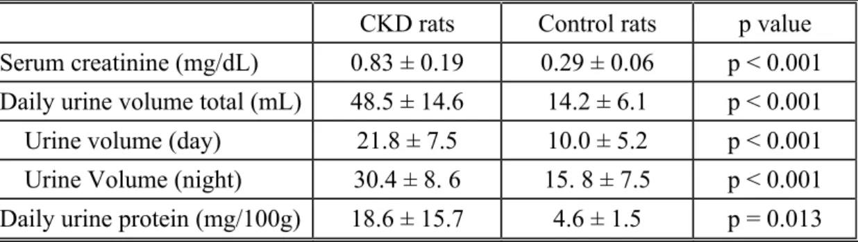
CKD modelThe laboratory data for the CKD and control rats is shown in table 1. Significant increases of mean serum creatinine level (0.83 ± 0.19 mg/dL in CKD vs. 0.29 ± 0.06 mg/dL in control, p < 0.001), total daily urine output (48.5 ± 14.6 mL in CKD vs. 14.2 ± 6.1 mL in control, p < 0.001), urine output of daytime (21.8 ± 7.5 mL in CKD vs. 10.0 ± 5.2 mL in control, p < 0.001) and nighttime (30.4 ± 8. 6 mL in CKD vs. 15. 8 ± 7.5 mL in control, p < 0.001) and daily urinary secretion of protein (18.6 ± 15.7 mg in CKD vs. 4.6 ± 1.5 mg in control, p = 0.013) were observed in the CKD rats.
Alterations of s leep-wake activities in the CKD rats
The EEG results are presented in figure 1. There was a trend of decreased wakefulness and increased SWS and REM sleep from ZT17 to ZT24 in the dark period. Significant increase of SWS (31.1 4.5% in CKD vs. 20.1 1.7% in control, p = 0.040) and REM sleep (7.7 1.7% in CKD vs. 2.1 0.9% in control, p = 0.014) with a mirror effect of decreased wakefulness (61.2 6.1% in CKD vs. 77.8% 2.4% in control, p = 0.027) were found in the ZT22-24 zeitgeber time point of the CKD rats compared with the control rats. On the other hand, there was no significant difference between CKD and normal rats in wakefulness, SWS and REM sleep throughout the light period.
G
ene expression analysis of clock genes
Clock gene expression in the hypothalamus is demonstrated in figure 2. There was a trend of up-regulation for rPer1 with significant findings in ZT2 (0.063 ± 0.018 vs. 0.118 ± 0.007, p = 0.005), ZT6 (0.035 ± 0.007 vs. 0.050 ± 0.007, p = 0.021) and ZT14 (0.081 ± 0.013 vs. 0.109 ± 0.009, p = 0.014) and a trend of up-regulation for rPer2 with significant findings in ZT2 (0.009 ± 0.001 vs. 0.012 ± 0.001, p = 0.001) and ZT14 (0.010 ± 0.002 vs. 0.015 ± 0.002, p = 0.018). There was also a trend of up-regulation for rBMAL 1b without reaching statistical significance.
Discussion
First of all, we established a rat model of CKD which was evidenced by a significant increase in urine output and serum creatinine level associated with significant
proteinuria. The EEG recording showed significantly decreased wakefulness and increased SWS and REM sleep in the late phase of the dark period (ZT22-24) in rats with CKD. These observations simulate the sleepiness happened earlier before usual sleep onset in patients with CKD. Per1 and Per2 are important components of the pacemaker (Zheng et al. 1999, Zheng et al. 2001). We further showed that this CKD-related alteration of sleep-wake architecture might be induced by an up-regulation of rPer1 and rPer2 clock genes in the hypothalamus particularly in the early phase of the dark period (ZT14).
Enhanced Per 1 and Per2 expression can be found following sleep deprivation
(Franken et al. 2007). In our study, increased sleep propensity of the CKD rats in the late phase of dark period could not be explained by a homeostatic process, that is, a consequence of sleep disturbance in the preceding light period. Enhanced Per1 and Per2 expression are also involved in resetting the biological clock in response to light pulse (Albrecht et al. 2001). In our study, although we could not show a complete phase shift of the circadian rhythm in the CKD rats, a better explanation might be partial disruption of circadian rhythm in the hypothalamus caused by certain uremic
toxins. To our knowledge this is the first animal study combining electrophysiological and molecular methods and focusing on the relationship between circadian rhythm and CKD. As nocturnal hemodialysis improves excessive daytime sleepiness (EDS) in patients with CKD, uremic toxins and other factors associated with impaired renal function might be important causes for the disrupted circadian rhythm in patients with CKD .
The patients with CKD often suffer from both nocturnal sleep disturbance and impaired daytime function. Many common sleep disorders are observed in CKD patients, particularly insomnia, restless legs syndrome (RLS), PLMS and obstructive sleep apnea (OSA) . EDS is one of the major daytime symptoms of OSA and also accompanies in patients with insomnia, RLS and PLMS. Therefore, clinical studies must deal with many confounding factors in order to clarify the pathogenesis of EDS in patients with CKD. This is why most clinical studies regarding CKD and sleep disorders only focused on nocturnal sleep disorders. Patients with CKD usually have EDS which is always considered as distressing as nocturnal symptoms and have an impact on quality of life . In our previous report, we emphasized not only nocturnal sleep quality but also EDS which make difference in CKD patients under different dialysis schedule . Thus our animal model of CKD is more helpful than clinical studies to clarify this issue.
The causes of EDS might be the adverse effects of dialysis, including activation of cytokines, compensatory cooling effect of the body temperature, disequilibrium syndrome, stressors and change of daily activities based on dialysis schedule . These confounding factors related with dialysis were controlled in our animal model of CKD. There is high prevalence of obstructive sleep apnea in patients with CKD. Although clinical presentation of the symptoms of OSA differs between CKD patients and general population, the prevalence of self-reported EDS is similar . Although rodent models of chronic intermittent hypoxia have been developed to investigate the effect of sleep apnea-mimicking condition, there is still no reliable device developed to measure the severity and pattern of sleep apnea . The serum level of melatonin and core body temperature which are indicators of circadian rhythm were not determined in current study. Change of melatonin fluctuation might be involved although it only has a minor influence on SCN supposing that the reninohypothalamic tract is intact . The core body temperature is more reliable as a physiological indicator of circadian rhythm which we did not measure in our animal study .
Disruption of circadian rhythm is important in CKD patients. The endogenous rhythm of the most important circadian hormone in human body is melatonin which is
impaired in patients with CKD and relates to the degree of disease . The mortality of dialysis patients tends to happen in the morning . This phenomenon could be partially
explained by the variation of circadian blood pressure . Modified circadian gene expression both centrally and peripherally has been shown in an animal model of hypertensive rats . Treatment of CKD from chronobiological points of view might be beneficial to patients with early CKD. Although current result is an association study indicationg that CKD-induced sleep disruption was associated with up-regulation of rPer1 and rPer2 mRNA expression in the hypothalamus , it provided the possible pathogenesis of sleep disturbance in patients with CKD. B lockade of rPer1 and rPer2 expression by pharmacology or genetically knock - down will further provide direct evidence of the circadian rhythm disruption in CDK-induced sleep disturbance. We will further investigate this issue in future .
Conclusions
The clinical studies investigating the disruption of circadian rhythm in patients with CKD were mostly biased by incomplete research design which needs to control daily physical activity, eating behavior and light exposure, a model called constant routine. The major contribution of present animal study, based on a homogenous subjects and easily controlled environmental factors, is to support the hypothesis that disruption of circadian rhythm at least play a role in EDS of CKD patients.
Conflict of interests:
The financial support of this animal study was from the annual research funding of Kaohsiung Medical University Hospital (KMUH98-8G17). There was absence of off-label or investigational use and no conflict of interest for all authors.
Referenes
Albrecht U, Zheng B, Larkin D, Sun ZS, Lee CC. mPer1 and mPer2 are essential components for normal resetting of the circadian clock. J Biol Rhythms 2001;16:100- 4.
Beecroft JM, Pierratos A, Hanly PJ. Clinical presentation of obstructive sleep apnea in
patients with end-stage renal disease. J Clin Sleep Med 2009;5:115-21.
Benstaali C, Mailloux A, Bogdan A, Auzeby A, Touitou Y. Circadian rhythms of body
temperature and motor activity in rodents their relationships with the light-dark cycle.
Life Sci 2001;68:2645-56.
Benz RL, Pressman MR, Hovick ET, Peterson DD. Potential novel predictors o mortality in end-stage renal disease patients with sleep disorders. Am J Kidney Dis 2000;35:1052-60.
De Santo RM, Bartiromo M, Cesare MC, Di Iorio BR. Sleeping disorders in early chronic
kidney disease. Semin in Nephrol 2006; 26: 64-7.
De Santo RM, Lucidi F, Violani C, Di Iorio BR. Sleep disorders in hemodialyzed patients--the role of comorbidities. Int J Artif Organs 2005;28:557-65.
Elung-Jensen T, Strandgaard S, Kamper AL. Longitudinal observations on circadian blood pressure variation in chronic kidney disease stages 3-5. Nephrol Dial Transplant
2008;23:2873-8.
Farre R, Rotger M, Montserrat JM, Calero G, Navajas D. Collapsible upper airway segment to study the obstructive sleep apnea/hypopnea syndrome in rats. Respir Physiol Neurobiol 2003;136:199-209.
Franken P, Dijk DJ. Circadian clock genes and sleep homeostasis. Eur J Neurosci 2009;
29:1820-9.
Franken P, Thomason R, Heller HC, O'Hara BF. A non-circadian role for
clock-genes in sleep homeostasis: a strain comparison. BMC Neurosci 2007;8:87. Hanly PJ, Gabor JY, Chan C, Pierratos A. Daytime sleepiness in patients with CRF: impact of nocturnal hemodialysis. Am J Kidney Dis 2003;41:403-10.
Herichova I, Mravec B, Stebelova K, Krizanova O, Jurkovicova D, Kvetnansky R, Zeman M. Rhythmic clock gene expression in heart, kidney and some brain nuclei involved in blood pressure control in hypertensive TGR(mREN-2)27 rats. Mol
Cell
Biochem 2007;296:25-34.
Hsu CY, Lee CT, Lee YJ, Huang TL, Yu CY, Lee LC, Lam KK, Chien YS, Chuang FR,
Hsu KT. Better sleep quality and less daytime symptoms in patients on evening hemodialysis: a questionnaire-based study. Artif Organs 2008;32:711-6.
Koch BC, van der Putten K, Van Someren EJ, Wielders JP, Ter Wee PM, Nagtegaal JE,
Gaillard CA. Impairment of endogenous melatonin rhythm is related to the degree of
chronic kidney disease (CREAM study). Nephrol Dial Transplant 2010;25:513-9. Koch BC, Hagen EC, Nagtegaal JE, Boringa JB, Kerkhof GA, Ter Wee PM. Effects of
nocturnal hemodialysis on melatonin rhythm and sleep-wake behavior: an uncontrolled trial. Am J Kidney Dis 2009;53:658-64.
Koch BC, Nagtegaal JE, Kerkhof GA, ter Wee PM. Circadian sleep-wake rhythm disturbances in end-stage renal disease. Nat Rev Nephrol 2009;5:407-16. Korf HW, von Gall C. Mice, melatonin and the circadian system. Mol Cell Endocrinol
2006; 252:57-68.
Liu S, Cai Y, Sothern RB, Guan Y, Chan P. Chronobiological analysis of circadian patterns in transcription of seven key clock genes in six peripheral tissues in mice. Chronobiol Int 2007;24:793-820.
Lee CT, Lien YH, Lai LW, Chen JB, Lin CR, Chen HC. Increased renal calcium and magnesium transporter abundance in streptozotocin-induced diabetes mellitus. Kidney
Int 2006;69:1786-91.
Lu CY, Yi PL, Tsai CH, Cheng CH, Chang HH, Hsiao YT, Chang FC. TNF-NF-kappaB
signaling mediates excessive somnolence in hemiparkinsonian rats. Behav Brain Res
2010;208:484-96.
Merlino G, Piani A, Dolso P, Adorati M, Cancelli I, Valente M, Gigli GL. Sleep disorders in patients with end-stage renal disease undergoing dialysis therapy. Nephrol Dial Transplant 2006;21:184-90.
Parker KP. Sleep disturbances in dialysis patients. Sleep Med Rev 2003; 7:131-43. Parker KP, Kutner NG, Bliwise DL, Bailey JL, Rye DB. Nocturnal sleep, daytime sleepiness, and quality of life in stable patients on hemodialysis. Health Qual Life
Outcomes 2003;1,68.
Shobeiri N, Adams MA, Holden RM. Vascular calcification in animal models of CKD: A
review. Am J Nephrol 2010;31:471-81.
Schulz P, Steimer T. Neurobiology of circadian systems. CNS drugs 2009;23 (Suppl 2):
3-13.
Shayamsunder AK, Patel SS, Jain V, Peterson RA, Kimmel PL. Sleepiness, sleeplessness, and pain in end-stage renal disease: distressing symptoms for patients.
Semin Dial 2005;18:109-18.
Tisler A, Logan AG, Akocsi K, Tornoci L, Kiss I. Circadian variation of death in hemodialysis patients. Am J Kidney Dis 2008;51:53-61.
Yi PL, Tsai CH, Lu MK, Liu HJ, Chen YC, Chang FC. Interleukin-1beta mediates sleep
alteration in rats with rotenone-induced parkinsonism. Sleep 2007;30:413-25. Zheng B, Albrecht U, Kaasik K, Sage M, LuW, Vaishnav S, Li Q, Sun ZS, Eichele G, Bradley A, Lee CC. Nonredundant roles of the mPer1 and mPer2 genes in the mammalian circadian clock. Cell 2001;105:683-694.
Zheng B, LarkinDW, Albrecht U, Sun ZS, Sage M, Eichele G, Lee CC, Bradley A. The
mPer2 gene encodes a functional component of the mammalian circadian clock. Nature 1999;400:169-173.
Zuber AM, Centeno G, Pradervand S, Nikolaeva S, Maquelin L, Cardinaux L, Bonny O,
Firsov D. Molecular clock is involved in predictive circadian adjustment of renal function. Proc Natl Acad Sci U S A 2009;106:16523-8.
Figure captions
Figure 1: 24-h panel of sleep-wake cycle between CKD and control rats. Value on y-axis represents percentage of each sleep stage. Value on x-y-axis represents circadian time points.
SWS: slow wave sleep; REM: REM sleep; awake: waking period. * p < 0.05
Figure 2: Clock gene mRNA expression in the hypothalamus between CKD and control rats. Value on y-axis represents normalized ratio of mRNA expression of genes of interest for CKD and control. Value on x-axis represents circadian time points.
Table 1 Laboratory data for the model of CKD rats vs. control rats
CKD rats Control rats p value
Serum creatinine (mg/dL) 0.83 ± 0.19 0.29 ± 0.06 p < 0.001 Daily urine volume total (mL) 48.5 ± 14.6 14.2 ± 6.1 p < 0.001 Urine volume (day) 21.8 ± 7.5 10.0 ± 5.2 p < 0.001 Urine Volume (night) 30.4 ± 8. 6 15. 8 ± 7.5 p < 0.001 Daily urine protein (mg/100g) 18.6 ± 15.7 4.6 ± 1.5 p = 0.013
Time (h) 2 4 6 8 10 12 14 16 18 20 22 24 S W S ( % ) 0 20 40 60 80 Time (h) 2 4 6 8 10 12 14 16 18 20 22 24 R E M ( % ) 0 10 20 30 40 Time (h) 2 4 6 8 10 12 14 16 18 20 22 24 A W A K E ( % ) 0 20 40 60 80 100 control group
4wks after kidney surgery
*
*
*
Figure 2
rPER1 mRNA expression
ZT2 ZT6 ZT10 ZT14 ZT18 ZT22 0.02 0.04 0.06 0.08 0.10 0.12 0.14
rPER2 mRNA expression
ZT2 ZT6 ZT10 ZT14 ZT18 ZT22 0.004 0.006 0.008 0.010 0.012 0.014 0.016 0.018 control rat CKD rat rBMAL1b ZT2 ZT6 ZT10 ZT14 ZT18 ZT22 0.00 0.01 0.02 0.03 0.04 0.05