Journal of Ethnopharmacology 179 (2016) 432 – 442
Anti-melanoma activity of Bupleurum chinense, Bupleurum kaoi
and nanoparticle formulation of their major bioactive compound
saikosaponin-d
Stephen Chu-Sung Hu
a,b,1, I-Ta Lee
c,1, Ming-Hong Yen
d, Chun-Ching Lin
d,
Chiang-Wen Lee
e,f,n, Feng-Lin Yen
g,h,nn aDepartment of Dermatology, College of Medicine, Kaohsiung Medical University, Kaohsiung, Taiwan
b
Department of Dermatology, Kaohsiung Medical University Hospital, Kaohsiung, Taiwan
c
School of Medicine, College of Medicine, China Medical University, Taichung, Taiwan
d
School of Pharmacy, College of Pharmacy, Kaohsiung Medical University, Kaohsiung, Taiwan
e
Division of Basic Medical Sciences, Department of Nursing, Chang Gung Institute of Technology and Chronic Diseases and Health Promotion Research Center, Chiayi, Taiwan
f
Research Center for Industry of Human Ecology, Chang Gung University of Science and Technology, Kweishan, Taoyuan, Taiwan
g
Department of Fragrance and Cosmetic Science, College of Pharmacy, Kaohsiung Medical University, Kaohsiung, Taiwan
h
Institute of Biomedical Sciences, National Sun Yat-Sen University, Kaohsiung, Taiwan
a r t i c l e i n f o Article history:
Received 24 June 2015 Received in revised form 27 October 2015
Accepted 29 December 2015 Available online 31 December 2015 Keywords: Bupleurum chinense Bupleurum kaoi Saikosaponin Nanoparticle Melanoma Apoptosis
Chemical compounds studied in this article: Saikosaponin-a (PubChem CID: 167928) (CAS number: 20736-09-8)
Saikosaponin-c (CAS number: 20736-08-7) Saikosaponin-d (PubChem CID: 107793) (CAS number: 20874-52-6)
a b s t r a c t
Ethnopharmacological relevance: Bupleurum chinense is a traditional Chinese medicinal herb which has
been used to treat various inflammatory and infectious diseases, while Bupleurum kaoi is an endemic plant in Taiwan. We determined whether B. chinense and B. kaoi and their biologically active saikosa-ponin compounds possess anti-melanoma activity. In addition, we developed a novel saikosasaikosa-ponin-d nanoparticle system to improve its solubility, and evaluated its antiproliferative effects and molecular mechanisms in melanoma cells.
Materials and methods: Ethanolic extracts from B. chinense and B. kaoi were prepared, and their
saiko-saponin contents were determined by high performance liquid chromatography analysis. Saikosaiko-saponin- Saikosaponin-d nanoparticles were synthesizeSaikosaponin-d, anSaikosaponin-d their physicochemical properties were evaluateSaikosaponin-d by particle size analyzer, transmission electron microscopy, differential scanning calorimetry, X-ray diffractometry, and Fourier transform infrared spectroscopy. Human A375.S2 melanoma cells were cultured, and cell viability determined by the MTT assay. Apoptosis was evaluated by determination of mitochondrial membrane potential, and signal transduction pathways investigated by Western blotting.
Results: Ethanolic extracts from B. kaoi showed more potent antiproliferative effect on human A375.S2
melanoma cells compared to B. chinense. The saikosaponin-a, -c and -d contents were higher in B. kaoi compared to B. chinense. Saikosaponin-d was the most potent compound in terms of anti-melanoma activity, and saikosaponin-d nanoparticles exhibited increased water solubility due to lowered particle size, amorphous transformation and intermolecular hydrogen bond formation with the excipient. Fur-thermore, saikosaponin-d nanoparticles showed enhanced antiproliferative activity against melanoma cells, and induced apoptosis through the mitochondrial pathway. The anti-melanoma activity was mediated by phosphorylation of JNK and p38, phosphorylation of p53, increased level of cytochrome c, and activation of caspase 9.
Conclusions: B. kaoi contains higher saikosaponin content and shows greater anti-melanoma activity
than B. chinense. Saikosaponin-d nanoparticles have improved solubility, and may have potential use in the future as a form of treatment for melanoma.
& 2016 Elsevier Ireland Ltd. All rights reserved.
n Corresponding author at: Department of Nursing, Division of Basic Medical Sciences, Chronic Diseases and Health Promotion Research Center, Chang Gung University of
Science and Technology, Chiayi, Taiwan.
nn Corresponding author at: Department of Fragrance and Cosmetic Science, College of Pharmacy, Kaohsiung Medical University, 100, Shih-Chuan 1st Road, Kaohsiung 807,
Taiwan.
E-mail addresses: [email protected] (C.-W. Lee), fl[email protected] (F.-L. Yen).
1
These authors contributed equally to this work.
http://dx.doi.org/10.1016/j.jep.2015.12.058
0378-8741/& 2016 Elsevier Ireland Ltd. All rights reserved.
Contents lists available at ScienceDirect
Journal of Ethnopharmacology
journal homepage: www.elsevier.com/locate/jep1. Introduction
Melanoma is the most aggressive form of skin cancer with a high mortality rate. It is locally invasive and frequently metasta-sizes early to regional lymph nodes and visceral organs, including lung, brain, liver and bone (Damsky et al., 2014). The risk factors for melanoma include ultraviolet radiation, dysplastic nevi, im-munosuppression, family history of melanoma, and people with white skin (Lo and Fisher, 2014; Thompson et al., 2005). Currently, the main form of treatment for melanoma is wide surgical exci-sion, combined with sentinel lymph node biopsy or elective lymph node dissection. Adjuvant chemotherapy, radiotherapy or im-munotherapy may be required in patients with advanced stage disease (Gray-Schopfer et al., 2007; Guadagnolo and Zagars, 2009). However, the prognosis for melanoma remains extremely poor despite treatment, and most people die within a few months to a few years following diagnosis (Eggermont et al., 2014). Therefore, the search for new medicinal herbs and biologically active com-pounds which may be effective for the treatment of melanoma is of great clinical importance.
The traditional Chinese medicinal herb Bupleurum chinense DC. belongs to the family Umbelliferae, and has been used for cen-turies in China, Japan and other Asian countries for the treatment of various inflammatory and infectious diseases (Ashour and
Wink, 2011). Bupleurum kaoi Liu, Chao and Chuang is an endemic
plant in Taiwan which has been used locally for its hepatopro-tective properties (Yen et al., 2005). In addition, B. chinense, B. kaoi and various Bupleurum-containing herbal prescriptions have been traditionally used in different countries to treat various cancers (Ashour and Wink, 2011; Hsu et al., 2004b; Kang et al., 2008; Law et al., 2014; Li et al., 2014; Kato et al., 2000; Yano et al., 1994). The major bioactive compounds in Bupleurum plants are triterpene saponins, which include saikosaponin-a, -c, and -d. Among these compounds, saikosaponin-d is believed to be the most biologically active (Wong et al., 2009). In recent years, saikosaponin-d has been shown to exhibit various pharmacological properties, in- cluding anti-inflammatory (Wong et al., 2009), antioxidant (Zhanget al., 2014), immunomodulatory (Ying et al., 2014), hepatopro- tective (Liu et al., 2014) and antiviral activities (Cheng et al., 2006). A number of studies have also demonstrated that saikosaponins exhibit anticancer activity in various cancer cell lines (Hsu et al.,
2004a; Liu and Li, 2014). However, the anti-melanoma activity of
B. chinense and B. kaoi and their biologically active saikosaponin
compounds have not been previously explored. In this study, we seek to determine whether ethanolic extracts from B. chinense and
B. kaoi and their constituent saikosaponin compounds have
anti-proliferative effects on human melanoma cells. Moreover, we compare the saikosaponin contents of these two Bupleurum species.
The pharmacological use of saikosaponins is limited by their low water solubility and poor bioavailability. Recently, micro-particulate formulations (such as nanoparticles and liposomes) have been demonstrated to increase a drug's water solubility and bioavailability, enhance therapeutic efficacy, decrease adverse ef-fects, and reduce the dosage of drug required (Mignet et al., 2013;
Dicheva and Koning, 2014; Dinda and Pattnaik, 2013). Nano-particles are stable colloidal Nano-particles which vary in size from 1 to 1000 nm, and are especially useful as a drug delivery system for compounds with low water solubility. In this study, we seek to develop a novel nanoparticle saikosaponin-d system to improve its water solubility, and evaluate its antiproliferative effects and mo-lecular mechanisms in human melanoma cells.
2. Materials and methods
2.1. Plant materials
The roots of B. kaoi Liu, Chao and Chuang were purchased from Kuang Hsing Biotech Co., Ltd. and the roots of B. chinense DC. were obtained from a local Chinese medicine grocery. B. chinense and B.
kaoi were authenticated by Professor Chun-Chin Lin (College of
Pharmacy, Kaohsiung Medical University). Voucher specimens of the plant samples were deposited at the Graduate Institute of Natural Products, Kaohsiung Medical University, Kaohsiung, Tai-wan. Saikosaponin-a, -c, and -d were obtained from Fusol material Co., Ltd.
2.2. Preparation of ethanolic extracts from Bupleurum chinense and Bupleurum kaoi
One hundred grams of powders from the roots of B. chinense or
B. kaoi were respectively placed in triangular flasks containing
1000 ml of 95% ethanol extraction solvent, heated to 90 °C for one hour and refluxed three times. The solution was filtered, con-centrated using rotary vacuum evaporation and subsequently lyophilized with a freeze-dryer. The extracted powders were stored at — 20 °C till further use.
2.3. Analysis of saikosaponin contents in Bupleurum chinense and Bupleurum kaoi
The saikosaponin contents in the ethanolic extracts of B.
chi-nense and B. kaoi were determined by high performance liquid
chromatography (HPLC) analysis with ultraviolet (UV) detector system (L-2100, Hitachi, Japan). HPLC was carried out using the reversed-phase C18 column (Mightysil, 250 ~ 4.6 mm2 i.d., 5 μm)
at room temperature. The mobile phase was composed of water and acetonitrile, the flow rate was set at 1.0 mL/min, and the wavelength of detection was 210 nm. HPLC was performed with 30–35% acetonitrile for the first 10 min, followed by 35–55% acetonitrile for the next 20 min. Stock solutions of saikosaponin-a, -c and -d (1000 μg/ml) were prepared, and serially diluted to 500, 100, 50, 10, and 5 μg/ml for analysis. Standard calibration curves were obtained by performing linear regression analysis with dif-ferent concentrations of saikosaponins. The calibration curves of the saikosaponins were linear (r 40.99) within the range 5– 500 μg/ml. The B. chinense and B. kaoi extracts (2 mg) were then subjected to HPLC analysis, and the saikosaponin contents were calculated using the calibration curves.
2.4. Preparation of saikosaponin-d nanoparticles
For preparation of d nanoparticles, saikosaponin-d ansaikosaponin-d polyvinylpyrrolisaikosaponin-done (PVP) (1:99, w/w) were placesaikosaponin-d in an agate jar, and then inserted into a planetary ball mill (PM 100, Retsch GmbH, Germany). The mixture was homogenized at 400 rpm for 10 min. The agate jar was then removed and the contents were placed in a clean flask at — 20 °C until use.
2.5. Analysis of saikosaponin-d nanoparticle size and morphology
The nanoparticle samples were prepared at a concentration of 10 mg/ml, diluted 50 times in distilled water, and particle size and polydispersity index (distribution broadness of the particle size) were determined using a submicron particle size analyzer (Mal-vern Zetasizer 3000 HSa, Mal(Mal-vern Instruments, UK). The analyzer was first warmed up for 30 min, and then the samples were placed into cuvettes for analysis.
In addition, the morphology and particle size of the
nanoparticles were evaluated with transmission electron micro-scopy. Nanoparticle samples (1 ml) were stained with 1 ml of 0.5% phosphotungistic acid solution, fixed on copper grids, dried, and then imaged with a transmission electron microscope (JEM-2000EXII TEM, JEOL, Tokyo, Japan).
2.6. Drug loading efficiency of saikosaponin-d nanoparticles
In order to measure the drug loading efficiency of the saiko-saponin-d nanoparticle system, 10 mg of saikosaiko-saponin-d nano-particles were dissolved in methanol, and subjected to HPLC analysis as described above using the Mightysil (250 ~ 4.6 mm2 i.
d., 5 μm) C18 column at room temperature. The bound and un-bound portions of saikosaponin-d from the nanoparticle for-mulation were separated using centrifugal filter devices (Microcon YM-10, Millipore) at 10,000 rpm for 10 min. The drug loading ef-ficiency of saikosaponin-d nanoparticles can be calculated by the following equation:
Drug loading efficiency (%) ¼ CSai/WSai ~ 100
CSai ¼ actual amount of saikosaponin-d in the nanoparticle
for-mulation; WSai ¼ theoretical amount of saikosaponin-d added.
2.7. Solubility study
10 mg of saikosaponin-d nanoparticles or pure saikosaponin-d were added to 1 ml of distilled water, and stirred with a shaker for 5 min. The solutions were then filtered through a 0.45 μm filter, and the saikosaponin content determined by HPLC analysis as described above. The solubility of saikosaponin-d nanoparticles and pure saikosaponin-d in water were compared.
2.8. Differential scanning calorimetry
The solubility of a drug improves when it is converted from a crystalline form into an amorphous form. Therefore, amorphous transformation is a possible mechanism for the improvement in solubility. Analysis of thermograms using differential scanning calorimetry (DSC-7, Perkin-Elmer, Norwalk, CT, USA) was per-formed. Around 3 mg of each sample was heated in aluminum pans from 100 to 300 °C, and the scanning rate was performed at 10 °C/min.
2.9. X-ray diffractometry
In order to confirm the amorphous transformation of saikosa-ponin-d nanoparticles, the pure and nanoparticle formulations of saikosaponin-d were subjected to X-ray diffractometry (Siemens D5000, Germany) performed using Ni-filtered Cu Kα radiation. The measurements were conducted at a voltage of 40 kV and current of 25 mA. The scanning angle (2θ) was set from 2° to 50°, and the
scan rate was 1 °/min.
2.10. Fourier transform infrared spectroscopy
To determine whether the improved solubility of saikosaponin-d nanoparticles is saikosaponin-due to the formation of intermolecular hysaikosaponin-drogen bonds between saikosaponin-d and PVP, the Fourier transform infrared spectra of the samples were evaluated on a Perkin-Elmer 2000 spectrophotometer (Perkin-Elmer, Norwalk, CT, USA). Each sample (1 mg) was mixed with potassium bromide (9 mg), grounded by an agate mortar, compressed into thin plates, and then analyzed by infrared spectroscopy. The scanning range was 400–4000 cm— 1
and the scanning speed was 1 cm— 1
/s.
2.11. Cell viability assay
Human A375.S2 melanoma cells were obtained from Bior-esource Collection and Research Center (Taiwan), and were cul-tured in Dulbecco's Modified Eagle Medium with 10% fetal bovine serum (FBS), 100 units/mL penicillin G, 100 μg/mL streptomycin and 0.25 μg/mL amphotericin B. The A375.S2 cells were incubated at 37 °C with 5% CO2. For cell viability assay, A375.S2 cells (1 ~
104)
were seeded in 96-well plates. After treatment of cells with the required drug for 24 h, MTT (3-(4,5-dimethylthiazol-2-yl)-2,5-di-phenyltetrazolium bromide) was added to each well. Plates were then incubated for three hours, and read with a microplate reader at a wavelength of 550 nm to determine the optical density.
2.12. Determination of mitochondrial membrane potential
A375.S2 melanoma cells (1 ~ 104) were grown in 96-well plates
at 37 °C and 5% CO2 for 24 h. The medium was then removed, and
new medium (90 μl) with different concentrations of drugs (10 μl) was added. Subsequently, 10 μg/ml JC-1 (5,5′,6,6′-tetrachloro-1,1′,3,3′-tetraethylbenzimidazolylcarbocyanine iodide) was added to each well. Plates were incubated at 37 °C for 30 min, and then read with a fluorescent spectrophotometer (excitation 490 nm, emission 590 nm for red fluorescence, and emission 540 nm for green fluorescence). The change in mitochondrial membrane po-tential (ΔΨm) is the ratio between the red fluorescence emission
and the green fluorescence emission.
2.13. Western blotting
A375.S2 melanoma cells (1 ~ 105) were cultured in 6-well
plates for 12 h, and then treated with drugs for different periods of time. Subsequently, cells were harvested and lysed with sample buffer, and proteins were denatured by heating to 95 °C for 10 min. Proteins were subjected to sodium dodecyl sulfate-polyacrylamide gel electrophoresis (SDS-PAGE), and then transferred onto poly-vinylidene difluoride (PVDF) membranes. Primary antibodies (Cell Signaling Technology, Danvers, MA, USA) were added and mem-branes were incubated overnight at 4 °C. Alkaline phosphatase-conjugated secondary antibody (British Biocell International Ltd., UK) was added for 3–4 h at room temperature, and the im-munoreactivity was visualized with the enhanced chemilumines-cence (ECL) kit. Anti-GAPDH antibody was used to check for equal loading of proteins between lanes.
2.14. Statistical analysis
All data were expressed as mean 7standard deviation (SD). To compare continuous data from multiple groups, analysis of var-iance (ANOVA) with Scheffe's post-hoc test was performed. Sta-tistical analysis was conducted with SPSS Version 17 software (SPSS Inc., Chicago, IL, USA). Results were regarded to be statisti-cally significant if the P value was o0.05.
3. Results
3.1. Bupleurum kaoi contained higher amounts of saikosaponins compared to Bupleurum chinense
The yields of ethanolic extracts from B. chinense and B. kaoi were 12.18% and 8.03%, respectively. Since previous studies had shown that saikosaponin-a, -c and -d are the major bioactive compounds in Bupleurum species (Ashour and Wink, 2011), we analyzed the contents of these compounds in the Bupleurum ex-tracts using the HPLC–UV system. The chromograms for the
Fig. 1. HPLC–UV chromograms for saikosaponin-a, -c and -d (A), Bupleurum chi-nense (B) and Bupleurum kaoi (C). SSA¼ saikosaponin-a; SSC¼ saikosaponin-c; SSD¼ saikosaponin-d. Results are representative of three separate experiments.
saikosaponins are shown in Fig. 1A, demonstrating that the re-tention times for saikosaponin-a, -c and -d are 16.88, 13.97 and 21.40 min, respectively. The chromograms for B. chinense and B.
kaoi are shown in Fig. 1B and C, which demonstrated that
saiko-saponin-a, -c and -d could be detected at the same retention times.
B. chinense ethanolic extract (2 mg) contained saikosaponin-a
(14.9370.94 μg/ml), -c (7.2871.27 μg/ml) and -d (13.9771.79 μg/ ml), while B. kaoi ethanolic extract (2 mg) contained saikosaponin-a (70.5877.00 μg/ml), -c (12.2872.09 μg/ml) saikosaponin-and -d (44.4973.98 μg/ml). Therefore, our results showed that B. kaoi contained higher amounts of saikosaponins compared to tradi-tionally more widely-used B. chinense (P o0.05).
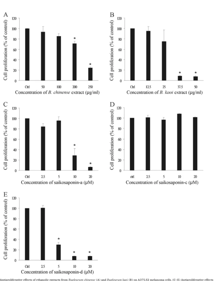
3.2. Ethanolic extracts from Bupleurum chinense and Bupleurum kaoi and saikosaponin-a and -d showed antiproliferative effects on human melanoma cells
As shown in Fig. 2A and B, ethanolic extracts from both B.
chinense and B. kaoi showed dose-dependent antiproliferative
ef-fects on human A375.S2 melanoma cells. Ethanolic extracts from
B.
kaoi showed more potent antiproliferative effect on A375.S2 cells
(effective concentration 50 μg/ml) compared to B. chinense (effec-tive concentration 250 μg/ml).
The bioactive compounds saikosaponin-a, -c and -d were also individually tested for antiproliferative activity on A375.S2 cells. As shown in Fig. 2C–E, saikosaponin-a (10 μM) and -d (5 μM) were cytotoxic to A375.S2 cells, while saikosaponin-c did not exhibit antiproliferative activity up to a concentration of 20 μM. Therefore, saikosaponin-d was the most potent compound in terms of anti-melanoma activity.
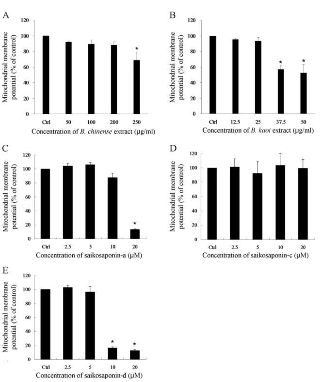
3.3. Ethanolic extracts from Bupleurum chinense and Bupleurum kaoi and saikosaponin-a and -d decreased mitochondrial membrane potential in human melanoma cells
As shown in Fig. 3, treatment of A375.S2 melanoma cells with ethanolic extracts from B. chinense and B. kaoi resulted in de-creased mitochondrial membrane potential. B. kaoi was more po-tent in reducing mitochondrial membrane popo-tential in A375.S2 cells (effective concentration 50 μg/ml) compared to B. chinense (effective concentration 250 μg/ml). In addition, the bioactive compounds saikosaponin-a and -d also effectively decreased mi-tochondrial membrane potential in A375.S2 cells, while saikosa-ponin-c did not have a significant effect on mitochondrial mem-brane potential. These results indicate that B. chinense, B. kaoi, and saikosaponin-a and -d induced human melanoma cell apoptosis through the mitochondrial pathway.
3.4. Physical properties of saikosaponin-d nanoparticles
Saikosaponin-d nanoparticles were synthesized using PVP as the excipient (carrier) and by grinding through a planetary ball mill. As shown in Table 1, the average size of the nanoparticles was 373.6748.7 nm with polydispersity index of 0.76, and the drug loading efficiency was 80.57%. The pure form saikosaponin-d compound exhibited significantly greater average particle size (11,066.77647.6 nm) and polydispersity (1) in water compared to the nanoparticle formulation. In addition, saikosaponin-d nano-particles showed 7.98 fold greater water solubility (360.26735.51 μg/ml) compared to the pure compound (45.16716.50 μg/ml). Therefore, the transformation of saikosapo-nin-d into a nanoparticle formulation effectively decreased parti-cle size and improved water solubility.
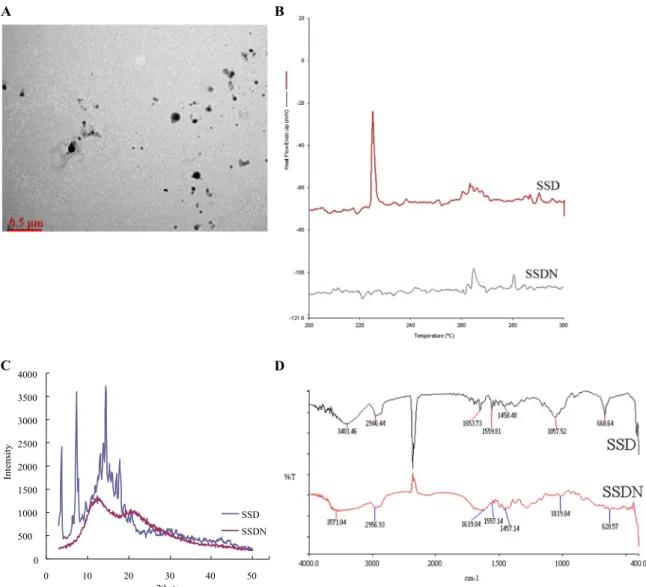
The transmission electron microscopy image of saikosaponin-d nanoparticles is shown in Fig. 4A. It can be seen that the nano-particles synthesized in this study were spherical in morphology with small uniform sizes.
3.5. Differential scanning calorimetry analysis
The result of the differential scanning calorimetry analysis is shown in Fig. 4B. It can be seen that the endothermic peak for pure saikosaponin-d occurred at 224 °C, indicating that this is the melting point for saikosaponin-d. On the other hand, there was no endothermic peak for the nanoparticle formulation of saikosapo-nin-d. This indicates that during the nanoparticle engineering process, saikosaponin-d had been dispersed throughout the ex-cipient, and transformed into a high-energy amorphous form.
3.6. X-ray diffractometry analysis
The result of the X-ray diffractometry analysis for saikosapo-nin-d and its nanoparticle formulation is shown in Fig. 4C. It can be seen that at diffraction angles of 3.7°, 7.3°, 14.5° and 17.8°,
saikosaponin-d showed characteristic diffraction peaks, indicating that pure saikosaponin-d has a highly crystalline structure. On the
Fig. 2. (A, B) Antiproliferative effects of ethanolic extracts from Bupleurum chinense (A) and Bupleurum kaoi (B) on A375.S2 melanoma cells. (C–E) Antiproliferative effects of saikosaponin-a (C), -c (D) and -d (E) on A375.S2 melanoma cells. Results are shown as mean 7 SD. Data are from 3 independent experiments with 3 replicates in each. *P o 0.05 versus control.
other hand, the nanoparticle formulation of saikosaponin-d had no diffraction peaks. This indicates that during the process of nano-particle formation, saikosaponin-d had been dispersed throughout the PVP polymers, transforming it from a crystalline into an amorphous form.
3.7. Fourier transform infrared spectroscopy analysis
Fourier transform infrared spectroscopy was used to evaluate the molecular interactions between saikosaponin-d and PVP. As shown in Fig. 4D, there were absorption bands for pure
Fig. 3. (A, B) Reduction of mitochondrial membrane potential in A375.S2 melanoma cells by ethanolic extracts from Bupleurum chinense (A) and Bupleurum kaoi (B). (C–E) Reduction of mitochondrial membrane potential in A375.S2 melanoma cells by saikosaponin-a (C), -c (D) and -d (E). Results are shown as mean7 SD. Data are from 3 independent experiments with 3 replicates in each. *P o 0.05 versus control.
Table 1
The size, polydispersity, drug loading efficiency and water solubility of saikosapo-nin-d nanoparticles (SSDN) compared to the pure saikosaposaikosapo-nin-d compound (SSD). Results are shown as mean 7SD. Data are from 3 independent experiments with 3 replicates in each.
saikosaponin-d at 3401 cm— 1
(indicating the presence of OH functional group), 2946 cm— 1
(CH2 functional group), 1653 cm— 1
(C¼ C(Alkene) functional group) and 668 cm— 1 (C–H on the
al-kene). On the other hand, saikosaponin-d nanoparticles showed
Size (nm) Polydispersity Drug loading
effi-ciency (%) Solubility (μg/ml) displacements of the absorption bands of these functional groups,
such as the OH functional group at 3571 cm— 1
. This indicates that PVP formed molecular bonds with the OH functional groups on
SSD 11,066.77647.6 1 – 45.16716.50
C
4000D
3500 3000 2500 2000 1500 1000 500 SSD SSDN 0 0 10 20 30 40 50A
B
2thetaFig. 4. (A) Morphology of saikosaponin-d nanoparticles under transmission electron microscopy. (B) Differential scanning calorimetry curves of saikosaponin-d and its nanoparticle formulation. (C) X-ray diffractometry patterns of saikosaponin-d and its nanoparticle formulation. (D) Fourier transform infrared spectra of saikosaponin-d and its nanoparticle formulation. Data are representative of three independent experiments. SSD¼ pure saikosaponin-d; SSDN¼ saikosaponin-d nanoparticles.
3.8. Effects of saikosaponin-d and its nanoparticle formulation on cell viability and mitochondrial membrane potential in human A375. S2 melanoma cells
As demonstrated in Fig. 5A, pure saikosaponin-d dissolved in PBS did not have any significant antiproliferative effect, while pure saikosaponin-d dissolved in DMSO and saikosaponin-d nano-particles dissolved in PBS showed antiproliferative effects on hu-man A375.S2 melanoma cells.
As shown in Fig. 5 B, pure saikosaponin-d dissolved in PBS did not have any significant effect on mitochondrial membrane po-tential, while pure saikosaponin-d dissolved in DMSO and saiko-saponin-d nanoparticles dissolved in PBS effectively decreased mitochrondrial membrane potential in A375.S2 cells. This in-dicates that saikosaponin-d nanoparticles may induce melanoma cell apoptosis through the mitochondrial pathway. Therefore, the transformation of saikosaponin-d into a nanoparticle formulation may enhance its antiproliferative and apoptotic effects on human melanoma cells, compared to the pure saikosaponin-d water (PBS) preparation.
3.9. Effects of saikosaponin-d and its nanoparticle formulation on expression of cell apoptosis proteins in human A375.S2 melanoma cells
In order to confirm whether the antiproliferative effect of sai-kosaponin-d may be due to induction of mitochondrial damage and cell apoptosis, we analyzed the effects of saikosaponin-d and its nanoparticle formulation on cell apoptosis proteins in A375.S2 melanoma cells. As shown in Fig. 6, saikosaponin-d nanoparticles dissolved in PBS (10 μM) induced p53 phosphorylation, and in-creased the levels of cytochrome c and cleaved caspase 9 in a time-dependent manner. Similar results were seen for pure saikosapo-nin-d dissolved in DMSO. Therefore, our results indicate that sai-kosaponin-d nanoparticles may mediate melanoma cell apoptosis by activation of p53, increased level of cytochrome c, and activa-tion of caspase 9.
3.10. Effects of saikosaponin-d and its nanoparticle formulation on MAPK signaling pathways in human A375.S2 melanoma cells
The activation of mitogen-activated protein kinase (MAPK)
In
te
ns
it
Fig. 5. Effects of pure saikosaponin-d (dissolved in PBS or DMSO) and saikosapo-nin-d nanoparticles (dissolved in PBS) on cell viability (A) and mitochondrial membrane potential (B) in A375.S2 melanoma cells. Results are shown as mean 7 SD. Data are from 3 independent experiments with 3 replicates in each. SSD¼ pure saikosaponin-d; SSDN¼ saikosaponin-d nanoparticles. *P o 0.05 versus control.
signaling pathways may be associated with cell apoptosis. As de-monstrated in Fig. 7, saikosaponin-d nanoparticles dissolved in PBS can effectively increase the phosphorylation of JNK and p38 in A375.S2 melanoma cells, but showed no effect on ERK phosphor-ylation. Similar results were seen for pure saikosaponin-d dis-solved in DMSO. Therefore saikosaponin-d nanoparticles induced JNK and p38 activation in human melanoma cells.
4. Discussion
Bupleurum species and their constituent saikosaponin com-pounds have been previously demonstrated to exhibit anticancer activity in various malignancies, including hepatocellular carci-noma (He et al., 2014; Jia et al., 2012), lung cancer (Hsu et al.,
2004a,b), thyroid cancer (Liu and Li, 2014) and prostate cancer
(Yao et al., 2014). However, their effects on skin cancers such as melanoma have not been previously investigated. In addition, the saikosaponin contents of various Bupleurum species and their relationship with anticancer activity have not been determined.
In this study, we demonstrated for the first time that both the traditional Chinese medicinal herb B. chinense and the Taiwanese endemic herb B. kaoi possess antiproliferative activity against human A375.S2 melanoma cells. In particular, B. kaoi shows more potent anticancer effect on melanoma cells compared to B.
chi-nense. In addition, we showed that the major bioactive compounds
in terms of anti-melanoma activity are saikosaponin-a and -d, while saikosaponin-c does not exhibit anti-melanoma activity up to a concentration of 20 μM. Therefore, Bupleurum species and saikosaponin-a and -d may have potential clinical use in the future as a form of treatment for melanoma.
We also showed that ethanolic extracts from B. chinense and B.
kaoi and saikosaponin-a and -d can effectively decrease
mi-tochondrial membrane potential in melanoma cells, while saiko-saponin-c has no significant effect. Previously, it has been de-monstrated that damaged mitochrondria can release cytochrome c into the cytoplasm, which combines with Apaf-1 in the presence of dATP to form the apoptosome, leading to activation of procas-pase 9 into casprocas-pase 9 (Adams and Cory, 2002, 2007). Therefore, the reduction in mitochrondrial membrane potential associated with mitochrondrial damage may induce cell apoptosis and explain the anti-melanoma activity of Bupleurum extracts and their biologi- cally active saikosaponin compounds.
Fig. 6. Effects of pure saikosaponin-d (dissolved in PBS or DMSO) and saikosaponin-d nanoparticles (dissolved in PBS) on proteins involved in cell apoptosis in A375.S2 melanoma cells, as determined by Western blotting. Control Western blots for DMSO and PVP (nanoparticle drug carrier) are also shown. The blots are representative of three independent experiments. The relative protein expression levels were quantified with densitometry (nindicates P o 0.05 versus time 0hr control). SSD¼pure
Fig. 7. Effects of pure saikosaponin-d (dissolved in PBS or DMSO) and saikosaponin-d nanoparticles (dissolved in PBS) on MAPK signaling pathways in A375.S2 melanoma cells, as determined by Western blotting. Control Western blots for DMSO and PVP (nanoparticle drug carrier) are also shown. The blots are representative of three independent experiments. The relative protein expression levels were quantified with densitometry (nindicates P o 0.05 versus time 0min control). SSD¼pure
saikosaponin-d; SSDN¼saikosaponin-d nanoparticles.
In order to determine the underlying reasons for the difference in anti-melanoma activity between B. chinense and B. kaoi, we analyzed the saikosaponin contents in the two Bupleurum species. HPLC analysis in this study demonstrated that B. kaoi contains 4.7 times greater amount of saikosaponin-a and 3.2 times greater amount of saikosaponin-d compared to B. chinense. This may ex-plain why B. kaoi shows more potent anti-melanoma activity compared to B. chinense. Therefore, the content of saikosaponin-a and -d may be used as an index to evaluate the anti-melanoma activity of different Bupleurum species.
Since saikosaponin-d is the most biologically active compound in terms of anti-melanoma activity, we performed further in-vestigations using this compound. Saikosaponin-d belongs to a class of triterpene saponins, which is characterized by poor water
solubility and dissolution properties. This may lead to poor oral bioavailability and thereby limit its future development for clinical applications (Mallick et al., 2007). In recent years, nanoparticle systems have emerged as a highly promising technology to im-prove a drug's water solubility and enhance drug delivery (Hu
et al., 2004; Devalapally et al., 2007; Brigger et al., 2002;
Merisko-Liversidge et al., 2003). Nanoparticles are stable colloidal particles
varying from 1 to 1000 nm in size, and are especially useful as a drug delivery system for poorly water-soluble compounds in-cluding various traditional Chinese herbal medicines (Yen et al., 2008, 2009; Huang et al., 2010). During the nanoparticle en-gineering process, polymers are used to entrap the free drug and act as carriers (excipients) for the drug. Polyvinylpyrrolidone (PVP) is a strongly hydrophilic polymer that is non-toxic and readily
dissolves in water (Karavas et al., 2007; Wu et al., 2009; Yen et al., 2010), and is therefore suitable as a nanoparticle drug carrier. In this study, we developed a novel saikosaponin-d nanoparticle system using PVP as the excipient.
Previous investigations have shown that the solubility of hy-drophobic compounds may be enhanced by improvement of their physical and chemical properties, including reduction of particle size and amorphous transformation (Pouton, 2006). We therefore performed further physicochemical characterization of the saiko-saponin-d nanoparticle system. Our results showed that nanoni-zation changed and improved the physicochemical properties of saikosaponin-d, including a decrease in particle size, conversion from crystalline into amorphous state, and formation of inter-molecular hydrogen bonds between drug and excipient. All these factors improve the water solubility and enhance the pharmaco-logical efficacy of the compound. Similar findings have also been reported for curcumin and quercetin nanoparticles (Yen et al., 2010; Wu et al., 2008).
In order to determine whether the nanoparticle formulation of saikosaponin-d possesses anti-melanoma activity, we performed cell viability studies in human melanoma cells. Our results re-vealed that pure saikosaponin-d dissolved in PBS did not show a significant anti-melanoma effect, which is likely due to its poor water solubility. On the other hand, saikosaponin-d nanoparticles dissolved in PBS showed significant antiproliferative effects on melanoma cells. Therefore, saikosaponin-d nanoparticles exhibit greater anti-melanoma activity compared to the pure saikosapo-nin-d water preparation. Dimethyl sulfoxide (DMSO) is often used in the laboratory to improve the solubility of hydrophobic drugs. Although pure saikosaponin-d dissolved in DMSO showed a si-milar antiproliferative effect on melanoma cells, this solvent is rarely used in the clinical setting due to its toxicity to human tissues (Windrum et al., 2005; Swanson, 1985; Willhite and Katz, 1984). Therefore, saikosaponin-d nanoparticles may be a poten-tially viable drug formulation for future clinical use as a treatment for melanoma.
To elucidate the molecular mechanisms for the antiproliferative effect of saikosaponin-d nanoparticles on human melanoma cells, we investigated whether the compound may reduce mitochon-drial membrane potential, which has been shown to be associated with mitochondrial damage and induction of apoptosis. Our re-sults revealed that saikosaponin-d nanoparticles can effectively decrease mitochondrial membrane potential in melanoma cells. In addition, we demonstrated by Western blotting that saikosaponin-d nanoparticles can insaikosaponin-duce phosphorylation of p53, ansaikosaponin-d increase the levels of cytochrome c and cleaved caspase 9. Previously, p53 has been shown to be a key regulator of the apoptotic cascade, and may activate the mitochondrial pathway (Chipuk and Green, 2006;
Murphy et al., 2004; Goh et al., 2014). During the process of apoptosis, cytochrome c is released from the mitochondria into the cytoplasm, leading to formation of the apoptosome and the con-version of procaspase 9 into caspase 9. Caspase 9 is an apoptosis initiator which can activate apoptosis effectors such as caspase 3, 6 and 7 (Hengartner, 2000). Therefore, our results indicate that saikosaponin-d nanoparticles may induce melanoma cell apopto-sis through the mitochondrial pathway.
The mitogen-activated protein kinase (MAPK) signaling path-way is involved in the control of cell proliferation, apoptosis, mi-gration and differentiation (Johnson and Lapadat, 2002). There are three major groups of MAPKs in humans: ERK, JNK and p38. ERK is activated in response to mitogenic stimuli, and usually mediates cell growth and survival. On the other hand, JNK and p38 can be activated by various forms of cellular stress, and may regulate downstream p53 expression and induce cell apoptosis (Wu, 2004;
Lin, 2003; Davis, 2000). In this study, we found that treatment with saikosaponin-d nanoparticles leads to activation of JNK and
p38 in melanoma cells, which is consistent with the drug's apoptotic effects. In contrast, saikosaponin-d nanoparticles had no significant effect on ERK phosphorylation.
5. Conclusions
In conclusion, this study has shown for the first time that ethanolic extracts from B. kaoi have more potent antiproliferative effect on human melanoma cells compared to B. chinense. This is likely due to the higher contents of saikosaponin-a and -d in B.
kaoi compared to B. chinense. Saikosaponin-d is the most potent
compound in Bupleurum species in terms of anti-melanoma ac-tivity, and the novel saikosaponin-d nanoparticle system devel-oped in this study shows improved physicochemical properties and increased water solubility due to lowered particle size, amorphous transformation and intermolecular hydrogen bond formation with the excipient. Furthermore, saikosaponin-d nano-particles have enhanced anti-melanoma activity, and induce apoptosis of melanoma cells through the mitochondrial pathway. This may be mediated by activation of JNK and p38, activation of p53, increased level of cytochrome c, and activation of caspase 9. Therefore, Bupleurum species and saikosaponin-d nanoparticles may have potential use in the future as a form of treatment for melanoma.
Conflict of interest
The authors have no conflict of interest to declare.
Acknowledgments
This work was supported by a grant from the Ministry of Sci-ence and Technology, Taiwan (MOST 102-2313-B-037-001-).
References
Adams, J.M., Cory, S., 2002. Apoptosomes: engines for caspase activation. Curr. Opin. Cell Biol. 14, 715 – 720 .
Adams, J.M., Cory, S., 2007. The Bcl-2 apoptotic switch in cancer development and therapy. Oncogene 26, 1324 – 1337 .
Ashour, M.L., Wink, M., 2011. Genus Bupleurum : a review of its phytochemistry, pharmacology and modes of action. J. Pharm. Pharmacol. 63, 305 – 321 . Brigger, I., Dubernet, C., Couvreur, P., 2002. Nanoparticles in cancer therapy and
diagnosis. Adv. Drug Deliv. Rev. 54, 631 – 651 .
Cheng, P.W., Ng, L.T., Chiang, L.C., Lin, C.C., 2006. Antiviral effects of saikosaponins on human coronavirus 229E in vitro. Clin. Exp. Pharmacol. Physiol. 33, 612 – 616 . Chipuk, J.E., Green, D.R., 2006. Dissecting p53-dependent apoptosis. Cell Death
Differ. 13, 994 – 1002 .
Damsky, W.E., Theodosakis, N., Bosenberg, M., 2014. Melanoma metastasis: new concepts and evolving paradigms. Oncogene 33, 2413 – 2422 .
Davis, R.J., 2000. Signal transduction by the JNK group of MAP kinases. Cell 103, 239 – 252 .
Devalapally, H., Chakilam, A., Amiji, M.M., 2007. Role of nanotechnology in phar- maceutical product development. J. Pharm. Sci. 96, 2547 – 2565 .
Dicheva, B.M., Koning, G.A., 2014. Targeted thermosensitive liposomes: an attractive novel approach for increased drug delivery to solid tumors. Expert Opin. Drug Deliv. 11, 83 – 100 .
Dinda, S.C., Pattnaik, G., 2013. Nanobiotechnology-based drug delivery in brain targeting. Curr. Pharm. Biotechnol. 14, 1264 – 1274 .
Eggermont, A.M., Spatz, A., Robert, C., 2014. Cutaneous melanoma. Lancet 383, 816 – 827 .
Goh, B.H., Chan, C.K., Kamarudin, M.N., Abdul Kadir, H., 2014. Swietenia macro- phylla King induces mitochondrial-mediated apoptosis through p53 upregu- lation in HCT116 colorectal carcinoma cells. J. Ethnopharmacol. 153, 375 – 385 .
Gray-Schopfer, V., Wellbrock, C., Marais, R., 2007. Melanoma biology and new targeted therapy. Nature 445, 851 – 857 .
Guadagnolo, B.A., Zagars, G.K., 2009. Adjuvant radiation therapy for high-risk nodal metastases from cutaneous melanoma. Lancet Oncol. 10, 409 – 416 .
Saikosaponin-d suppresses the expression of cyclooxygenase-2 through the phospho-signal transducer and activator of transcription 3/hypoxia-inducible factor-1 α path- way in hepatocellular carcinoma cells. Mol. Med. Rep. 10, 2556 – 2562 . Hengartner, M.O., 2000. The biochemistry of apoptosis. Nature 407, 770 – 776 . Hsu, Y.L., Kuo, P.L., Lin, C.C., 2004a. The proliferative inhibition and apoptotic
me-chanism of Saikosaponin D in human non-small cell lung cancer A549 cells. Life Sci. 75, 1231 – 1242 .
Hsu, Y.L., Kuo, P.L., Weng, T.C., Yen, M.H., Chiang, L.C., Lin, C.C., 2004b. The anti- proliferative activity of saponin-enriched fraction from Bupleurum Kaoi is through Fas-dependent apoptotic pathway in human non-small cell lung can- cer A549 cells. Biol. Pharm. Bull. 27, 1112 – 1115 .
Hu, J., Johnston, K.P., Williams 3rd, R.O., 2004. Nanoparticle engineering processes for enhancing the dissolution rates of poorly water soluble drugs. Drug Dev. Ind. Pharm. 30, 233 – 245 .
Huang, Q., Yu, H., Ru, Q., 2010. Bioavailability and delivery of nutraceuticals using nanotechnology. J. Food Sci. 75, R50 – R57 .
Jia, X., Dang, S., Cheng, Y., Zhang, X., Li, M., Li, Y., Li, S., 2012. Effects of saikosaponin- d on syndecan-2, matrix metalloproteinases and tissue inhibitor of metallo- proteinases-2 in rats with hepatocellular carcinoma. J. Tradit. Chin. Med. 32, 415 – 422 .
Johnson, G.L., Lapadat, R., 2002. Mitogen-activated protein kinase pathways mediated by ERK, JNK, and p38 protein kinases. Science 298, 1911 – 1912 . Kang, S.J., Lee, Y.J., Kim, B.M., Kim, Y.J., Woo, H.D., Jeon, H.K., Chung, H.W., 2008.
Effect of Bupleuri Radix extracts on the toxicity of 5- fluorouracil in HepG2 he- patoma cells and normal human lymphocytes. Basic Clin. Pharmacol. Toxicol. 103, 305 – 313 .
Karavas, E., Georgarakis, M., Docoslis, A., Bikiaris, D., 2007. Combining SEM, TEM, and micro-Raman techniques to differentiate between the amorphous mole- cular level dispersions and nanodispersions of a poorly water-soluble drug within a polymer matrix. Int. J. Pharm. 340, 76 – 83 .
Kato, M., Isobe, K., Dai, Y., Liu, W., Takahashi, M., Nakashima, I., 2000. Further characterization of the Sho-saiko-to-mediated anti-tumor effect on melanoma developed in RET-transgenic mice. J. Investig. Dermatol. 114, 599 – 601 . Law, B.Y., Mo, J.F., Wong, V.K., 2014. Autophagic effects of Chaihu (dried roots of
Bupleurum Chinense DC or Bupleurum scorzoneraefolium WILD). Chin. Med. 9,
21 .
Li, X., Sun, M., Zhao, Z., Yang, J., Chen, K., 2014. Research on effect of minor Bu- pleurum decoction of proliferation and apoptosis of esophageal cancer cell strain eca-109 cell. Pak. J. Pharm. Sci. 27 (Suppl. 5), S1675 – S1679 . Lin, A., 2003. Activation of the JNK signaling pathway: breaking the brake on
apoptosis. Bioessays 25, 17 – 24 .
Liu, R.Y., Li, J.P., 2014. Saikosaponin-d inhibits proliferation of human un- differentiated thyroid carcinoma cells through induction of apoptosis and cell cycle arrest. Eur. Rev. Med. Pharmacol. Sci. 18, 2435 – 2443 .
Liu, A., Tanaka, N., Sun, L., Guo, B., Kim, J.H., Krausz, K.W., Fang, Z., Jiang, C., Yang, J., Gonzalez, F.J., 2014. Saikosaponin d protects against acetaminophen-induced hepatotoxicity by inhibiting NF- κ B and STAT3 signaling. Chem. Biol. Interact. 223C, 80 – 86 .
Lo, J.A., Fisher, D.E., 2014. The melanoma revolution: from UV carcinogenesis to a new era in therapeutics. Science 346, 945 – 949 .
Mallick, S., Pattnaik, S., Swain, K., De, P.K., 2007. Current perspectives of solubili- zation: potential for improved bioavailability. Drug Dev. Ind. Pharm. 33, 865 – 873 .
Merisko-Liversidge, E., Liversidge, G.G., Cooper, E.R., 2003. Nanosizing: a formula- tion approach for poorly-water-soluble compounds. Eur. J. Pharm. Sci. 18, 113 – 120 .
Mignet, N., Seguin, J., Chabot, G.G., 2013. Bioavailability of polyphenol liposomes: a challenge ahead. Pharmaceutics 5, 457 – 471 .
Murphy, M.E., Leu, J.I., George, D.L., 2004. p53 moves to mitochondria: a turn on the path to apoptosis. Cell Cycle 3, 836 – 839 .
Pouton, C.W., 2006. Formulation of poorly water-soluble drugs for oral adminis- tration: physicochemical and physiological issues and the lipid formulation classi fi cation system. Eur. J. Pharm. Sci. 29, 278 – 287 .
Swanson, B.N., 1985. Medical use of dimethyl sulfoxide (DMSO). Rev. Clin. Basic Pharm. 5, 1 – 33 .
Thompson, J.F., Scolyer, R.A., Kefford, R.F., 2005. Cutaneous melanoma. Lancet 365, 687 – 701 .
Willhite, C.C., Katz, P.I., 1984. Toxicology updates. Dimethyl sulfoxide. J. Appl. Tox- icol. 4, 155 – 160 .
Windrum, P., Morris, T.C., Drake, M.B., Niederwieser, D., Ruutu, T., EBMT Chronic Leukaemia Working Party Complications Subcommittee, 2005. Variation in dimethyl sulfoxide use in stem cell transplantation: a survey of EBMT centres. Bone Marrow Transplant 36, 601 – 603 .
Wong, V.K., Zhou, H., Cheung, S.S., Li, T., Liu, L., 2009. Mechanistic study of saiko- saponin-d (Ssd) on suppression of murine T lymphocyte activation. J. Cell. Biochem. 107, 303 – 315 .
Wu, G.S., 2004. The functional interactions between the p53 and MAPK signaling pathways. Cancer Biol. Ther. 3, 156 – 161 .
Wu, K., Li, J., Wang, W., Winstead, D.A., 2009. Formation and characterization of solid dispersions of piroxicam and polyvinylpyrrolidone using spray drying and precipitation with compressed antisolvent. J. Pharm. Sci. 98, 2422 – 2431 . Wu, T.H., Yen, F.L., Lin, L.T., Tsai, T.R., Lin, C.C., Cham, T.M., 2008. Preparation, phy-
sicochemical characterization, and antioxidant effects of quercetin nano- particles. Int. J. Pharm. 346, 160 – 168 .
Yano, H., Mizoguchi, A., Fukuda, K., Haramaki, M., Ogasawara, S., Momosaki, S., Kojiro, M., 1994. The herbal medicine sho-saiko-to inhibits proliferation of cancer cell lines by inducing apoptosis and arrest at the G0/G1 phase. Cancer Res. 54, 448 – 454 .
Yao, M., Yang, J., Cao, L., Zhang, L., Qu, S., Gao, H., 2014. Saikosaponin-d inhibits proliferation of DU145 human prostate cancer cells by inducing apoptosis and arresting the cell cycle at G0/G1 phase. Mol. Med. Rep. 10, 365 – 372 . Yen, F.L., Wu, T.H., Lin, L.T., Cham, T.M., Lin, C.C., 2008. Nanoparticles formulation
of Cuscuta chinensis prevents acetaminophen-induced hepatotoxicity in rats.
Food Chem. Toxicol. 46, 1771 – 1777 .
Yen, F.L., Wu, T.H., Lin, L.T., Cham, T.M., Lin, C.C., 2009. Naringenin-loaded nano- particles improve the physicochemical properties and the hepatoprotective effects of naringenin in orally-administered rats with CCl(4)-induced acute liver failure. Pharm. Res. 26, 893 – 902 .
Yen, F.L., Wu, T.H., Tzeng, C.W., Lin, L.T., Lin, C.C., 2010. Curcumin nanoparticles improve the physicochemical properties of curcumin and effectively enhance its antioxidant and antihepatoma activities. J. Agric. Food Chem. 58, 7376 – 7382 . Yen, M.H., Weng, T.C., Liu, S.Y., Chai, C.Y., Lin, C.C., 2005. The hepatoprotective effect
of
Bupleurum kaoi , an endemic plant to Taiwan, against dimethylnitrosamine-induced hepatic fibrosis in rats. Biol. Pharm. Bull. 28, 442 – 448 .
Ying, Z.L., Li, X.J., Dang, H., Wang, F., Xu, X.Y., 2014. Saikosaponin-d affects the differentiation, maturation and function of monocyte-derived dendritic cells. Exp. Ther. Med. 7, 1354 – 1358 .
Zhang, B.Z., Guo, X.T., Chen, J.W., Zhao, Y., Cong, X., Jiang, Z.L., Cao, R.F., Cui, K., Gao, S.S., Tian, W.R., 2014. Saikosaponin-D attenuates heat stress-induced oxidative damage in LLC-PK1 cells by increasing the expression of anti-oxidant enzymes and HSP72. Am. J. Chin. Med. 42, 1261 – 1277 .