行政院國家科學委員會專題研究計畫 成果報告
雙層仿生骨組織支架材之設計及分析(第 3 年) 研究成果報告(完整版)
計 畫 類 別 : 個別型
計 畫 編 號 : NSC 98-2221-E-040-006-MY3
執 行 期 間 : 100 年 08 月 01 日至 101 年 07 月 31 日 執 行 單 位 : 中山醫學大學口腔科學研究所
計 畫 主 持 人 : 丁信智 共 同 主 持 人 : 陳震漢
計畫參與人員: 博士班研究生-兼任助理人員:何佳哲 博士班研究生-兼任助理人員:魏忠楷
報 告 附 件 : 出席國際會議研究心得報告及發表論文
公 開 資 訊 : 本計畫涉及專利或其他智慧財產權,2 年後可公開查詢
中 華 民 國 101 年 09 月 27 日
中 文 摘 要 : 組織工程的目的是為克服因捐贈者短缺的移植難題,及突破 人工材料不能夠取代所有受傷或缺損的器官或組織。人工合 成支架材料必須具備生物相容性,且與骨組織相近的結構及 機械性。骨組織是一種複合材,主要是由有機相(膠原蛋 白)與無機相(HA)構成。是以成功的取代材設計應以骨組 織本質為藍圖。本計劃的主要目標是結合天然高分子及矽酸 鈣,製造骨組織工程用之新式仿生支架,研究支架設計對仿 生複合材機械及生物性能之影響。成功製造出機械強度高達 140 MPa 與彈性係數 2.3 GPa 的仿生矽酸鈣-明膠複合體(明 膠含量 10 wt%)。分析生物活性將材料浸泡到模擬體液之 後,1 天內即形成似骨磷灰石,然複合體逐漸損失強度。180 天之後,明膠含量 5 及 10 wt%的複合體分別損失 30 及 47%的 強度。僅管如此,明膠提供有利的細胞環境及減少免疫表 現。牙髓細胞培養於複合體後其增生、分化與礦化增加。整 體而言,明膠含量 10 wt%的仿生矽酸鈣-明膠複合體具高強 度、無毒性及骨形成性等特質,可作為承載負荷得骨移植 材。
中文關鍵詞: 骨組織工程、矽酸鈣、明膠、支架
英 文 摘 要 : Tissue engineering has emerged as a promising approach without the limitations of donor shortage and of synthetic prostheses that are not able to replace all the functions of a damaged or lost organ or tissue. A synthetic material used as scaffold must be biocompatible and it should exhibit some
structural and mechanical equivalence to bone. Bone is a composite material and consists mainly of an organic matrix (collagen) and a mineral phase
(hydroxyapatite). Thus, the successful design of bone substitute materials should be an analogue of the bone structure. In this project, we aim at
fabricating an advanced biomimetic scaffold that combines natural polymers and calcium silicates
(CaSi) for use in bone tissue engineering. The effect of design strategies on mechanical and biological performances of the hybrid scaffold is investigated.
We successfully fabricated the biomimetic 10 wt%
gelatin-containing CaSi composites with high compressive strength up to 140 MPa and elastic modulus of 2.3 GPa. The materials precipitated a
‘bone-like’ apatite after soaking in SBF for 1 day.
However, all bone grafts gradually lost their strengths with the increase in soaking time. The composites containing 5 and 10 wt% gelatin lost 30 and 47% in compressive strength, respectively, after 180-day immersion. Nevertheless, the presence of gelatin promoted greater cell attachment and proliferation on the composite bone grafts. Pulp cells on the calcium silicate-gelatin bone grafts expressed higher levels of osteocalcin, osteopontin, and bone sialoprotein. The inhibition of inducible nitric oxide synthase and interleukin-1 expression and the activation of interleukin-10 were increased with increasing gelatin content. Overall, these findings provide evidence that composite bone grafts containing 10 wt % gelatin with a high initial
strength were bioactive, nontoxic, and osteogenic and may be able to promote bone healing for load-bearing applications.
英文關鍵詞: Bone tissue engineering, calcium silicate, gelatin, scaffold
目錄
1. 中文摘要 --- 3
2. 英文摘要 --- 3
3. 緣由與目的 --- 3
4. 實驗方法--- 4
4.1. Preparation of the composite --- 4
4.2. Preparation of SBF --- 5
4.3. Evaluation of physicochemical properties --- 5
4.4. Measurement of mechanical properties --- 6
4.5 In vitro bioactivity and degradation --- 6
4.6. Cytotoxicity --- 7
4.7. Primary cell culture --- 7
4.8. Statistical analysis --- 8
5. 結果與討論--- 8
5.1. Gelatin distribution and porosity --- 8
5.2. Phase composition --- 8
5.3. Morphology --- 9
5.4. Compressive properties --- 9
5.5. Weibull analysis ---11
5.6. Hardness --- 11
5.7. In vitro fatique --- 12
5.8. In vitro bioactivity and degradation --- 12
5.9. Cytotoxicity --- 14
5.10. Cell attachment and proliferation--- 15
5.11. Cell differentiation--- 15
5.12. Osteogenesis --- 16
5.13. Immune response ---17
6. 結 論--- 17
7. 成果自評--- 18
8. 參考文獻--- 18
Table 1 --- 19
Figure 1--- 20
Figure 2--- 20
Figure 3--- 20
Figure 4--- 21
Figure 5--- 21
Figure 6--- 22
Figure 7--- 22
Figure 8--- 23
Figure 9--- 24
Figure 10 --- 25
Figure 11--- 25
Figure 12--- 25
Figure 13--- 26
Figure 14--- 26
Figure 15--- 26
Figure 16--- 27
Figure 17--- 28
1. 中文摘要
組織工程的目的是為克服因捐贈者短缺的移植難題,及突破人工材料不能夠取代所 有受傷或缺損的器官或組織。人工合成支架材料必須具備生物相容性,且與骨組織相近 的結構及機械性。骨組織是一種複合材,主要是由有機相(膠原蛋白)與無機相
(HA)構成。是以成功的取代材設計應以骨組織本質為藍圖。本計劃的主要目標是結 合天然高分子及矽酸鈣,製造骨組織工程用之新式仿生支架,研究支架設計對仿生複合 材機械及生物性能之影響。成功製造出機械強度高達140 MPa與彈性係數2.3 GPa的仿生 矽酸鈣-明膠複合體(明膠含量10 wt%)。分析生物活性將材料浸泡到模擬體液之後,1 天內即形成似骨磷灰石,然複合體逐漸損失強度。180天之後,明膠含量5及10 wt%的 複合體分別損失30及47%的強度。僅管如此,明膠提供有利的細胞環境及減少免疫表 現。牙髓細胞培養於複合體後其增生、分化與礦化增加。整體而言,明膠含量10 wt%
的仿生矽酸鈣-明膠複合體具高強度、無毒性及骨形成性等特質,可作為承載負荷得骨 移植材。
關鍵詞:骨組織工程、矽酸鈣、明膠、支架 2. 英文摘要
Tissue engineering has emerged as a promising approach without the limitations of donor shortage and of synthetic prostheses that are not able to replace all the functions of a damaged or lost organ or tissue. A synthetic material used as scaffold must be biocompatible and it should exhibit some structural and mechanical equivalence to bone. Bone is a composite material and consists mainly of an organic matrix (collagen) and a mineral phase (hydroxyapatite). Thus, the successful design of bone substitute materials should be an analogue of the bone structure. In this project, we aim at fabricating an advanced biomimetic scaffold that combines natural polymers and calcium silicates (CaSi) for use in bone tissue engineering. The effect of design strategies on mechanical and biological performances of the hybrid scaffold is investigated. We successfully fabricated the biomimetic 10 wt% gelatin- containing CaSi composites with high compressive strength up to 140 MPa and elastic modulus of 2.3 GPa. The materialsprecipitated a‘bone-like”apatiteaftersoaking in SBF for 1 day. However, all bone grafts gradually lost their strengths with the increase in soaking time.
The composites containing 5 and 10 wt% gelatin lost 30 and 47% in compressive strength, respectively, after 180-day immersion. Nevertheless, the presence of gelatin promoted greater cell attachment and proliferation on the composite bone grafts. Pulp cells on the calcium silicate-gelatin bone grafts expressed higher levels of osteocalcin, osteopontin, and bone sialoprotein. The inhibition of inducible nitric oxide synthase and interleukin-1 expression and the activation of interleukin-10 were increased with increasing gelatin content. Overall, these findings provide evidence that composite bone grafts containing 10 wt % gelatin with a high initial strength were bioactive, nontoxic, and osteogenic and may be able to promote bone healing for load-bearing applications.
Keywords: Bone tissue engineering, calcium silicate, gelatin, scaffold 3. Introduction and purpose
A variety of bone replacement materials have been developed and the majority has been limited to non-load bearing applications. Bone grafts are necessary to provide support, fill voids, and enhance biologic repair of skeletal defects. Calcium phosphates, particularly
hydroxyapatite (HA), have been used as implant materials for bone repair and regeneration due to their chemical compositions being similar to the inorganic component of bone. However, the poorly compressive strength and fatigue failure limit its applicability to the low or non load-bearing sites in human body. On the other hand, it must be emphasized at this point that the successful design of a bone substitute material requires an appreciation of the structure of bone. Bone is a composite structure consisting of nanocrystals incorporated within a collagen matrix. Thus, the use of a hybrid composite that comprises polymer and ceramic most aptly resembles the morphology and properties of natural bone. This may be one way to solve the problem of ceramics, such as brittleness, without reducing mechanical properties, in addition to possessing good biocompatibility, high bioactivity, and great bone-bonding capability.
Extensive research has been carried out in this regard and composite materials based on calcium phosphate and a variety of polymers have been worked out. To design biomimetic materials, it is important to understand the mechanical properties of bone materials and the structural relationship between them at the various levels of hierarchical structural organization. At the macrostructure level, bone is distinguished into the cortical (or compact) and cancellous (or trabecular) types. The compressive strength and modulus of cortical bone are about 100–230 MPa and 7–30 GPa, respectively [1]. Cortical bone has a solid structure with a series of voids that result in 3–12% porosity [2]. Since the bone tissue is a two-phase composite comprised mainly of apatite and collagen, the amount of mineral is usually thought to determine the stiffness of the bone material. The properties and geometrical arrangement of the two components might have a much larger influence on the properties than traditionally assumed.
In previous studies [3−7], a fast-setting calcium silicate cement (CSC) consisting of powders containing one or more solid compounds of calcium silicate and water or a phosphate buffer solution have been developed. The cement, which was initially derived from an equimolar mixture of tetraethyl orthosilicate and calcium nitrate via a sol-gel method, hardened in about 20 min after mixing the powder with water. The purpose of this three-year project is to develop strong scaffolds by incorporating gelatin into calcium silicate. In particular, efforts have been oriented toward calcium silicates where we added gelatin to reinforce the mechanical properties. The appropriate amount of gelatin added to the composite cements provided the greater resistance to disintegration in simulated physiological solution.
Following the first year of this project, the biological properties of biomimetic gelatin- containing calcium silicate composites were evaluated in the 2nd-year progress.
4. Experimental
4.1. Preparation of the composite
The sol-gel method has been described elsewhere [7]. Reagent-grade tetraethyl orthosilicate (Si(OC2H5)4, TEOS, 98.0%) (Sigma-Aldrich, St. Louis, MO) and calcium nitrate (Ca(NO3)2·4H2O, 98.0%) (Showa, Tokyo, Japan) were used as precursors for SiO2 and CaO, respectively. Nitric acid was used as the catalyst and ethanol was used as the solvent. The general sol-gel procedure, including hydrolysis and aging, was adopted. Briefly, TEOS was hydrolyzed by the sequential addition of 2 M HNO3 and absolute ethanol, with 1 h of stirring after each addition. Ca(NO3)2·4H2O was added to the TEOS solution in an equimolar ratio, and the mixture was stirred for an additional 1 h. The molar ratio of (HNO3 + H2O)-TEOS- ethanol was 10:1:10. The sol solution was sealed and aged at 60ºC for 1 day. After vaporization of the solvent in an oven at 120C, the dried gel was heated in air to 800C at a heating rate of 10C/min for 2 h using a high-temperature furnace and then cooled to room
temperature in the furnace to produce a powder. The sintered powders were then ball-milled for 12 h in ethyl alcohol using a Retsch S 100 centrifugal ball mill (Hann, Germany) and dried in an oven at 60C. Type B gelatin (isoelectric point at pH = 4.7−5.2) from bovine skin (Sigma-Aldrich) was weighed and dissolved in distilled water at 60C until a homogeneous gelatin solution was obtained. To fabricate the organic-inorganic composite, the calcium silicate (CaSi) powder was mixed with different gelatin solutions (10%, 20%, and 30%) at a powder-to-liquid ratio of 2 mg mL-1 using a conditioning mixer (ARE-250, Thinky, Tokyo, Japan), and then the mixture was dried at room temperature for 12 h. Herein, the ratios of gelatin to CaSi were approximately 5, 10, and 15% by weight (Table 1). After grinding the dried powders the dense bulks were obtained by molding the specimens with an aspect ratio of 2:1 (6 mm in diameter 12 mm in length) in a cylindrical stainless steel mold under the applied pressure of 500 MPa for 1 min using a uniaxial press, followed by being soaked in deionized water at 60C for 1 h for hydrothermal processing prior to being dried at 60C for 2 days in an oven, unless described otherwise. For comparison purposes, specimens without hydrothermal processing in water at 60C for 1 h were also prepared.
4.2. Preparation of SBF
The simulated body fluid (SBF) solution, an extracellular solution with an ionic composition similar to that of human blood plasma, was used as the supporting solution for the compressive property measurement and fatigue tests. It consists of 7.9949 g of NaCl, 0.3528 g of NaHCO3, 0.2235 g of KCl, 0.147 g of K2HPO4, 0.305 g of MgCl2·6H2O, 0.2775 g of CaCl2, and 0.071 g of Na2SO4 in 1000 mL of distilled H2O and was buffered to pH 7.4 with hydrochloric acid (HCl) and trishydroxymethyl amino methane (Tris, CH2OH)3CNH2). All of the chemicals used were of reagent grade and used as obtained.
4.3. Evaluation of physicochemical properties
The uniformity of the gelatin distribution in the composites was measured by burnout in air at 600ºC for 2 h. Each composite bulk was cut into five parts using a low-speed diamond saw (Isomet, Buehler, Lake Bluff, IL). The specimens were dried at 120ºC for 2 h and then were weighed before and after burnout using a four-digital balance (AE 240S, Mettler-Toledo AG, Greifensee, Switzerland). Five samples were tested for each condition. The measurement of the density and porosity was conducted using a liquid displacement technique. In this method, ethanol was used as the displacement liquid because water was the setting liquid. A total of five specimens were used in each measurement to enhance the precision. The average value of six measurements was taken as the porosity and density of the specimens. Before the measurement, the set specimens were dried in an oven at 120C for 2 h. The specimen was immersed in a graduated cylinder containing a known volume (V1) of ethanol. Afterwards, it was ultrasonically stirred for at least 3 min to force the ethanol into the pores of the specimens until no air bubbles were observed emerging from the specimen. The total volume of ethanol and the ethanol-impregnated specimen was then recorded as V2. The ethanol-impregnated specimen was removed from the cylinder, and the residual ethanol volume was recorded as V3.
The volume differences, (V1 –V3) and (V2 −V3), were the pore volume and total volume of the specimen, respectively. Thus, the porosity of the specimen was obtained by the following equation:
Porosity = (V1 − V3)/(V2 − V3) (1)
The density can be expressed by the equation: d = W/(V2 − V3), where W is the weight of the specimen. Phase analysis of the specimens was performed using an X-ray diffractometer
(XRD, Shimadzu XD-D1, Kyoto, Japan) operated at 30 kV and 30 mA at a scanning speed of 1/min. Micro-Raman measurements were taken with a DXR instrument (Thermo Scientific, Waltham, MA) equipped with a laser at 780 nm, a microscope with ×10 magnification, and an electrically refrigerated CCD camera. The laser spot size was approximately 5 μm. The specimens for the interior structure examination were prepared by cutting with a diamond saw.
The interiors and surfaces of the specimens were dried with liquid CO2 using a critical point dryer device (LADD 28000, LADD, Williston, VT) and then coated with gold using a JFC- 1600 (JEOL, Tokyo, Japan) coater and examined under a scanning electron microscope (SEM, JSM-7401F, JEOL) operated in the lower secondary electron image (LEI) mode at 3 kV of accelerating voltage.
4.4. Measurement of mechanical properties
The compressive strength (CS) was measured in an SBF at a crosshead speed of 1 mm min-1 using a static mechanical testing machine AG-1000E (Shimadzu, Kyoto, Japan) with a 10 kN load cell. The CS value of each specimen was calculated using the relationship defined in the equation CS = P/πr2, where P is the peak load (Newtons, N) and r is the radius (mm) of the specimen. The maximal compression load at failure was obtained from the recorded load- deflection curves.Young’smodulusofthespecimens was determined from the slope of the initial linear elastic portion of the load-deflection curve. The work-of-fracture (toughness, kJ m-2) was calculated by taking the area under the load-deflection curve. For the statistical significance of the two-parameter Weibull analysis, twenty specimens from each group were tested. A digital microhardness tester (HMV-2000, Shimaduz, Kyoto, Japan) with a four-sided diamond pyramid was used to evaluate the hardness of the various specimens. A load of 4.9 N for 15 s in an air atmosphere was used. The indentation impressions were also examined using SEM to observe the change in the damage associated with indentation. The average value was determined from twenty collections. For in vitro fatique test the specimen was placed in a polystyrene container, to which SBF in a dynamic (continuous exchange of solution) condition using a peristaltic pump at 37ºC was added to completely cover the specimen. A fatigue cyclic loading lower than the maximal compression loading at failure with a stress ratio of Smin/Smax
= 0.1 was imposed at 5 Hz until fracture was achieved using a Shimadzu servopulser 48000 system (Kyoto, Japan). The number of cycles to failure under a cyclic compression condition was promptly recorded upon the specimen rupture. Three specimens from each group were tested.
4.5 In vitro bioactivity and degradation
To evaluate the in vitro physicochemical activity, the specimens were immersed in a 37 ºC 30 mL SBF solution, equivalent to a specimen surface-to-volume ratio of 0.1 cm-1, which was arbitrarily chosen to completely cover each surface of the specimen. After soaking for specific time duration (15, 30, 90, and 180 days), specimens were removed from the vials and their properties were evaluated. Samples were removed from the vials and evaluated for CS or were dried in an oven at 60 ºC for the analysis of weight loss, phase composition, and morphology. To monitor the weight change of the samples, the dried specimens were weighed until reaching a constant weight before (day 0) and after immersion using a four-digit balance (AE 240S, Mettler-Toledo AG, Greifensee, Switzerland). Eight repeated specimens were examined for each of the materials investigated at each time point. At each time point, the pH values of the SBF solutions were measured using a pH meter (SP-701, Suntex Instruments, Taipei, Taiwan). Eight measurements were used.
4.6. Cytotoxicity
The cytotoxicity of the bone grafts were evaluated by incubating the specimens with L929 mouse fibroblast cells (BCRC 60279, Hsinchu, Taiwan) for 12, 24, and 48 h. Prior to cell incubation, the specimens were sterilized by soaking each in a 75% ethanol solution and exposing to UV lightfor2 h.TheL929 cellsweresuspended in Dulbecco’sModified Eagle’s medium (DMEM; Gibco, Langley, OK) containing 10% fetal bovine serum (FBS; Gibco) and 1% penicillin/streptomycin solution (Gibco). Cell suspensions (5 103 cells per well) were directly seeded over each of the specimens in a 96-well plate. The segmented polyurethane films containing 0.1% zinc diethyldithiocarbamate (RM-A) were used as positive standard reference materials, and a high-density polyethylene sheet (RM-C) was used as the negative standard reference material, based on ISO 10993-5. The two standard reference materials were purchased from Hatano Research Institute, Food and Drug Safety Center (Kanagawa, Japan).
After the established L929-cell incubation period, the cytotoxicity was examined using the alamarBlue assay (Invitrogen, Grand Island, NY), which is based on the detection of mitochondrial activity. One microliter of alamarBluesolution and 100 μL of DMEM were added to each well followed by 2 h of incubation. After incubation, the solution in each well was transferred to a new 96-well ELISA plate. Plates were read in a Sunrise microtiter plate reader (Tecan Austria Gesell- schaft, Salzburg, Austria) at 570 nm with a reference wavelength of 600 nm. Optical density (OD) results were obtained from three separate experiments.
4.7. Primary cell culture
Human dental pulp cells were freshly derived from an intact caries-free premolar that was extracted for orthodontic treatment purposes. The tooth was split sagittally with a chisel, and the periodontal ligament tissue was then immersed in phosphate-buffered saline (PBS) solution. The pulp tissue was cut into fragments and immersed in DMEM containing 0.1%
collagenase (Sigma) and 0.1% Dispase (Sigma) for 1 h. The pulp cells were collected from the medium by centrifugation at 1500 rpm for 5 min. The cell pellet was resuspended in DMEM containing 20% FBS, 100 units/mL penicillin G, and 100 mg/mL streptomycin in 5% CO2 at 37 ºC. Cells were subcultured by successive passaging at a 1:3 ratio. Cell cultures between the fourth and eighth passages were used. Pulp cell suspensions (2 104 cells per well) were directly seeded over each sterilized specimen, which was placed in a 24-well plate.
The reagent alamarBlue was used for real-time and repeated monitoring of cell attachment and proliferation. To assess the attachment, the cells were cultured for 3, 6, and 12 h. Cell proliferation was assessed on days 1, 3, and 7. Briefly, at the end of the culture period, the medium was discarded and the wells were washed twice with PBS. Each well was filled with 100 µL of solution at a ratio 1:100 of alamarBlue to fresh medium and were incubated at 37 °C for 2 h. The solution in each well was transferred to a new 96-well tissue culture plate.
Plates were read in a Sunrise Microtiter reader at 570 nm with a reference wavelength of 600 nm. The OD results were obtained in six independent measurements.
To evaluate the effect of gelatin amount on earlier cell differentiation, the alkaline phosphatase (ALP) activity assay was performed using a TRACP & ALP assay kit (Takara, Shiga, Japan) according to the manufacturer’sinstructions.TheALP catalyzes the hydrolysis of the colorless organic phosphate ester substrate, p-nitrophenyl phosphate (pNPP), to p- nitrophenol, a yellow product, and phosphate. To perform the assay, after incubation, the cells were washed with physiological saline (150 mM NaCl) and lysed in 50 μL oflysisbuffer(1%
NP40 in 150 mM NaCl). For measurement purposes, 50 μL ofthesubstratesolution (20 mM Tris−HCl,1 mM MgCl2, 12.5 mM p-nitrophenyl phosphate, pH = 9.5) was added to each well and allowed to react at 37 ºC for 30 min. The reaction was stopped by theaddition of50 μL of 0.9 N NaOH and read at 405 nm using a Sunrise Microplate reader. The experiments were carried out in triplicate. The pH of the media was also monitored with an IQ120 miniLab pH meter (IQ Scientific Instruments, San Diego, CA). Triplicate measurements were used.
Reverse transcriptase-polymerase chain reaction (RT-PCR) was used to determine gene expression in the human dental pulp cells cultured on the various specimens. The matrixes were washed with PBS on days 1, 3, 7, and 15, and the total RNA from the cells was isolated using Trizol (Invitrogen) according to the manufacturer’s protocol. The total RNA concentration was quantified by the OD at 260 nm, and the OD260/OD280 ratio was calculated using a Beckman DU640B spectrophotometer (Fullerton, CA). PCR primers were designed for various bone-formation genes: osteocalcin (OC), collagen type I (COL I), alkaline phosphatase (ALP), bone sialoprotein (BSP), and actin (AC). These primers were designed on the basis of published gene sequences (NCBI and PubMed). The mRNA was converted to cDNA using a thermal cycler (GeneAmp PCR System 9700, Applied Biosystems, Foster City, CA). Each PCR product was analysed by separation with 2% agarose (in TrisacetateEDTA buffer) gel electrophoresis and visualized after ethidium bromide staining. The stained bands were photographed using a Syngene bioimaging system (Frederick, MD). For semiquantitative analysis of the genes, the photographed bands were analyzed with Scion Image software (Scion Corp., Frederick, MD), and the intensity of each band was normalized to that of AC.
The semiquantitative analysis was carried out in three separate sets of experiments.
4.8. Statistical analysis
The one-way analysis of variance (ANOVA) was used to evaluate significant differences between means in the measured data. Scheffe’s multiple comparison testing was used to determine the significance of standard deviations in the measured data from each specimen under different experimental conditions. In all cases the results were considered statistically significant with a p-value of less than 0.05.
5. Results and Discussion
5.1. Gelatin distribution and porosity
The burnout test was used to confirm the distribution extent of gelatin in the composites because of the gelatin combustion at 600ºC. The actual gelatin content agrees well with the theoretical values (Fig. 1). Additionally, different parts of the same composite had almost identical gelatin content, indicating that the gelatin dispersed uniformly in the calcium silicate phase and that there was no phase separation between the two phases.
The porosity was 16, 12, 10, and 11%, respectively, for the specimens containing 0, 5, 10, and 15 wt% gelatin. The control without gelatin was significantly (p < 0.05) higher than the gelatin-containing composites (Table 1). The porosity of the three bio-inspired hybrid composites was close to that of cortical bone (312%). No significant difference (p > 0.05) was found in the density (approximately 1.8 g cm-3) of the specimens, as listed in Table 1.
5.2. Phase composition
The XRD patterns of the as-prepared CaSi powders sintered at 800ºC, indicating that the majordiffraction peaksat2θ between 32and 34 areattributed to theβ-Ca2SiO4(β-dicalcium silicate) phase (Fig. 2a). For the bulk composites with and without gelatin, an obvious
diffraction peak near2θ = 29.4,corresponding to the calcium silicate hydrate (C–S–H) gel overlapped with calcite (CaCO3),and incompletely reacted inorganiccomponentphasesofβ- Ca2SiO4were found. The hydration reaction of the β-Ca2SiO4powder is initiated upon contact with water, which was featured the present study. The hydration reaction connected the originally hydrophilic particles together, resulting in a C–S–H gel that developed bonding properties and calcium hydroxide (Ca(OH)2) that may transform into the calcite phase. It is clear that the peak intensity of C–S–H formed in the gelatin-containing specimen was lower, in particular for the 15 wt% gelatin, when compared to the control. The diffraction peaks of the gelatin- containing composites resulted mainly from poorly crystalline C––S–H, CaCO3, and β-Ca2SiO4, confirming that the amorphous gelatin filler did not affect the ceramic phase, although the gelatin did contribute to the lower peak intensities of the C–S–H and β-Ca2SiO4 phases.
In the Raman spectra the broad peak between 590 and 735 cm-1 is assigned to the SiOSi symmetric bending mode of the silicate species [8], corresponding to CSH (Fig.
2b). The observed Raman activity at 860 cm-1 for the symmetric vibration of silicate was ascribed to β-Ca2SiO4.31 Furthermore, the broad band at 984 cm-1 was attributed to the antisymmetric vibration of the dimeric silicate species [9]. A wavenumber of 1086 cm-1 was assigned to the symmetric stretching of the carbonate ion [10], whose appearance is due to the rapid uptake of CO2 from air. Carbonation led to the formation of calcium carbonate at the expense of Ca(OH)2, whose characteristic Raman peak disappeared at 356 cm-1 [8], consistently with the XRD results. Additionally, the peaks at 1392 and 822 cm-1 were suggested to be the functional group of gelatin in the Raman spectrum [11], which indicated the existence of gelatin in the CaSi-gelatin composites.
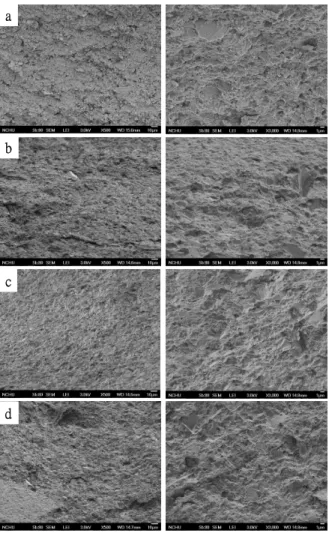
5.3. Morphology
Fig. 3 shows the surfaces of specimens with different ratios of gelatin. The control without gelatin has an appearance of entangled particles and exhibited several pores (Fig. 3a).
When adding gelatin, the hybrid composite becomes a heterogeneous structure with a homogenous distribution of gelatin within the CaSi matrix, and there are many filament crystals surrounding the CaSi body, which become more obvious with increasing gelatin content (Figs. 3bd). In sharp contrast, it seems that the morphology of the hybrid composites was more compact than the control, consistent with the porosity.
The analysis of the fractured surface using fractographic principles is a well-established analytical tool to determine the failure behavior of brittle materials. The control presents a sandy, smooth, and loose structure, while the gelatin-containing specimens are of solid and dense features (Fig. 4); this finding is similar to another study [12]. The added gelatin, which was examined in this study, may play a crucial role in the properties of the bio-inspired composites. A colloidal gel facilitates the formation of a dense structure of hydrated composites that entangle the original C–S–H structure. Such dense structures of gelatin- containing composites might be due to the existence of negatively charged gelatin. Type B gelatin with an isoselectric point of 5 has a high density of carboxyl groups, which makes the gelatin negatively charged [13,14]. The carboxyl groups might bind calcium ions on the surface of the CaSi particles. Hence, the gelatin structure not only serves as the filler incorporating to the CaSi matrix but also provides an anchoring site for the CaSi particles in the structure, binding them together to form a composite.
5.4. Compressive properties
Table 1 lists the relationships between the compressive strength, modulus, work-of- fracture, or hardness of the composites and the gelatin content. The specimens showed non- monotonous changes in strength and a significant difference (p < 0.05) among the specimens was found. Composites containing 5 wt% gelatin (CSG5) had a compressive strength value of 105.0 MPa on average, significantly (p < 0.05) higher than the CSG0 control (86.1 MPa). The addition of gelatin up to 10 wt% (CSG10) achieved a significantly (p < 0.05) increased compressive strength of up to 141.7 MPa (p < 0.05), which is within the reported compressive strength for cortical bone [15]. However, higher gelatin content at 15 wt% (CSG15) adversely affected the mechanical strength with a reduction of up to 31% of the highest value.
Although a high isostatic pressure of 500 MPa was applied to the specimens, which partly contributed to the strength measured, the obtained high strength could result from the hydrothermal factor. To confirm the speculation, the CaSi powders containing 0, 5, 10, and 15 wt% gelatin were directly pressed by cold isostatic pressing at 500 MPa without the further hydrothermal treatment in water at 60ºC. The compressive strength values of the directly pressed compact specimens only were 6.3, 4.2, 11.4, and 14.3 MPa, respectively, indicating significantly (p < 0.05) lower values than the corresponding specimens prepared by the dual pressing-hydrothermal method. The hydrothermal treatment-induced increase in strength is attributable to the more complete hardening during soaking in solution, which has also been observed in other studies [5,13,16]. The development of mechanical properties of the present materials is mainly the result of the reaction of liquid phases, such as water, with the components. Nevertheless, high applied pressure is beneficial via the densification mechanisms of nanoparticle rearrangement and sliding, plastic deformation, and pore shrinkage. More importantly, such densification procedures using a simple pressing- hydrothermal route can provide the possibility of incorporating proteins and drugs without damaging their biological activity, featuring a drug delivery system.
One concern is that the incorporation of gelatin into CaSi ceramics might result in degradation of the mechanical strength of the composites. However, in this study, this concern did not occur. When increasing the gelatin, to the result is an increase in the compressive strength; however, once it reaches the maximum value (i.e., 10% of gelatin), the compressive strength decreases drastically. The resulting high strength of the hybrid composites are due to a combination between the progressive hardening originating from the main CaSi reactant and reinforcementeffectfilling thedefectsby thegelatin phase,which servesasa‘glue’to fusethe particles together, as confirmed by SEM. Some surface chemistry properties unique to the gelatin enhance the interactions between the filler and the matrix [17]. It is also possible that the gelatin in the composites reduces the effect of the flaws and the porosity on the mechanical behavior of these hybrid composites, as indicated by the results of the porosity testing.
Porosity has a deleterious effect on the properties of ceramic materials, acting as a stress raiser and reducing the mechanical strength [18]. More recently, Martínez-Vázquez et al. suggested that the presence of polymer within the macropores of the ceramic modified the stress acting on the ceramic during the uniaxial compression tests for strengthening [19]. The proper stress transfer occurring between the reinforcement and the matrix may govern the mechanical characteristics of the hybrid composites [20]. Concerning the present study, chemical and mechanical interlocking between the CaSi and gelatin accounts for the efficient stress transfer in the composite system. It could be expected that excessive gelatin might disrupt the entanglement structure, resulting in weaker interfacial bonding between the CaSi matrix and gelatin. Moreover, the loose connection throughout the excessive gelatin might be another factor that decreases the strength. Additionally, the decrease in the strength of the CSG15 group can be attributed to the poor mechanical strength of the excessive amorphous phase that
lacks a crystalline configuration.
The values of the mean elastic moduli obtained in the range of 2.7 to 3.1 GPa during the measurement of compressive strength are also listed in Table 1 with the respective standard deviations. The amount of added gelatin influenced the elastic modulus of the composites;
however, the trend presented was not similar to the changes in compressive strength. The modulus decreased somewhat after the incorporation of either 5 wt% or 10 wt% gelatin to the control, but there was no significant difference (p > 0.05) after Scheffe’smultiplecomparison testing. A reason for the observed decrease in the compressive modulus of the composites is that gelatin is not stiffer than the surrounding ceramic matrix. Hence, it can be expected that the increased contents of the gelatin lead to a lower modulus, as confirmed by the result of CSG15. It is noted that the modulus of all of the bio-inspired composites is slightly lower than thelowerbound forthoseofthehuman corticalbone.When thematerialhasahigherYoung’s modulus than bone, they can cause stress shielding and lead to bone resorption. Regarding the work-of-fracture, various specimens increased with increasing gelatin content up to 10 wt%, indicating there was a significant difference (p < 0.05). After that, the work-of-fracture began to decrease, eliciting a trend similar to the variations in the strength. Although the detailed mechanism of this change is not fully understood, enhanced plastic deformation is achieved when the small organic molecules are uniformly dispersed throughout the ceramics and interacted strongly with the ceramic matrix.
5.5. Weibull analysis
To understand the level of the structural reliability of the materials, a Weibull analysis is commonly used to analyze historical failure data and produce failure distributions. The twenty measurements of failure strength from each group were ranked in order from the weakest to the strongest. Fig. 5 shows a double logarithmic plot of the experimental compressive strength data. It can be seen that the data points fell approximately along a straight line, which indicated that the two-parameter Weibull distribution is a reasonable assumption for the compressive strength distribution of the four specimens. The description of the Weibull distribution is given by the formula [21]:
F(σ)= 1-exp [-(σ/σ0)m] (2)
whereF(σ)isthefailureprobability (defined by therelation ofF(σ)= i/(N+1),in which iisthe rank orderofthecompressivestrength and N isthenumberofspecimens),σ isthestrength at a given F(σ),σ0 is the characteristic strength (scale parameter) at the fracture probability of 63.2%,and m istheWeibullmodulus(shapeparameter).Largervaluesofσ0 imply greater strength, and larger values of m imply more reliably reproducible specimens in a narrower distribution of values. The characteristic strengths of the composites were 90.1, 106.4, 144.7, and 102.4 MPa for the 0, 5, 10, and 15 wt% gelatin, respectively, and the corresponding dependent behaviors. The 10 wt% gelatin-containing composite had a higher Weibull modulus (17.5) than the other three specimens, indicating more uniform strength distribution and Weibull moduli were 12.2, 8.9, 17.5, and 8.1, respectively, which were calculated from the slope of the fitting curves. The Weibull characteristic strength and Weibull modulus did display gelatin-higher structural reliability. In general, ceramics have wide variability in failure strength because of the flaws incorporated during processing. Most ceramics are reported to have m values in the range of 5 to 15 [22].
5.6. Hardness
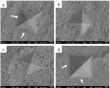
Hardness is intended to be a measure of the resistance to plastic deformation, which may include effects such as material displacement and fracture. Table 1 shows that CSG5 has a
significantly (p < 0.05) higher Vickers’microhardnessvalue(120.8)than thatoftheCSG0 control (99.1). The microhardness value of CSG10 (103.5) was not much higher than that of the CSG0 control, indicating no significant difference (p > 0.05). Different from the trend in the compressive strength, when the composite contained 15 wt% gelatin (CSG15), the specimen hardness (82.0) was significantly (p < 0.05) lower than that of the control. Such a large decrease in hardness might be related to the soft feature of the gelatin.
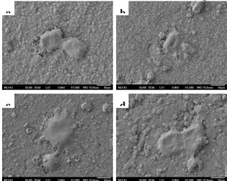
Fig. 6 shows the typical indentations produced by Vickers’ microhardness tester, indicating the appearance of well-defined features. A number of surface cracks of varying lengths were associated with the impression of the indentation on the CSG0 control and CSG15 surfaces. The observed surface cracks are believed to be from the mismatch between the ceramic matrix and polymer additive. In contrast to the findings, the CSG5 and CSG10 composites had no signs of crack motion at the impression tip. The amorphous gelatin phase of the composites during the compression could be reorienting up to breakage. The high resistance to crack-induction makes the present CSG10 composite highly interesting in both fundamental and practical aspects.
5.7. In vitro fatique
The load-bearing implant materials must be evaluated not only by their strength as described previously but also by their resistance to fatigue in solution because of the cyclic nature of in vivo loading. However, to the knowledge of the authors, relatively little information is available in the published literature concerning fatigue behavior in a physiological solution for bio-inspired ceramic/polymer composites. The results of fatigue experiments are presented as S-N diagrams (the so-called fatigue life diagram), where S is the maximum stress in a cyclic loading and N is the number of cycles until fracture. Fig. 7 is a plot of the maximum stress applied in the compressive cyclic fatigue against the number of cycles to failure at 37ºC in SBF. The graph showed that the stability of the composites was apparently affected by the cyclic loading with a remarkable decrease in the strength as the number of cycles increased. For example, the control fatigued in SBF for 2 ×10³cycles had a significant degradation down to 35% of the original strength. When a loading stress of 37 MPa was applied, the specimen containing 10 wt% gelatin (CSG10) lasted approximately 40 min in the in vitro fatigue test until failure occurred. As for the 0, 5, and 15 wt% gelatin-containing specimens, 40-min fatigue-induced failure in SBF solution required only 27, 32, and 26 MPa, respectively.
When subjected to fatigue testing in SBF under a dynamic condition, the detrimental effect on the specimens is seen, consistent with a previous study [23]. In addition to the crack growth that occurs under stress, the decrease in strength of the bio-inspired materials is also caused by environmental factors, such as the penetration of water/ions [23, 24]. Water/ions can easily infiltrate the inner portion of the specimens through structural imperfections, particularly under applied stress, resulting in weakened bond strength due to particle dissolution. The addition of gelatin can also increase the ratio of water absorption of the composite, which reduced the retention of the mechanical properties of the CaSi-gelatin composite fatigued under moisture conditions. The conclusion may be drawn that the present specimens have a limited resistance to fatigue failure. However, it provides that the fatigue properties of composites should be a noticeable factor.
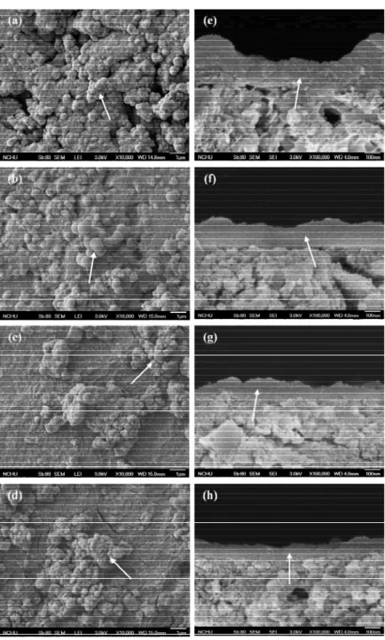
5.8. In vitro bioactivity and degradation
Broad face and cross-sectional SEM micrographs of the bone grafts after soaking in a SBF solution for 1 day are shown in Fig. 8. The surface morphology of CSG0 immersed for 1
day was similar to those of the other three specimens (Fig. 8 a-d). It is clear that precipitation took place on all specimen surfaces which were covered with clusters of precipitated spherulites. From the cross-sectional SEM micrograms (Fig. 8 e-h), it can be seen that such a precipitated layer with a distinct contrast was observed in all specimens, confirming the broad face examination. The precipitated apatite layer was smoother and denser than the original specimen structure. The average thickness of precipitated apatite layers was approximately 220, 200, 160, 100 nm, respectively, for CSG0, CSG5, CSG10, and CSG15. To further confirm that the observed layer was indeed ascribed to apatite precipitated from the SBF solution, SEM/EDS analyses were performed on the 1-day-immersed specimens, in addition to the XRD analysis. Ca/P ratios of the SBF-immersed specimens were 4.0, 4.5, 5.8, and 10.6 for CSG0, CSG5, CSG10, and CSG15, respectively, which significantly (p < 0.05) increased with increasing gelatin content. The reason for the much lower apatite precipitation rate on CSG15 was possibly due to the presence of gelatin in its as-prepared material. Nevertheless, all the bonegraftsshowed astrong tendency for“attracting”apatiteprecipitateonto theirsurfaces, as evidenced by the Ca/P molar ratio. The much higher Ca/P ratio (compared to the 1.67 stoichiometric Ca/P ratio of apatite) on 1-day-immersed surfaces was not surprising due to the fact that a large quantity of calcium originating from the underlying specimens was detected.
The lower Ca/P ratio on the surface of the CSG0 control without gelatin was possibly due to the faster apatite precipitation rate, consistent with the thickness of the precipitated layer. The in vitro bioactivity of the SiO2–CaO-based materials indicates that the presence of PO43
ions in the composition is not an essential requirement for the development of an apatite layer, which consumes the calcium and phosphate ions. This is because PO43
ions originate from the in vitro assay solution. An increase in the pH of SBF at different time intervals was attributed to the release of Ca(OH)2, which is conducive for the formation of apatite precipitation. The results of the higher pH value in the CSG0 control-immersed SBF paralleled the apatite precipitation rate.
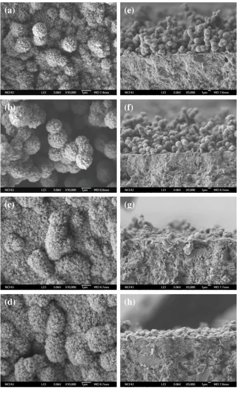
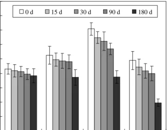
It was of interest to immerse specimens in an SBF solution for extended time (up to 180 days) to investigate the variations in the activity and degradation of the composites. After soaking for 180 days, the surface morphology of the specimens was significantly altered in the presence of the etching-induced micropores on the apatite layer (Fig. 9). It appears that during the immersion test dissolution of the surface had taken place. To further understand the etching effects, porosity measurements were conducted using a liquid displacement technique.
Before soaking in SBF, the porosities were 16%, 12%, 10%, and 11% for the specimens containing 0, 5, 10, and 15 wt % gelatin, respectively. On day 180, the porosities became 17%, 22%, 23%, and 28%, respectively. Significant differences (p < 0.05) between the porosities before and after soaking were found in the gelatin-containing composites.
Four soaking regimes up to 180 days were selected for testing compressive strength of the specimens, as shown in Fig. 10. The results revealed that all the four different types of bone grafts gradually lost their strengths with the increase in soaking time. After immersion for 180 days, the strength values of immersed specimens were in the range of 77−39 MPa, lower than respective strength values on day 0. It is worth noting that the gelatin-containing composites had a significantly lower strength (p < 0.05) compared to corresponding as-prepared composites, but not for the CSG control. Additionally, CSG0, CSG5, and CSG10 had a similar strength at day 180. It is surprising that the CSG0 control has insignificantly bond degradability (p > 0.05) when immersed in SBF solution up to 180 days. The statistical analysis using Scheffé multiple comparison test showed that the strength of CSG15 significantly declined from 98 MPa, the as-prepared strength, down to 39 MPa with a
reduction of approximately 60% after immersion for 180 days (p < 0.05). CSG5 and CSG10 composites lost 30 and 47% in compressive, respectively, after 180-day immersion. This deterioration in strength seems unavoidable for gelatin-containing composites immersed in SBF.
The effect of the soaking time on the changes in the weight of the bone graft specimens are presented in Fig.11. After soaking for 15 days, gelatin-containing composites showed a small amount of weight loss (~2%), whereas the CSG0 control gained weight. All the specimens exhibited an increased weight loss with an increase in the soaking time, reaching a weight loss of up to about 1–5% after 90 days depending on the type of specimen. At the end of the soaking experiment (180 days), weight loss of approximately 6%, 8%, 10%, and 18%
were observed for CSG0, CSG5, CSG10, and CSG15, respectively, which indicated a significant difference (p < 0.05). The degradation of the current bone graft systems was a slow process with the exception of CSG15, and the degree of the degradation was time dependent.
When soaked in SBF, the three gelatin-containing bone graft systems were associated with a relatively small degree of weight loss of about 2% after a 30-day soaking time; the CSG0 control even exhibited a weight increase. The few changes in sample weight may be explained by the formation of apatite, which was consistent with the morphology results. The four specimens continued to dissolve without stopping accompanied by continued weight loss after 90 days. This may be due to the release of soluble fractions (mainly gelatin). Because gelatin is biocompatible and has been classified as a bioresorbable material, its presence or dissolution is not expected to cause biological problems. Immersed CSG15 reached its maximal weight loss of 18% on day 180. It is important to note that 10 wt % gelatin (CSG10) led to a weight loss of 10% even after soaking in an SBF solution for 180 days. Moreover, the trend in weight loss of gelatin-containing composites was similar to the changes in the compressive strength. High physiochemical activity and low degradability were the characteristics of the present SiO2– CaO-based material, which are of utmost importance and a necessity for successful results of bone graft substitutes for vertebroplasty applications [25]. The designed composites were expected to have an optimal mechanical performance, a controllable degradation rate, and eminent bioactivity, which will be of great importance for bone remodeling and growth.
However, the degradation rate and mechanical strength may be improved to support large defect sites for long-lasting and permanent implantation applications.
While soaking in the SBF solution, all bone grafts caused the pH of the solution to increase during the first 15 days, as shown in Fig. 12. By day 30, the pH of the solution approached a steady state value ranging from 8.4 to 9.0 depending on the type of bone grafts.
A greater amount of gelatin in the composite produced a lower pH value of the SBF solution.
On day 180, the pH of the solution containing CSG0 (pH 9.1) was significantly (p < 0.05) higher than the pH of the solution containing the gelatin-containing composites (pH 8.48.7).
5.9. Cytotoxicity
A common objective in orthopedic and dental fields is the design of biomaterials that supports cell and tissue growth, improving fracture healing and bone defect filling. Cell viability and function on a bone graft are closely related to the physical, chemical and biological characteristics of the materials used. This study supports the hypothesis that a natural polymer such as gelatin successfully improves the mechanical properties of calcium silicate ceramics. More specifically, this investigation will show that the newly developed composites enhance the cell functions.
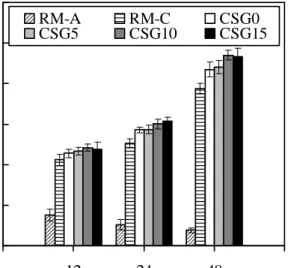
The results of the alamarBlue assay for cytotoxicity are shown in Fig.13. The cells on the
positive control (RM-A) showed a high degree of cytotoxicity with an increase in the culture time while OD values decreased. In contrast, the negative control (RM-C) exhibited an increased OD level of mitochondrial activity with an increasing culture time. When the cells were seeded on different bone grafts, no depression of cellular activity occurred. More importantly, the absorbance values of all the tested bone grafts were significantly (p < 0.05) higher than those obtained from the negative control after 24 and 48 h of culture.
5.10 Cell attachment and proliferation
Figure 14 shows that a higher attachment of pulp cells was observed for the gelatin- containing specimen surfaces than that for the surface of the control specimen after 3 h of culture. CSG15 indicated an increase of approximately 13% (p < 0.05) in the OD value compared to the CSG0 control during the initial 12 h incubation. Similarly, cell proliferation was notably higher in the gelatin-containing specimens than the cells in the control. Moreover, the optical density values increased with an increase in the culture time.
The number of cells initially attached was different between the bone grafts with and without gelatin. Cell attachment and proliferation increased after the incorporation of either 10% or 15% gelatin to the control, although there was no significant difference (p > 0.05) after Scheffe’smultiplecomparison testing. When gelatin was present on the specimen surfaces, cellular attachment took place rapidly, possibly via interactions between the functional OH, COOH, and NH2 groups of the gelatin and the integrins [26]. The interactions may consequently influence the spreading, proliferation, and differentiation of the cells.
5.11. Cell differentiation
One possibility is that this trend in the elevated pH value, caused when soaking materials in an SBF solution, would be expected with cell culture. Fig. 15A shows the variations of the pH in culture medium during the culture interval of pulp cells seeded on the specimens. It can be clearly seen that the pH values significantly (p < 0.05) increased from initially 7.3 to 7.67.8 after 1 h of assay, reaching a higher value after 3 days. The pH revealed a decrease with an increased gelatin amount of the composites. There are significant (p < 0.05) differences in extracellular pH between CSG0 control and CSG15 at all incubation times.
In addition to the role of gelatin in cell functions, another point of interest in the changes of the pH is its relation with the cell behavior. Cellular mechanisms involved in bone formation and resorption may be responsive to the acidbase balance. An alkalinization of the extracellular medium with the bone grafts rather than the normal pH (7.4) during cell culture was indeed determined. Alkalinization of the medium caused by calcium silicate-based materials was not unexpected because reactions which the material underwent in physiological solutions involved a mandatory disappearance of protons. More importantly, the alkalinization of the medium is relevant to the enhancement of osteoblastic function, since it is accompanied by a shift in the intracellular pH in the same direction in the culture systems of osteoblastic clone cells and cell lines [27].
The intracellular ALP level was measured to observe the functional activity of cells, as shown in Fig. 15B. The intracellular ALP level increased with the gelatin content of the composites at all incubation times. On day 3, a significant 15% increment (p < 0.05) in ALP level was measured for CSG15 compared to the CSG0 control. The increment became 12%
and 9% after 7 and 15 days, respectively, together with the significant (p < 0.05) differences.
Cell-differentiation studies, like cell-proliferation assay results, showed a significant impact of gelatin, with an emphasis on the importance of material composition. ALP activity was seen to
increase up to day 15 on the materials, with enzyme activity increasing with amount of gelatin.
It is generally accepted that an increase in the specific activity of ALP in bone cells reflects a shift to a more differentiated state [28]. ALP enzyme activity is also associated with bone ormation, and it is produced in high levels during the bone formation phase [29].
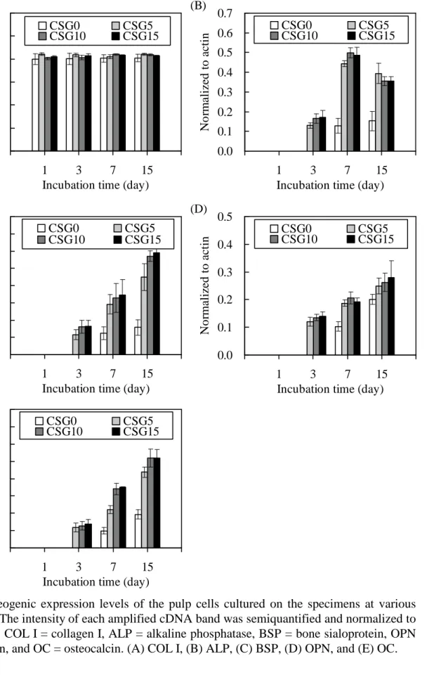
5.12. Osteogenesis
Changes in the phenotype marker expression of the pulp cells for varying culture durations are shown in Fig. 16. AC was used as an internal control and was produced at a comparable level in control cells and in the cells grown on the test specimens. Collagen is the major constituent of organic bone matrix, and its expression was similar in the control and experimental groups at all culture time points (Fig. 16A). On day 1, the gene expression levels of all osteogenic differentiation biomarkers including ALP, BSP, OPN, and OC were not detected. ALP expression peaked in the gelatin-containing cells after 7 days of incubation, which was significantly higher (p < 0.05) than in cells adhered to the CSG0 control, but the expression was all down- regulated after 15 days of culture except in the control (Fig. 16B). In contrast, BSP secretion on days 3, 7, or 15 was enhanced in cells cultured on the higher gelatin-containing specimen surfaces compared to those on the lower gelatin specimens (Fig.
16C). Similar to BSP, as the content of the gelatin increased, the level of OPN increased (Fig.
16D). OC production was greatest in the pulp cells cultured on CSG10 and CSG15 compared to the cells on the other two specimens (Fig. 16E).
A synchronized sequence of genes must be activated in the osteoblasts so that they undergo cell division and then synthesize an extracellular matrix that is capable of mineralizing to become bone. The cellular response of osteoblasts to foreign materials is genetically controlled. Osteoblast differentiation is generally accompanied by ALP expression, the production of BSP, OPN, OC, and COL I, and in vitro mineralization. There were no significant differences in COL I expression among the four bone grafts. The secretion of ALP from pulp cells was initiated earlier for cultures on the gelatin-containing composite surfaces than on the control surface. ALP appears to play a crucial role in the initiation of matrix mineralization, and ALP gene expression is down-regulated after the start of mineralization, as shown in all gelatin-containing composites on day 15. ALP gene expression was coincident with ALP enzyme activity on days 3 and 7 of incubation. On the contrary, after 15 days, this profile of the translated protein, measured here as ALP enzymatic activity, differed from the expression profile observed at the mRNA level. A factor may account for disparities between measured gene expression and enzyme activity levels on day 15. The translational machinery can translate a given mRNA many times into polypeptides which are then transformed into active enzyme [30]. Thus, this would lead to a profile in which there is an initial increase in mRNA that is lagged by an enzyme activity. In a word, degradation of the mRNA could begin at a certain time point and enzyme activity would reach a plateau. The potential explanation for the low number of increments seen in ALP enzyme activity results with incubation time may be an indicator of the cell progressing in mineralization.
OC is the most abundant noncollagenous bone-matrix protein characteristic of osteoblast synthetic function. The synthesis of OC is recognized at the late stage in osteoblast differentiation, and its expression increases rapidly as mineralization increases. BSP is specific to mineralized tissues, and strong mRNA expression of BSP is observed in fully differentiated osteoblasts associated with the initial formation of bone, as is the distribution of OC. OPN is one of the major phosphoproteins of mammalian bone, and its expression occurs later during bone formation [31]. Pulp cells on the gelatin-containing composites showed significantly increased BSP, OPN, and OC expression levels with increased culture time compared to the
control without gelatin. The results of the current study consistently indicated that the presence of gelatin was effective in supporting the proliferation of pulp cells and actively stimulating a biological response in these cells through the production of bone-specific proteins. The presence of gelatin provided a favorable environment for pulp cells, resulting in a large amount of mineralized tissue formation.
5.13. Immune response
The role of gelatin on the immune expression of IL-1 and iNOS in human dental pulp cells was also exaimed. IL-1 (Fig. 17A) and iNOS (Fig. 17B) production on day 1 were higher in the pulp cells grown on the CSG0 control compared to all the other gelatin-containing composites. Significant differences (p < 0.05) in IL-1 and iNOS expression were detected between the bone grafts at all time points. With increasing incubation time, the two pro- inflammatory cytokines had a decreased expression. In contrast, pulp cells grown on CSG15 had the highest level of anti-inflammatory cytokine IL-10 expression among all of the specimens (Fig. 17C), resulting in a significant difference (p < 0.05) compared to the other specimens.
IL-1isthefirst“immune”cytokinepositively identified to be involved in the control of bone turnover and stimulates the proliferation of osteoclast precursors. It is also a typical example of multifunctional cytokines involved in the regulation of immune responses, hematopoiesis, and inflammation [32]. iNOS is only expressed in responses to inflammatory stimuli. IL-1 causes activation of the iNOS pathway in bone cells [33]. It was found that both IL-1 and iNOS expression of pulp cells on the bone grafts decreased with increasing incubation time. Most importantly, the lowest levels of the pro-inflammatory cytokine IL-1 and iNOS were expressed by pulp cells cultured on the specimen with the highest gelatin contentatallculturetimepoints.Interestingly,Scheffe’smultiplecomparison testing indicated that gelatin-containing specimens had significantly (p < 0.05) higher IL-10 values than the CSG0 control at all culture time points, with the exception of the 1-day expression of which there was no reaction. The greater the amount of gelatin in the bone grafts, the more iNOS and IL-1 expression levels were inhibited and the more IL-10 was activated.
6. Conclusions
Our principal objective was to develop synthetic biomimetic bone analogs with improved mechanical and osteoconductive properties based on the combination of bioactive calcium silicate and naturally polymeric gelatin. The hard calcium silicate ceramic matrix provides structural consistency and hardness, while the soft polymeric gelatin filler acts as a binder and provides a certain degree of ductility and fracture resistance. A suitable calcium silicate/gelatin ratio of 10% by mass-to-mass in the composite achieved the highest bending strength of 141.7 MPa, which is strong enough to be used in load-bearing sites of bone tissue. The pulp cells cultured on gelatin-enriched materials exhibited higher levels of osteogenic differentiation biomarkers, such as BSP, OPN, and OC. Moreover, gelatin effectively inhibited iNOS and IL-1 expression and activated IL-10 expression. Taking the high initial mechanical strength and biological functions into account, the 10 wt % gelatincalcium silicate composites appear to be promising for the use in load-bearing applications such as dental and orthopedic repair.
More importantly, it is more suitable for a small bone defect or fast healing trauma site.
The biomimetic composites with high-strength will open up the possibility of calcium silicate-type implant materials.
7. Evaluation
The three-year project has successfully developed biomimetic calcium silicate-based composite scaffolds for applications in load-bearing bone tissue engineering. Two SCI papers have been published and the others results have been prepared to submit to SCI journal. The application of patent to USA and Taiwan is pending.
本計畫目前衍生的成效
項目 成果
專利 已申請美國與台灣專利各 1 件:
1. 丁信智,“雙層骨移植裝置”,中華民國專利,100123987,July 7,
2011,申請。
2. 2. Ding SJ, “Bilayered bone graft device”, US PatentApplication 13/166,090, June 22, 2011.
SCI 論文 已有 2 篇論文被接受,第 3 及 4 篇準備中
1. Ding SJ*, Shie MY, Wei CK. In vitro physicochemical properties, osteogenic activity, and immunocompatibility of calcium silicate-gelatin bone grafts for load-bearing applications. ACS Applied Materials &
Interfaces 2011;3(10):41424153. (Impact factor: 2.925-10; Ranking:
38/225)
2. Ding SJ*, Wei CK, Lai MH. Bio-inspired calcium silicate-gelatin bone grafts for load-bearing applications. Journal of Materials Chemistry 2011;21(34):1279312802.(Impact factor: 5.968-11; Ranking: 17/231),
國際會議 1. 2011 年第 11 屆亞洲生物陶瓷研討會邀請演講
2. 2012 年第 9 屆世界生物材料大會場次主持人 8. 參考文獻
[1] W. J. Landis, Bone 1995, 16, 533.
[2] D. M. Cooper, J. R. Matyas, M. A. Katzenberg and B. Hallgrimsson, Calcif. Tissue Int.
2004, 74, 437.
[3] S. J. Ding, M. Y. Shie and C. Y. Wang, J. Mater. Chem. 2009, 19, 1183.
[4] C. C. Chen, C. C. Ho, C. H. Chen and S. J. Ding, J. Endod. 2009, 35, 1288.
[5] C. C. Chen, C. C. Ho, C. H. Chen, W. C. Wang and S. J. Ding, J. Endod. 2009, 35, 1554.
[6] C. C. Chen, W. C. Wang and S. J. Ding, J. Biomed. Mater. Res. B 2010, 95, 456.
[7] S. J. Ding, M. Y. Shie, T. Hoshiba, K. Kawazoe, G. Chen and H. C. Chang, Tissue Eng. A 2010, 16, 2343.
[8] M. Tarrida, M. Madon, B. Le Rolland and P. Colombet, Adv. Cem. Based Mater. 1995, 2, 15.
[9] L. Black, C. Breen, J. Yarwood, K. Garbev, P. Stemmermann and B. Gasharova, J. Am.
Ceram. Soc. 2007, 90, 908.
[10] S. S. Potgieter-Vermaak, J. H. Potgieter and R. Van Grieken, Cem. Concr. Res. 2006, 36, 656.
[11] J. D. Pasteris and B. Wopenka, Biomaterials 2004, 25, 229.
[12] T. Y. Chiang, C. C. Ho, C. H. Chen, M. H. Lai and S. J. Ding, Mater. Chem. Phys. 2010, 120, 282.
[13] M. Y. Shie, C. H. Chen, C. Y. Wang, T. Y. Chiang and S. J. Ding, Acta Biomater. 2008, 4, 646.
[14] Y. Tabata and Y. Ikada, Adv. Drug Deliv. Rev. 1998, 31, 287.
[15] W. J. Landis, Bone 1995, 16, 533.
[16] M. Y. Shie, T. H. Huang, C. T. Kao, C. H. Huang and S. J. Ding, J. Endod. 2009, 35, 98.
[17] A. J. McManus, R. H. Doremus, R. W. Siegel and R. Bizios, J. Biomed. Mater. Res. A 2005, 72, 98.
[18] H. L. R. Alves, L. A. dos Santos and C. P. Bergmann, J. Mater. Sci. Mater. Med. 2008, 19, 2241.
[19] F. J. Martínez-Vázquez, F. H. Perera, P. Miranda, A. Pajares and F. Guiberteau, Acta Biomater 2010, 6, 4361.
[20] G. S. Sailaja, S. Velayudhan, M. C. Sunny, K. Sreenivasan, H. K. Varma and P. Ramesh, J. Mater. Sci. 2003, 38, 3653.
[21] M. Albakry, M. Guazzato and M. V. Swain, J. Dent. 2004, 32, 91.
[22] S. Karakoca and H.Yılmaz,J. Biomed. Mater. Res. B 2009, 91, 930.
[23] S. J. Ding, Dent. Mater. J. 2006, 25, 706.
[24] S. J. Ding, C. W. Wang, C. H. Chen and H. C. Chang, Ceram. Int. 2005, 31, 691.
[25] P. F. Heini and U. Berlemann, Eur. Spine J. 2001, 10, S205.
[26] S. J. Ding and M. Y. Shie, Adv. Eng. Mater. 2011, 13, B246.
[27] A. Ehara, K. Ogata, S. Imazato, S. Ebisu, T. Nakano and Y. Umakoshi, Biomaterials 2003, 24, 831.
[28] L. Malaval, F. Liu, P. Roche and J. E. Aubin, J. Cell. Biochem. 1999, 74, 616.
[29] A. Sabokbar, P. Millett, B. Myer and N. Rushton, Bone Miner. 1994, 27, 57.
[30] C. Glanemann, A. Loos, N. Gorret, L. B. Willis, X. M. O’Brien,P. A. Lessard and A.
Sinskey, J. Appl. Microbiol. Biotechnol. 2003, 61, 61.
[31] J. Chen, H. S. Shapiro and J. Sodek, J. Bone Miner. Res. 1992, 7, 987.
[32] S. Akira, T. Hirano, T. Taga and T. Kishimoto, FASEB J. 1990, 4, 2860.
[33] R. J. van’tHofand S. T. Ralston, Immunology 2001, 103, 255.
Table 1 Nominal composition, physicochemical properties, and Weibull parameters of various specimens
Composition ratio (wt:
vol) Sample
CaSi: gelatin
Gelatin amount (wt%)
Porosity (%)
Density (g cm-3)
Compressive strength (MPa)
Modulus (GPa)
Work-of fracture (kJ m-2)
Hardness (Hv)
CSG0 100:0 0 16.3±3.8a 1.8±0.1c 86.1±7.4d 3.1±0.1g 24.6±2.2i 99.1±9.1l CSG5 90:10 5 11.6±1.4b 1.7±0.1c 105.0±12.5e 3.0±0.1g 35.3±3.2j 120.8±10.3m CSG10 80:20 10 10.0±1.5b 1.8±0.1c 141.7±8.6f 2.9±0.1g 52.1±4.8k 103.5±13.3l CSG15 70:30 15 11.1±2.4b 1.8±0.1c 97.8±12.6e 2.7±0.1h 40.4±5.3j 82.0±6.3n Values are mean ± standard deviation.
Mean values followed by the same superscript letter were not significantly different (p >
0.05) according to Scheffé post hoc multiple comparisons.
0 2 4 6 8 10 12 14 16
10 20 30
Gelatin concentration (%)
Gelatincontent(%)
300 500 700 900 1100 1300 1500
Wavenumber (cm-1)
Ramanintensity
CSG15
CSG0 CSG5 CSG10
20 25 30 35 40 45 50
2θ
Diffractionintensity
CSG15
CaSi powder CSG0 CSG5 CSG10
a
d c
b
Fig. 1 The actual gelatin content and distribution of the composites.
(a) (b)
Fig. 2 (a) XRD patterns of CaSi powder and composites with and without gelatin; (b) Raman spectra for various specimens.
Fig. 3 Surface SEM micrographs of various specimens. (a) CSG0 control, (b) CSG5, (c) CSG10, and (d) CSG15.
-4 -3 -2 -1 0 1 2
4.0 4.2 4.4 4.6 4.8 5.0 5.2
lnσ
lnln(1/(1-F))
CSG0 CSG5 CSG10 CSG15
a
b
c
d
Fig. 4 SEM micrographs of the interior surface of the specimens. (a) CSG0 control, (b) CSG5, (c) CSG10, and (d) CSG15. The right pictures are at a higher magnitude.
Fig. 5 Weibull strength distribution plots of various specimens. F is the failure probability and σ isthestrength.The solid line represents the regression line.