行政院國家科學委員會專題研究計畫 期末報告
清胃散主成分黃連萃取之小檗鹼的抗單純皰疹病毒ˋ抗口 腔癌及免疫調節功效(第 3 年)
計 畫 類 別 : 個別型
計 畫 編 號 : NSC 99-2320-B-040-004-MY3
執 行 期 間 : 101 年 08 月 01 日至 102 年 07 月 31 日
執 行 單 位 : 中山醫學大學醫學檢驗暨生物技術學系(所)
計 畫 主 持 人 : 楊繼江
計畫參與人員: 其他-兼任助理人員:李雅鈴
公 開 資 訊 : 本計畫涉及專利或其他智慧財產權,2 年後可公開查詢
中 華 民 國 102 年 10 月 29 日
中 文 摘 要 : 本研究進行清胃散與其組成生藥中的當歸、生地、黃連、牡 丹皮、升麻的免疫調節的特性。
活體內的研究顯示,清胃散於每天 1mL 的 25,000 mg/mL 濃 度,對老鼠 3 天的急性毒性測試肝功能指數與 28 天的亞急性 毒性測試肝功能指數與腎功能指數皆有意義增高。以 PCR 微 陣列以小檗鹼 5 倍 IC50 濃度處理 HT-29, T-47D, 與 BXPC3 細胞,顯示 BCL2L10, HRK, FASLG, LTA, TNF, 與 TNFRSF9 等為主要調控基因。而清胃散對 IL-2, IL-4, IFN-gamma,與 TNF-alpha 的平均細胞激素濃度與其組成生藥中的個別成分 效果並不相同。清胃散可上調 IL-2, IL-4 與 TNF-alpha,
但下調 IFN-gamma。這結果顯示清胃散的免疫調節的特性無 法由組成生藥中的單一個別成分所取代。
中文關鍵詞: 清胃散、黃連、小檗鹼、細胞激素
英 文 摘 要 : The cytokine modulating effects of a herbal medicine, Ching-Wei-San, and its individual herbal components, Coptidis rhizoma, Angelicae sinensis radix,
Rehmanniae radixet rhizom, Moutan radicis cortex, and Cimicifuga foetida, were evaluated in this study. The in vivo results suggested that one milliliter Ching- Wei-San at the 25,000 mg/mL concentration daily for the mice had significantly high levels in the liver function indexes in the 3-day acute toxicity test and in both the liver and kidney function indexes in the 28-day subacute toxicity test (P<0.01). By PCR array, the major genes, including BCL2L10, HRK, FASLG, LTA, TNF, and TNFRSF9, regulated by cells treated with berberine at a concentration of 5 times IC50 for three different cells, HT-29, T-47D, and BXPC3 were revealed. The mean cytokine ratios of IL-2, IL-4, IFN-, and TNF- in Balb/c mice treated with individual herbal components were shown to be different from each other. Ching-Wei-San modulated the immunity of mice, up-regulated IL-2, IL-4 and TNF-alpha, but down-regulated IFN-gamma . The effects of none of the individual herbal components alone can substitute for the cumulative effect of Ching-Wei-San.
英文關鍵詞: Ching-Wei-San、Coptidis rhizoma、berberine、cytokine
行政院國家科學委員會補助專題研究計畫 成果報告
□期中進度報告
清胃散主成分黃連萃取之小檗鹼的抗單純皰疹病毒、抗口腔癌及免 疫調節功效
計畫類別: 個別型計畫 □整合型計畫 計畫編號:NSC 99-2320-B-040-004-MY3
執行期間: 2010 年 8 月 1 日至 2013 年 7 月 31 日 執行機構及系所:中山醫學大學醫學檢驗暨生物技術學系
計畫主持人:楊繼江 共同主持人:
計畫參與人員:
成果報告類型(依經費核定清單規定繳交):□精簡報告 完整報告
本計畫除繳交成果報告外,另須繳交以下出國心得報告:
□赴國外出差或研習心得報告
□赴大陸地區出差或研習心得報告
□出席國際學術會議心得報告
□國際合作研究計畫國外研究報告
處理方式:除列管計畫及下列情形者外,得立即公開查詢
涉及專利或其他智慧財產權,□一年 二年後可公開查詢 中 華 民 國 102 年 10 月 29 日
附件一
中英文摘要及關鍵詞 中文
本研究進行清胃散與其組成生藥中的當歸、生地、黃連、牡丹皮、升麻的免疫調節的特性。
活體內的研究顯示,清胃散於每天 1mL 的 25,000 mg/mL 濃度,對老鼠 3 天的急性毒性測 試肝功能指數與 28 天的亞急性毒性測試肝功能指數與腎功能指數皆有意義增高。以 PCR 微陣列以小檗鹼 5 倍 IC50 濃度處理 HT-29, T-47D, 與 BXPC3 細胞,顯示 BCL2L10, HRK, FASLG, LTA, TNF, 與 TNFRSF9 等為主要調控基因。而清胃散對 IL-2, IL-4, IFN-, 與 TNF-的平均細胞激素濃度與其組成生藥中的個別成分效果並不相同。清胃散可上調 IL-2, IL-4 與 TNF-,但下調 IFN-。這結果顯示清胃散的免疫調節的特性無法由組成生藥中的 單一個別成分所取代。
關鍵詞: 清胃散、黃連、小檗鹼、細胞激素
英文
The cytokine modulating effects of a herbal medicine, Ching-Wei-San, and its individual herbal components, Coptidis rhizoma, Angelicae sinensis radix, Rehmanniae radixet rhizom, Moutan
radicis cortex, and Cimicifuga foetida, were evaluated in this study. The in vivo results suggested
that one milliliter Ching-Wei-San at the 25,000 mg/mL concentration daily for the mice had significantly high levels in the liver function indexes in the 3-day acute toxicity test and in both the liver and kidney function indexes in the 28-day subacute toxicity test (P<0.01). By PCR array, the major genes, including BCL2L10, HRK, FASLG, LTA, TNF, and TNFRSF9, regulated by cells treated with berberine at a concentration of 5 times IC50 for three different cells, HT-29, T-47D, and BXPC3 were revealed. The mean cytokine ratios of IL-2, IL-4, IFN-, and TNF- in Balb/c mice treated with individual herbal components were shown to be different from each other. Ching-Wei-San modulated the immunity of mice, up-regulated IL-2, IL-4 and TNF-, but down-regulated IFN-. The effects of none of the individual herbal components alone can substitute for the cumulative effect of Ching-Wei-San.關鍵詞: Ching-Wei-San、Coptidis rhizoma、berberine、cytokine
報告內容
Several lines of evidence have revealed that cytokines play important roles not only in tissue homeostasis but also in the pathogenesis of many infectious diseases. The known actions of cytokines suggest that these molecules hold a great potential for modulating the host immune response and enhancing tissue regeneration. Recent research on biological activities in the normal periodontium and the pathogenesis of periodontal diseases has clarified the involvement of various cytokines in the biological activities observed in the sites (Okada and Murakami, 1998). It has been shown by many investigators that IL-1, IL-1, IL-6, IL-8, and TNF- can be detected in gingival crevicular fluid (Rossomando et al., 1990; Geivelis et al., 1993; Payne et al., 1993). Several studies also have shown that there are more abundant Th2 than Th1 cells in various diseased periodontal tissues (Yamazaki et al., 1995, 1997; Sigusch et al., 1998; Lappin et al., 2001). Thus, directing or influcing Th1 vs. Th2 differentiation could have impact on the development and/or progression of the periodontal disease. Presently, it is not clear how the innate immune system interacts with acquired immune responses to enforce and/or generate overall anti-bacterial immunity and the subsequent cytokine profile involved at the onset of the periodontal disease or more stable lesions (i.e. gingivitis) (Taubman and Kawai, 2001; Teng, 2002). It may, however, be possible, once the technology is optimized, to apply cytokine therapy to resolve periodontal infections. Such therapeutic measures hold promise not only in the resolution of periodontal infections, but also in the treatment of other diseases of the oral cavity (Riccelli et al., 1995).
Chinese people have used traditional Chinese herbal medicines to treat periodontal diseases for hundreds of years. Some Chinese medicine can prevent periodontal diseases efficiently by enhancing the immune system of the host. It has been reported that three Chinese herbal composites, Conth Su, Chi Tong Ning, and Xi Gua Shuang, and the major components of these composites, can effectively inhibit periodontal bacterial growth without causing cell mutation (Chan et al., 2003). Our previous study suggested that predominantly TH2-pattern cytokines can be achieved in mice serum with a dietary combined dose-dependent herb-soaked solution, Yi-fey Ruenn-hou tea (Lin et al., 2004). A Chinese medicine, Ching-Wei-San, which is composed of
Coptidis rhizoma, Angelicae sinensis radix, Rehmanniae radixet rhizom, Moutan radicis cortex,
and Cimicifuga foetida, has been used for periodontal diseases since the Yuan dynasty in China (A.D. 1279-1368)(Zhang and Li, 2000). However, the mechanism remains unclear. In the present study, the antimicrobial and cytokine regulatory effects of different prepared (combined or individual decocted) Ching-Wei-San and their individual herbal components were studied and compared. The information revealed in the present study may be helpful in using Ching-Wei-San safely and across a wide spectrum of patients in the clinical resolution of periodontal diseases.Materials and Methods
Animals
In this study, the Institutional Guidelines for Animal Experiments (Animal Center, Chung Shan Medical University) were followed for animal care and use. All the SPF (specific pathogen-free) Balb/c female mice, 8–week-old, used in this study were purchased from the National Laboratory Animal Center, Taipei, Taiwan. These animals were raised at a temperature of 25 ± 1℃ and in 55 ± 5% relative humidity with 12 h light exposure daily in the animal center of Chung Shan Medical University.
Preparation of herbal medicines
The constituent proportion of Angelicae sinensis radix, Rehmanniae radixet rhizom, Coptidis
rhizoma, Moutan radicis cortex and Cimicifuga foetida was 0.6:0.6:0.6:1:2 (weight of dried
herbs), respectively, according to the component instructions of the Committee on Chinese Medicine and Pharmacy, Department of Health, Taiwan, R.O.C. These five components, individually or in combination, were added in a beaker with a 10-fold volume of water, and heated to 100℃ as individual or combined decocted, respectively. The hot complex was allowed to cool down and then filtered by Whatman filter papers for 4 times. In this way (Lin, 2002), the collected filtrate was concentrated using cryogenic dry to prepare a stock Ching-Wei-San extract at 25,000 mg/mL. This stock solution was then used to prepare various test solutions by dilution with distilled water. The individual decocted (ID) Ching-Wei-San extract of Angelicae sinensisradix (62.5 mg/mL), Rehmanniae radixet rhizom (62.5 mg/mL), Coptidis rhizome (62.5 mg/mL), Moutan radicis cortex (104 mg/mL), and Cimicifuga foetida (208 mg/mL) was decocted
separately in the similar way. The individual decoction of the herbs were combined and then diluted to 500 mg/mL for testing to compare with the traditionally combined preparation.Tetracycline (250 mg/mL) for reducing oral microbes and periodontal symptoms in clinical patients was used as the positive control. Distilled water was used as the normal control.
Acute toxicity test and subacute toxicity test
Eight-week-old Balb/C mice were used for acute and subacute toxicity tests treated with different concentrations (125, 250, 500, 1,000, 5,000, 10,000, and 25,000 mg/mL) of Ching-Wei-San, its individual herbal components, or distilled water (negative control) one milliliter daily for each of the groups of ten mice. The toxicity was investigated for 72 hours. For the subacute toxicity test, a similar approach was used for 28 days. Body weight was measured every week. The mice were sacrificed for blood routine index, including ALT, AST, creatinine, and BUN detection (Automatic analyzer, ARCO, Biotechnica Instruments, Italy) at the indicated time, respectively. The autopsy of the mice was examined by H&E staining.
H&E staining
All specimens were examined by routine fixation with H&E staining. All brains, hearts, livers, spleens, and kidneys were fixed immediately in a 10% formalin solution overnight at 4°C and embedded in paraffin. Serial sagittal sections 5
m thick were cut, and stained with H&E for
histological study.PCR array
The RT2 Profiler PCR Array was manipulated according to the manufacture's instruction (Qiagen, Hilden, German). Briefly, the experimental extracted RNA samples were converted into first strand cDNA, the template for the polymerase chain reaction. Then, template was combined with an instrument-specific and ready-to-use RT2 Real-Time™ SYBR Green PCR master mix. Equal aliquots of this mixture was added to each well of the same PCR Array plate containing the pre-dispensed gene-specific primer sets, and PCR was performed. The instrument’s software was used to calculate the threshold cycle (Ct) values for all genes on all PCR Arrays. Finally, fold-changes in gene expression for pair-wise comparison using the DDCt method were calculated. The regulated fold-changes were compared for “before” and “after”
treatment related to a housekeeping gene. The consistency in the housekeeping genes’ Ct values was examined by choosing the proper normalization method. The built-in ACTB titration checks the linearity of the assay results. The negative controls insure a lack of DNA contamination and set the threshold for the absent / present call.
Cytokine ELISA tests
Combined or individual decocted Ching-Wei-San at the concentration of 125, 250, 500, 1,000, 5,000, 10,000, or 25,000 mg/mL, its individual herbal components, or distilled water, were administered by gavages for one concentration, one milliliter daily for each of the twenty 8-week-old Balb/c mice. Five mice were sacrificed at the end of each week for four weeks after the regimen commenced. Sera were separated from the blood samples of sacrificed mice by centrifugation at 3,500×g for 15 min, and aliquoted and stored at -70℃ until required for cytokine assay. The cytokines (IL-2, IL-4, IFN-γand TNF-) were assayed in triplicate using commercial ELISA kits (Pharmingen, San Diego, CA, USA), following the manufacturer’s instructions as described before (Tsai et al., 2000; Lin et al., 2004; Ho et al., 2005; Huang et al., 2005; Lin et al, 2005a, 2005b). A standard curve was also derived for IL-2, IL-4, IFN-γand TNF-.
Statistical analysis
Data for the cytokines and biochemical index are shown as mean ± S.D. The Wilcoxson rank
sum test was used for comparing the weight difference and cytokine ratio between the experimental group and the control group. P values < 0.01 were taken as significant.
Results
Acute and subacute toxicity tests
For the acute and subacute toxicity test, Ching-Wei-San, its individual herbal components, or distilled water, were administered one milliliter daily for each of the groups of ten 8-week-old Balb/c mice. The mean body weight of all the mice before test was 28.8±0.9 gms. The concentrations of Ching-Wei-San used were 125, 250, 500, 1,000, 5,000, 10,000, and 25,000 mg/mL, the doses administered for each tested group therefore were approximately 4.3, 8.7, 17.4, 34.7, 173.6, 347.2, and 868.1 mg/kg, respectively. The weight and physiological characteristics of the mice were recorded for each group for 28 days. No mice in any group died or were wounded during the experimental period. Moreover, there was no significant change in body weight observed for the mice treated with Ching-Wei-San except for the 25,000 mg/mL group.
The mean weights at week 1, week 2, week 3, and week 4 were 28.81.1, 28.90.9, 29.20.9, and 29.91.2 gms, respectively, for the normal control (distilled water) group, while were 28.11.2, 26.60.8, 26.21.0, and 25.81.1 gms, respectively, for the 25,000 mg/mL group. The 25,000 mg/mL group showed a significant decrease (P<0.05) in weight beginning the second week, as well as in their appetite and moving activity.
The levels of liver function (ALT and AST) and kidney function (creatinine and BUN) indexes in the serum of the mice were also detected after 3-day and 28-day treatments (Table 1). There was no significant change (P>0.01) among the different groups in the acute toxicity test, except for the liver function indexes of the 25,000 mg/mL group (P<0.01). Moreover, in the subacute toxicity test, the 25,000 mg/mL group showed significantly high levels in both the liver and kidney function indexes (P<0.01). However, no pathological lesion or change was noted by H&E staining in the brain, heart, liver, spleen, or kidneys of the mice given a 28-day one milliliter daily 25,000 mg/mL Ching-Wei-San treatment (histology data not shown).
PCR array
The major genes regulated by cells treated with berberine at a concentration of 5 times IC50 for three different cells, HT-29, T-47D, and BXPC3 were detected by PCR array. The cells were treated for 24 hours and the regulated fold-changes were compared for “before” and “after”
treatment related to a housekeeping gene. The major genes, including BCL2L10, HRK, FASLG, LTA, TNF, and TNFRSF9, were revealed (Fig. 1).
Cytokine modulating effect of Ching-Wei-San and its individual herbal components on
healthy Balb/c mice
Mice were divided into several groups and treated as described in the Materials and Methods section. The mean cytokine values of the normal control (distilled water) group at week 1, week 2, week 3, and week 4 were 4.14 0.45, 4.610.51, 4.190.39, and 4.710.42 pg/mL, respectively, for IL-2, 0.390.12, 0.480.13, 0.430.09, and 0.510.12 pg/mL, respectively, for IL-4, 2.200.31, 2.020.26, 1.980.17, and 2.150.26 pg/mL, respectively, for IFN-, and 15.38
4.62, 16.315.51, 17.225.82, and 16.387.29 pg/mL, respectively, for TNF-. Comparing the
cytokine values of treated groups with control, the results demonstrated that the ratios of cytokines, i.e. IL-2, IL-4, IL-6, IFN-and TNF-, were different for different concentrations of the Ching-Wei-San and its individual herbal components. Table 2 shows the mean cytokine ratios of IL-2, IL-4, IFN-, and TNF- in Balb/c mice treated with Ching-Wei-San and its individual herbal components for 4 weeks, compared with a normal control group. The level of IL-2 was increased in the BALB/c mice in the first 2 weeks after treatment with both preparations of Ching-Wei-San and most of its individual herbal components, i.e. Angelicae sinensis radix,Rehmanniae radixet rhizom, Coptidis rhizoma, and Moutan radicis cortex. IL-2, IL-4, and
TNF- were up-regulated with the increase of the concentration of combined decocted Ching-Wei-San, compared with the normal control, however, the IFN-γ of mice in the Ching-Wei-San-treated groups and the 250 mg/mL tetracycline positive control group were down-regulated. IFN- was the only cytokine decreased by either combined or individual decocted Ching-Wei-San treatment. The effect of IFN-down-regulation was also demonstrated by Angelicae sinensis radix, Cimicifuga foetida, and tetracycline. The mean cytokine ratios of IL-2, IL-4, IFN-, and TNF- in Balb/c mice treated with individual herbal components were shown to be different from each other during the 4-week period. Angelicae sinensis radix was found to up-regulate IL-2 and TNF-but down-regulate IL-4 and IFN- serum concentrations.Rehmanniae radixet rhizome, Coptidis rhizoma, and Moutan radicis cortex up-regulated all of
the 4 tested cytokines. Cimicifuga foetida up-regulated IL-4 and TNF-, down-regulated IFN-, but showed no effect on IL-2. Tetracycline down-regulated IL-2, IL-4, and IFN-however, TNF-was the only cytokine not affected.Discussion
Ching-Wei-San has been used for periodontitis for many hundred of years in China (Zhang and Li, 2000). Immune regulation is associated with T-helper (Th) type cells and their cytokine production. IL-4 was secreted by Th2 cell; Th1 cells secreted IL-2 and IFN-γ. One report concluded that Th1 cells were associated with cell-mediated immunity and Th2 cells were related to humoral immunity (Mosmann and Coffman, 1989). Cytokines present during immune responses have a tremendous influence on resistance/susceptibility to oral diseases, including periodontal disease and oral infections, and may also play a role in preventing infection-induced
immunopathology (Okada and Murakami, 1998; Bartova et al., 2000; Ebersole et al., 2000;
Seymour and Gemmell, 2001; Yamazaki et al., 2003). Fujihashi et al. (1994) demonstrated the absence of mRNA for IL-2 and IL-4 production but significant levels mRNA for IL-5 and IL-6 production in gingival mononuclear cells isolated from lesional sites of adult periodontitis. The absence of IL-2 would be an indication of the absence Th0/Th1 CD4+ cells, and the absence of IL-4 would suggests that certain Th2 activities may not be occurring, such as the activation of resting B cells. Some report suggested that the cytokine levels in gingival crevicular fluid were closely associated with the the severity of inflammatory response and/or periodontal tissue destruction (Masada et al., 1990; Stashenko et al., 1991). Recently, it has been demonstrated that IL-4 concentrations were lower at sites of chronic periodontitis (Mogi et al., 1999) and IFN-
concentrations were significantly greater within giniva adjacent to 3 to 6 mm diseased sites (Johnson and Serio, 2005). Masada et al. (1990) demonstrated that IL-1 levels were elevated in gingival crevicular fluid at periodontis sites and that marked reductions of total IL-1 levels were observed following effective treatment. The Th1/Th2 picture is not clear at present, however, the possibility that the mechanism of the antimicrobial effect of Ching-Wei-San is related to the cytokine modulation of its individual herbal components cannot be excluded. In this study, the results suggested that Ching-Wei-San modulated the immunity of mice, up-regulated IL-2, IL-4 and TNF-, but down-regulated IFN-. The results also demonstrated that the IFN-down-regulation effect of Ching-Wei-San was achieved by Angelicae sinensis radix and
Cimicifuga foetida, although their antimicrobial effects were not impressive. Notably, it was
reported that minocycline, a tetracycline derivative, exerted an inhibitory effect on TNF-and IFN- production by stimulated Tcells, whereas the production of IL-6 remained unaffected; the addition of minocycline to lipopolysaccharide-stimulatedmonocytes led to a dose-dependent increase in TNF-and IL-6production, which was paralleled by an enhancement of TNF-mRNA synthesis (Kloppenburg et al., 1996). In this study, tetracycline was found to down-regulate IL-2, IL-4, and IFN- secretion in vivo, whereas the level of TNF- remained unaffected. The effects of tetracycline on IL-2 and IL-4, which were in contrast to those of Ching-Wei-San, require further study and an examination of their beneficial effects on periodontal diseases.
In summary, the results suggested that Ching-Wei-San is safe for use in vivo (Lu and Kacew, 2002), at least in a 28-day period. The results suggested that one milliliter Ching-Wei-San at the 25,000 mg/mL concentration daily for the mice had no toxic effect on organs, e.g. the brain, heart, liver, spleen, and kidneys, by pathological examination. However, for the 25,000 mg/mL group, a decrease in weight beginning the second week, as well as in their appetite and moving activity was observed. Moreover, there were significantly high levels in the liver function indexes of the 25,000 mg/mL group in the 3-day acute toxicity test and in both the liver and kidney function indexes in the 28-day subacute toxicity test (P<0.01). Both combined and
separately decocted preparations showed comparable antimicrobial and cytokine modulating effects, although minor differences could be observed. The antimicrobial and cytokine modulating effects of individual herbal components varied with each others to a great extent, and were not consistent with those of either combined or separately decocted preparations of Ching-Wei-San. With antimicrobial activity it appears that Coptidis Rhizoma is the most potent (Lin et al., 2006). However, when cytokine modulating effects were taken into consideration, none of the individual herbal components alone could substitute for the cumulative (antimicrobial and cytokine modulating) effects of Ching-Wei-San. Based on these results, the effects of none of the individual herbal components alone can substitute for the cumulative effects of Ching-Wei-San. Further investigation into the comparison of different preparations, different concentrations of Ching-Wei-San and its individual herbal components for clinical applications in periodontal therapy, or other bacterial infections, is encouraged.
參考文獻
Bartova, J., Kratka-Opatrna, Z., Prochazkova, J., Krejsa, O., Duskova, J., Mrklas, L., Tlaskalova, H., Cukrowska, B., 2000. Th1 and Th2 cytokine profile in patients with early onset periodontitis and their healthy siblings. Mediators Inflamm. 9:115-20.
Chan, Y., Lai, C.H., Yang, H.W., Lin, Y.Y., Chan, C.H., 2003. The evaluation of Chinese herbal medicine effectiveness on periodontal pathogens. Am. J. Chin. Med. 31, 751-761.
Ebersole, J.L., Cappelli, D., Holt, S.C., Singer, R.E., Filloon, T., 2000. Gingival crevicular fluid inflammatory mediators and bacteriology of gingivitis in nonhuman primates related to susceptibility to periodontitis. Oral Microbiol. Immunol. 15:19-26.
Fujihashi, K., McGhee, J. R., Yamamoto, M., Beagley, K. W. and Kiyono, H., 1994. Cytokine networks and immunoglobulin synthesis in inflamed gingival tissues. In: Molecular Pathogenesis of Periodontal Disease. Genco, R., Hamada, S., Lehner, T., McGhee, J. R. and Mergenhagen, S.
E. (eds.), ASM Press, pp. 135-145.
Geivelis, M., Turner, D.W., Pederson, E.D., Lamberts, B.L., 1993. Measurements of interleukin-6 in gingival crevicular fluid. from adults with destructive periodontal disease. J.
Periodontol. 64:980-983.
Gibbons, R.J., van Houte, J., 1980. Bacterial adherence and the formation of dental plaques. In:
Beachey E.H. (Ed.), Bacterial adherence. Chapman & Hall, London, pp62-104.
Havemose-Poulsen, A., Holmstrup, P., 1997. Factors affecting IL-1-mediated collagen metabolism by broblasts and the pathogenesis of periodontal disease: a review of the literature.
Crit. Rev. Oral Bio. Med. 8, 217-36.
Higaki, S., Nakamura, M., Morohashi, M., Yamagishi, T., 2004. Propionibacterium acnes biotypes and susceptibility to minocycline and Keigai-rengyo-to. Int. J. Dermatol. 43, 103-107.
Ho, C.C., Lin, S.S., Chou, M.Y., Chen, F.L., Hu, C.C., Chen, C.S., Lu, G.Y., Yang, C.C., 2005.
Effects of CAPE-like compounds on HIV replication in vitro and modulation of cytokines in vivo. J. Antimicrob. Chemother. 56, 372-379.
Hu, J.P., Takahashi, N., Yamada, T., 2000. Coptidis rhizoma inhibits growth and proteases of oral bacteria. Oral Dis. 6, 297-302.
Huang, T.H., Yang, C.C., Ding, S.J., Yen, M., Kao, C.T., Chou, M.Y., 2005. Inflammatory cytokines reaction elicited by root-end filling materials. J. Biomed. Mater. Res. 72B, 123-128.
Johnson, R.B., Serio, F.G., 2005. Interleukin-18 concentrations and the pathogenesis of periodontal disease. J. Periodontol. 76:785-790.
Kleinfelder, J.W., Muller, R.F., Lange, D.E., 1999. Antibiotic susceptibility of putative periodontal pathogens in advanced periodontitis patients. J. Clin. Periodontol. 26, 347-351.
Kloppenburg, M., Brinkman, B.M., de Rooij-Dijk, H.H., Miltenburg, A.M., Daha, M.R., Breedveld, F.C., Dijkmans, B.A., Verweij, C., 1996. The tetracycline derivative minocycline differentially affects cytokine production by monocytes and T lymphocytes. Antimicrob. Agents Chemother. 40, 934-940.
Lappin, D.F., Macleod, C., Kerr, A., Mitchell, T., Kinane, D.F., 2001. Anti-inflammatory cytokine IL-10 and T cell cytokine profile in periodontitis granulation tissue. Clin. Exp. Immunol.
123:294-300.
Lin, H.F., 2002. Experience of decoction of the traditional Chinese medicine soup. Modern TCM.
6, 60-61.
Lin, S.J., Tsai, J.H., Tsai, C.H., Hsu, H.T., Lin, Y.C., Xu, F.L., Yang, C.C., 2004. The in vivo effects of cytokines modulation for balb/c mice fed with a combined Chinese herb-soaked solution, Yifei-Ruennhou tea. Immunopharacol. Immunotoxicol. 26, 435-444.
Lin, S.J., Tsai, J.H., Tsai, C.H., Lin, Y.C., Hsu, H.T., Xu, F.L., Yang, C.C. 2005a.The in vivo effects of cytokine modulation for Balb/C mice given Canavalia ensiformis (L.) seeds with different heat treatment. Food Chem. 91, 139-145.
Lin, S.S., Chou, M.Y., Ho, C.C., Kao, C.T., Tsai, C.H., Wang, L., Yang, C.C., 2005b. Study of the viral infections and cytokines associated with recurrent aphthous ulceration. Microbes Infect. 7, 635-644.
Lin SJ, Chen CS, Ho CC, Lin SS, Shih HT, Lee IP, Chou MY, Kao CT, Chen FL, Ho YC, Hsieh KH, Huang CR,Yang CC. In vitro anti-microbial and in vivo cytokines modulation effects of different prepared Chinese herbal medicines. Food Chem Toxicol. 2006, 44;2078-2085.
Listgarten, M.A., Loomer, P.M., 1998. Microbial identification in the management of periodontal diseases. A systematic review. Ann. Periodontol. 8, 182-192.
Lu, F.C., Kacew, S., 2002. Basic toxicology: Fundamentals, target organs and risk assessment.
4th ed. Taylor & Francis Press,London-New York, pp.73-88.
Masada, M.P., Persson, R., Kenney, J.S., Lee, S.W., Page, R.C., Allison, A.C., 1990.
Measurement of interleukin-1 and -1 in gingival crevicular fluid: Implications for the
pathogenesis of periodontal disease. J. Periodont. Res. 25:156-163.
Mogi, M., Otogoto, J., Ota, N., Inagaki, H., Minami, M., Kojima, K., 1999. Interleukin 1beta, interleukin 6, beta2-microglobulin, and transforming growth factor-alpha in gingival crevicular fluid from human periodontal disease. Arch. Oral Biol. 44:535-539.
Mosmann, T.R., Coffman, R.L., 1989. Th1 and Th2 cells: different patents of lymphokine secretion lead to different functional properties. Ann. Rev. Immunol. 7, 145-173.
Okada, H., Murakami, S., 1998. Cytokine expression in periodontal health and disease. Crit. Rev.
Oral Bio. Med. 9, 248-266.
Page, R.C., Schroeder, H.E., 1997. Pathogenesis of chronic inflammatory periodontal disease: a summary of current work. Lab. Invest. 33, 235-249.
Payne, J.B., Reinhardt, R.A., Masada, M.P., DuBois, L.M., Allison, A.C., 1993. Gingival crevicular fluid IL-8: Correlation with local IL-1beta levels and patient estrogen status. J.
Periodont. Res. 28:451-453.
Rossomando, E.F., Kennedy, J.E., Hadjimichael, J., 1990. Tumour necrosis factor alpha in gingival crevicular fluid as a possible indicator of periodontal disease in humans. Arch. Oral Biol.
35:431–434.
Riccelli, A.E., Agarwal, S., Piesco, N.P., Hoffman, R.D., Suzuki, J.B., 1995. Role of cytokines in periodontal diseases. J. Calif. Dent. Assoc. 23, 48-51.
Seymour, G. J., Gemmell, E., 2001. Cytokines in periodontal disease: where to from here? Acta Odontol. Scand. 59, 167-173.
Sigusch, B., Klinger, G., Glockmann, E., Simon, H.U., 1998. Early-onset and adult periodontitis associated with abnormal cytokine production by activated T lymphocytes. J. Periodontol.
10:1098–1104.
Stashenko, P., Fujiyoshi, P., Obernesser, M.S., Prostak, L., Haffajee, A.D., Socransky, S.S., 1991.
Levels of interleukin 1 beta in tissue from sites of active periodontal disease. J. Clin. Periodontol.
18:548–554.
Takashiba, S., Naruishi, K., Murayama, Y., 2003. Perspective of cytokine regulation for periodontal treatment: fibroblast biology. J. Periodontol. 74, 103-110.
Taubman, M.A., Kawai,T., 2001 Involvement of T-lymphocytes in periodontal disease and in direct and indirect induction of bone resorption. Crit. Rev. Oral Biol. Med. 12:125-135.
Teng, Y-T.A. 2002. Mixed periodontal Th1/Th2 cytokine profile in Actinobacillus
actinomycetemcomitans-specific osteoprotegerin-ligand-mediated alveolar bone destruction in
vivo. Infect. Immun. 70:5269-5273.Tsai, C.H., Tsai, H.H., Yang, Y.Y., Ku, C.S., Hu, C.C., Lin, H.W., Yang, C.C., 2000. The in vivo effects of cytokines modulation in Balb/C mice fed with Canavalia ensiformis seeds. J. Biomed.
Lab. Sci. 12, 31-34.
Van Palenstein Helderman, W.H., 1986. Is antibiotic therapy justified in the treatment of human
chronic inflammatory periodontal disease? J. Clin. Periodontol. 13, 932-938.
Yamazaki, K., Nakajima, T., Hara, K., 1995. Immunohistological analysis of T cell functional subsets in chronic inflammatory periodontal disease. Clin. Exp. Immunol. 79:384–391.
Yamazaki, K., Nakajima, T., Kubota, Y., Gemmell, E., Seymour, G.J., Hara, K., 1997. Cytokine messenger RNA expression in chronic inflammatory periodontal disease. Oral Microbiol.
Immunol. 12:281-287.
Yamazaki, K., Yoshie, H., Seymour, G.J., 2003. T cell regulation of the immune response to infection in periodontal diseases. Histol. Histopathol. 18, 889-96.
Zhang, L.H., Li, X.L., 2000. Clinical application of “Qing Wei Powder”. J. Hebei TCM Pharmacol. 15, 20.
TABLE TITLES:
Table 1. The effect of Ching-Wei-San and its individual herbal components on biochemical indexes for liver and kidney function, in 3-day or 28-day treatment regimens (n=10). For individual decocted (ID) Ching-Wei-San, only a 500 mg/mL concentration was used in this study, to compare with the traditionally combined preparation.
Table 2. The ratio of the mean cytokines (IL-2, IL-4, IFN-, and TNF-concentration in BALB/c mice (n=5) treated with Ching-Wei-San or its individual herbal components for 4 weeks compared with the normal control (distilled water) group.
FIGURE LEGEND
Figure 1. The major genes regulated by cells treated with berberine at a concentration of 5 times IC50 for three different cells, HT-29, T-47D, and BXPC3 detected by PCR array. The cells were treated for 24 hours and the regulated fold-changes were compared for “before” and “after”
treatment related to a housekeeping gene. (+: up regulation; -: down regulation)
Table 1. The effect of Ching-Wei-San and its individual herbal components on biochemical indexes for liver and kidney function, with 3-day or 28-day treatments (n=10). For individual decocted (ID) Ching-Wei-San, only a 500 mg/mL concentration was used in this study, to compare with the traditionally combined preparation.
Reagents (mg/mL) AST(IU/L) ALT(IU/L) Creatinine(mg/dl) BUN(mg/dl)
3-day
Ching-Wei-San 125 68. 6±13.2 51.4±10.2 0.42±0.10 30.4±3.8
Ching-Wei-San 250 71.9±12.4 49.6±9.5 0.46±0.08 28.6±4.6
ID-Ching-Wei-San 500 62.5±11.3 50.8±7.6 0.42±0.09 27.4±6.1
Ching-Wei-San 500 66.7±10.5 53.0±8.3 0.45±0.11 26.8±6.3
Ching-Wei-San 1,000 58.2±9.4 43.0±4.3 0.50±0.07 30.9±2.7
Ching-Wei-San 5,000 59.0±7.6 44.2±3.6 0.46±0.04 26.9±1.8
Ching-Wei-San 10,000 69.5±14.5 49.8±6.9 0.42±0.07 27.8±4.0
Ching-Wei-San 25,000 109.2±12.1* 77.3±6.4* 0.48±0.04 32.5±4.2
Distilled water 89.2±8.8 55.4±3.7 0.48±0.06 29.6±5.6
28 days
Ching-Wei-San 125 88.6±14.4 64.4±10.6 0.51±0.09 38.4±7.8
Ching-Wei-San 250 94.6±15.2 59.8±11.2 0.50±0.11 39.5±8.2
ID-Ching-Wei-San 500 88.6±13.7 59.1±6.5 0.51±0.10 36.7±3.7
Ching-Wei-San 500 97.8±12.1 57.7±7.6 0.53±0.09 38.2±2.3
Ching-Wei-San 1,000 87.0±9.8 58.4±6.2 0.45±0.07 35.6±4.1
Ching-Wei-San 5,000 95.7±10.4 64.0±8.8 0.47±0.06 36.6±5.7
Ching-Wei-San 10,000 90.4±15.4 61.5±9.0 0.43±0.04 32.8±2.0
Ching-Wei-San 25,000 168.6±18.9* 93.6±10.5* 0.84±0.15* 62.0±12.6*
Distilled water 94.3±15.9 62.2±7.4 0.46±0.05 37.2±6.5
ID-Ching-Wei-San: individual decocted Ching-Wei-San;
*
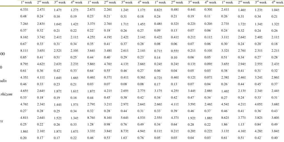
: statistically significant difference (P<0.01)Table 2. The ratio of the mean cytokines (IL-2, IL-4, IFN-, and TNF-concentration in BALB/c mice (n=5) treated with Ching-Wei-San or its individual herbal components for 4 weeks compared with the normal control (distilled water) group.
Reagents (mg/mL) IL-2 IL-4 IFN- TNF-
1st week 2nd week 3rd week 4th week 1st week 2nd week 3rd week 4th week 1st week 2nd week 3rd week 4th week 1st week 2nd week 3rd week 4th week
Ching-Wei-San 125 6.33±
0.48+
2.47±
0.24+
1.47±
0.16
1.27±
0.19
2.67±
0.23+
2.26±
0.21+
1.24±
0.31
1.17±
0.18
0.82±
0.24
0.48±
0.21-
0.44±
0.19-
0.30±
0.11-
2.41±
0.26+
1.46±
0.31
1.23±
0.34
1.04±
0.21
Ching-Wei-San 250 7.26±
0.37+
2.83±
0.32+
1.64±
0.21
1.42±
0.22
3.37±
0.22+
2.76±
0.18+
1.71±
0.26
1.45±
0.27
0.48±
0.09-
0.32±
0.13-
0.22±
0.07-
0.20±
0.06-
2.73±
0.24+
1.72±
0.32
1.34±
0.24
1.32±
0.26
Ching-Wei-San 500 8.16±
0.67+
3.74±
0.33+
2.41±
0.31+
2.11±
0.34+
4.25±
0.35+
4.19±
0.41+
2.42±
0.37+
2.14±
0.28+
0.42±
0.08-
0.41±
0.06-
0.21±
0.07-
0.11±
0.06-
3.11±
0.30+
2.64±
0.24+
2.40±
0.20+
2.11±
0.18+
ID-Ching-Wei-San 500 8.11±
0.85+
3.65±
0.41+
2.52±
0.51+
2.10±
0.25+
3.64±
0.44+
3.48±
0.40+
2.61±
0.29+
2.14±
0.23+
0.71±
0.14
0.55±
0.10
0.21±
0.06-
0.10±
0.05-
3.32±
0.51+
2.76±
0.34+
2.31±
0.27+
2.21±
0.28+
Ching-Wei-San 1,000 8.79±
0.61+
4.62±
0.36+
2.63±
0.42+
2.23±
0.33+
5.86±
0.64+
4.76±
0.55+
4.13±
0.46+
2.66±
0.27+
0.24±
0.06-
0.24±
0.04-
0.13±
0.05-
0.09±
0.03-
3.65±
0.38+
2.94±
0.41+
2.55±
0.31+
2.43±
0.32+
Angelicae sinensis radix 4.35±
0.46+
4.11±
0.33+
1.64±
0.23
1.66±
0.21
0.46±
0.03-
0.37±
0.07-
0.41±
0.08-
0.36±
0.08-
0.72±
0.17
0.46±
0.13-
0.12±
0.07-
0.07±
0.04-
2.38±
0.26+
2.46±
0.44+
3.24±
0.45+
2.86±
0.37+
Rehmanniae radixet rhizom 4.65±
0.33+
2.64±
0.18+
1.87±
0.19
1.61±
0.16
1.87±
0.44
4.21±
0.45+
2.65±
0.38+
2.77±
0.42+
3.17±
0.34+
4.25±
0.42+
3.44±
0.47+
2.88±
0.34+
1.46±
0.27
2.13±
0.24+
2.34±
0.33+
2.44±
0.31+
Coptidis Rhizoma 4.76±
0.27+
2.34±
0.28+
1.44±
0.25
1.57±
0.34
2.79±
0.32+
3.21±
0.28+
2.97±
0.44+
2.64±
0.31+
2.66±
0.33+
4.11±
0.39+
3.59±
0.46+
2.46±
0.37+
4.54±
0.46+
4.21±
0.41+
4.05±
0.36+
3.68±
0.43+
Moutan radicis cortex 4.81±
0.25+
2.64±
0.22+
1.52±
0.26
1.34±
0.33
8.76±
1.28+
8.16±
0.98+
5.64±
0.76+
4.53±
0.49+
2.55±
0.34+
4.37±
0.64+
1.92±
0.28
1.88±
0.22
8.62±
1.86+
3.77±
1.13+
3.82±
0.84+
3.60±
0.49+ Cimicifuga foetida 1.86±
0.20
2.16±
0.17+
1.87±
0.17
1.67±
0.22
3.33±
0.46+
3.84±
0.53+
8.73±
1.63+
4.94±
0.76+
0.11±
0.05-
0.21±
0.03-
0.20±
0.04-
0.22±
0.03-
3.13±
0.61+
4.16±
0.51+
4.28±
0.42+
3.84±
0.40+
Tetracyclin 4.85±
0.31+
4.25±
0.25+
4.31±
0.29+
4.13±
0.34+
0.48±
0.51-
0.42±
0.08-
0.33±
0.07-
0.37±
0.07-
0.23±
0.08-
0.21±
0.06-
0.18±
0.05-
0.18±
0.05-
1.12±
0.13
1.21±
0.16
1.34±
0.26
1.44±
0.22 Distilled water 1..00±
0.27+
1.00±
0.21+
1.00±
0.28+
1.00±
0.27+
1.00±
0.18-
1.00±
0.12-
1.00±
0.09-
1.00±
0.09-
1.00±
0.11-
1.00±
0.07-
1.00±
0.08-
1.00±
0.07-
1.00±
0.11
1.00±
0.13
1.00±
0.16
1.00±
0.15
ID-Ching-Wei-San: individual decocted Ching-Wei-San; +: ratio ≧ 2.00; -: ratio ≦ 0.50.
Gene symbol Fold up- or downregulation;
T-47D-B /T-47D-N
Fold up- or downregulation;
BXPC3-B /BXPC3-N
Fold up- or downregulation;
HT-29-B /HT-29-N BCL2L10
HRK FASLG LTA TNF TNFRSF9
7.6741 12.2101 5.579 15.455 7.21 3.2043
1.9053 -1.1019 1.1487 3.4822 21.1121 3.7064
2.6027 2.6574 16.6795 22.4711 27.8576 2.5847
Fig. 1. The major genes regulated by cells treated with berberine at a concentration of 5 times IC50 for three different cells, HT-29, T-47D, and BXPC3 detected by PCR array. The cells were treated for 24 hours and the regulated fold-changes were compared for “before” and “after”
treatment related to a housekeeping gene. (+: up regulation; -: down regulation)
國科會補助計畫衍生研發成果推廣資料表
日期:2013/10/29
國科會補助計畫
計畫名稱: 清胃散主成分黃連萃取之小檗鹼的抗單純皰疹病毒ˋ抗口腔癌及免疫調節功效 計畫主持人: 楊繼江
計畫編號: 99-2320-B-040-004-MY3 學門領域: 藥理及毒理
無研發成果推廣資料
99 年度專題研究計畫研究成果彙整表
計畫主持人:楊繼江 計畫編號:99-2320-B-040-004-MY3
計畫名稱:清胃散主成分黃連萃取之小檗鹼的抗單純皰疹病毒ˋ抗口腔癌及免疫調節功效 量化
成果項目 實際已達成
數(被接受 或已發表)
預期總達成 數(含實際已
達成數)
本計畫實 際貢獻百
分比
單位
備 註 ( 質 化 說 明:如 數 個 計 畫 共 同 成 果、成 果 列 為 該 期 刊 之 封 面 故 事 ...
等)
期刊論文 0 0 100%
研究報告/技術報告 0 0 100%
研討會論文 0 0 100%
論文著作 篇
專書 0 0 100%
申請中件數 1 1 100%
專利 已獲得件數 1 2 50% 件
件數 0 1 0% 件
技術移轉
權利金 0 0 100% 千元
碩士生 2 2 100%
博士生 0 0 100%
博士後研究員 0 0 100%
國內
參與計畫人力
(本國籍)
專任助理 0 0 100%
人次
期刊論文 2 3 67%
研究報告/技術報告 0 0 100%
研討會論文 0 0 100%
論文著作 篇
專書 0 0 100% 章/本
申請中件數 0 0 100%
專利 已獲得件數 0 0 100% 件
件數 0 0 100% 件
技術移轉
權利金 0 0 100% 千元
碩士生 0 0 100%
博士生 0 0 100%
博士後研究員 0 0 100%
國外
參與計畫人力
(外國籍)
專任助理 0 0 100%
人次
其他成果
(無法以量化表達之成
果如辦理學術活動、獲 得獎項、重要國際合 作、研究成果國際影響 力及其他協助產業技 術發展之具體效益事 項等,請以文字敘述填 列。)
無
成果項目 量化 名稱或內容性質簡述
測驗工具(含質性與量性) 0
課程/模組 0
電腦及網路系統或工具 0
教材 0
舉辦之活動/競賽 0
研討會/工作坊 0
電子報、網站 0
科 教 處 計 畫 加 填 項
目 計畫成果推廣之參與(閱聽)人數 0
國科會補助專題研究計畫成果報告自評表
請就研究內容與原計畫相符程度、達成預期目標情況、研究成果之學術或應用價 值(簡要敘述成果所代表之意義、價值、影響或進一步發展之可能性)、是否適 合在學術期刊發表或申請專利、主要發現或其他有關價值等,作一綜合評估。
1. 請就研究內容與原計畫相符程度、達成預期目標情況作一綜合評估
■達成目標
□未達成目標(請說明,以 100 字為限)
□實驗失敗
□因故實驗中斷
□其他原因 說明:
2. 研究成果在學術期刊發表或申請專利等情形:
論文:□已發表 □未發表之文稿 ■撰寫中 □無 專利:□已獲得 ■申請中 □無
技轉:□已技轉 □洽談中 ■無 其他:(以 100 字為限)
自古以來很多中草藥可藉由調控人體的免疫機制來達到治療的功用,或是調節免疫系統以 及加強細胞修補功能來達到抗癌的作用,先前的研究中我們探討了黃連及小蘗鹼的功用,是 具有相當好的抗癌能力,現進一步探討黃連及小蘗鹼是否有調控免疫系統的功能,PCR array 的結果顯示,也有著許多免疫調控的基因因為藥物處理之後造成了改變,往後可以更進一步 的去研究黃連及小蘗鹼免疫調控相關的機制。
3. 請依學術成就、技術創新、社會影響等方面,評估研究成果之學術或應用價 值(簡要敘述成果所代表之意義、價值、影響或進一步發展之可能性)(以 500 字為限)
1. 由小鼠體內細胞激素檢測發現,牙周病病態動物服用清胃散 7 日後 IL-4 呈現上升,IFN- γ呈現下降的趨勢,這顯示清胃散對於牙周病病態動物的抗發炎的功效可能與疫功能調節 作用有某種程度上的關係。而正常的實驗動物服用清胃散 7 日後 IL-2 則呈現上升趨勢亦 具有刺激免疫提升作用,這與傳統中醫的補脾說法相契合。我們由清胃散急性及亞急性毒 性安全性評估試驗中確認清胃散是一具有高度安全性的牙周病治療藥物,值得推廣與應 用。
2. 本研究之醫藥組合物可用以抑制習知引起口腔疾病之微生物,另可用於提高免疫系統 所分泌之細胞激素之量,以提升自身的免疫能力來達到預防或治療的作用。本研究係發現 中藥組合物於治療微生物引起之口腔疾病之新穎用途,並可以口服方式來調節免疫功能。