© 2007 The Electrochemical Society. 关DOI: 10.1149/1.2717501兴 All rights reserved.
Manuscript submitted September 22, 2006; revised manuscript received January 4, 2007. Available electronically April 3, 2007.
Recently, there has been extensive interest in the creation of po-rous materials with three-dimensional periodicity due to their poten-tial applications, such as catalysts,1 chemical2 and gas3 sensors, photonic4and optoelectronic devices,5thermal insulation materials,6 and membranes.7 In general, the atomic clusters,8 nanoparticles,9 and nanostructures10of materials exhibit size-dependent properties that are profoundly different from their corresponding bulk material counterparts. Hence, it can be anticipated that porous material in the nano or submicrometer scale with a larger surface area will exhibit improved performance in applications, such as those requiring trans-port through the pores or interaction with the surface. Moreover, it has been suggested that metals with well-ordered porous networks possess interesting photonic properties.11
For porous metals prepared using conventional methods, such as powder sintering, slip casting and fiber metallurgy, it is difficult to achieve materials with high surface areas.12 Although some microporous13or mesoporous14metals exhibit high surface areas, a significant fraction of the pores is not easily accessible to large fillers due to the relatively small pore diameters. Thus, well-ordered macroporous metals fabricated via a template-mediated method have attracted a great deal of attention recently due to their efficient process and wider variety of applications.
Template-mediated technology is an important process for form-ing special nanostructures, including one-dimensional 共1D兲 nanofiber15 and three-dimensional 共3D兲 well-ordered macroporous structures.16By utilizing a nanochannel-filling technology, a replica of the host template can be obtained. The entire process has two key factors. The first is the choice of filling technology. Many methods15,17-19 for nanochannel filling have been developed in which the filling for nanochannels is performed by chemical vapor deposition,17a dipping process,18and electrophoresis,15but the top of the template usually encounters the issue of jamming. However, the mechanism of filling nanochannels by using an electrodeposition20is from bottom to top, and the jamming of tem-plate surfaces can be avoided.21Hence, electrodeposition is a pow-erful tool for the fabrication of nanostructures with high structural quality and aspect ratio.22The fabrication of high-quality and appro-priate templates is the second factor for creating high-quality repli-cas via a template-mediated process.23-25For example, the colloidal crystal, in which monodispersed colloids self-assemble into an or-dered 3D artificial crystal structure, often acts as the template for forming macroporous structures.26-31Hence, by combining a
high-quality template and efficient nanochannel-filling technology, the template-mediated process can be easily manipulated.
In this report we prepared a well-ordered nickel macroporous structure via a template-mediated electrodeposition process. Be-cause the colloidal crystal template via the capillary-enhanced pro-cess exhibits the face-centered cubic 共fcc兲 structure, the nickel replica, which was constructed by using a template-mediated elec-trodeposition process, has the same structure. In addition, according to the relationship between current values and electrodeposition time, the behavior of nickel electrodeposition in the interstitial spaces of colloidal crystal template can be determined. Hence, well-ordered nickel nanorod, nanoporous arrays, and macroporous struc-ture can be fabricated via a one-step process. Further, the nickel macroporous structures covered with hollow sphere arrays on the surface are also constructed due to different interstitial-spaces-filling mechanisms.
Experimental
Preparation of monodispersed colloidal particles.— The
mono-dispersed P共St-co-MAA兲 microspheres were prepared by using an emulsifier-free-emulsion copolymerization of styrene共St兲 and meth-acrylic acid共MAA兲 based on the method proposed by Wang and Pan.32By controlling the concentration of MAA and reaction time, two batches with different diameters of colloidal particles共180 and 300 nm with a relative standard deviation smaller than 5%兲 were formed.
Fabrication of colloidal crystal template.— An efficient process
for preparing high-quality colloidal crystals, called capillary-enhanced method, was used in this study.31 First, the substrate coated with indium tin oxide共ITO兲 thin film was placed horizontally at the bottom of a container which was filled with an aqueous dis-persion of the P共St-co-MAA兲 microspheres obtained by ultrasonica-tion. The formation of colloidal crystals can be obtained by the self-assembly of microspheres at 45°C and under a high-humidity 共90%兲 condition. After evaporation was terminated in 24 h, the sub-strate was picked up with a colloidal crystal film thereon.
Synthesis of nickel macroporous structure.— Cathodic dc
elec-trodeposition was conducted in a conventional three-electrode cell. The aqueous solution used for Ni deposition33 was composed of NiSO4共2.13 M兲, NiCl2共0.35 M兲, and H3BO3共0.43 M兲. The work-ing electrode was a conductive indium-tin oxide共ITO兲-glass sub-strate with a deposited colloidal crystal film. A platinum plate and silver/silver chloride were used as the counter and reference elec-trodes, respectively. After finishing nickel electrodeposition, the col-z
loidal crystal template can be removed by calcinations at 450°C for 4 h. Finally, nickel well-ordered macroporous film was formed on the substrate.
Instrumentation and characterization.— Surface morphology
was observed using a Hitachi S4100 field emission scanning elec-tron microscope, and the quality of colloidal crystal template was gauged by using a UV-2001 UV-visible spectrometer from Hitachi. Electrochemical synthesis of nickel was performed by a potentiostat 共EG&G, model 263兲.
Results and Discussion
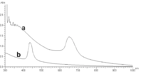
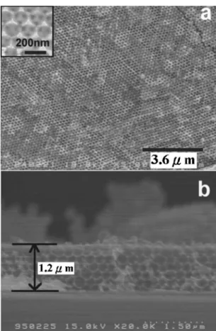
The colloidal crystal templates for the synthetic macroporous structure studied in this work were prepared using an efficient tech-nology described in Ref. 31. SEM images of the共111兲 surface of the synthetic reveal a close-packed arrangement, as shown in Fig. 1a, where some vacancies are found in this well-ordered array. A typical result is shown in Fig. 1b, where the different facets of square and hexagonal arrangements can be clearly observed at the facets共100兲 and共111兲, respectively. In fact, a square arrangement can only cor-respond to a兵100其-type surface of a fcc structure, instead of in the hexagonal close-packed共hcp兲. Hence, this colloidal crystal has a fcc structure. According to discussion of modified Bragg’s law,34a high-quality colloidal crystal in a large area can diffract light and exhibits a stop band in its transmission spectrum. Figure 2 shows that the colloidal crystals consisted of 180 and 300 nm colloidal particles and have absorptive peaks at 451 and 654 nm, respectively. The result means that the colloidal crystals are of good crystallinity.
Figure 3 shows the nickel electrochemically deposited into the colloidal crystal template from a nickel sulfate solution at −1.0 V 共Ag/AgCl兲. Comparing the two insets, the interstitial space of col-loidal crystal template had been filled. Because the filling behavior of the electrodeposition process is from bottom to top, surface
jam-Figure 1. SEM images of well-ordered colloidal crystals prepared by a
capillary-enhanced method:共a兲 top view and 共b兲 side view.
Figure 2. The absorptive spectra of
col-loidal crystals with the different colloid diameters of共a兲 300 and 共b兲 180 nm.
Figure 3. SEM surface images of colloidal crystal filled with nickel via an
electrodeposition process. The inset figures indicate that colloidal crystal templates are either unfilled or filled with nickel.
ming is avoided. After the nickel electrodeposition process, the well-ordered colloidal crystal was not destroyed due to the presintering process performed at 135°C for 2 h.
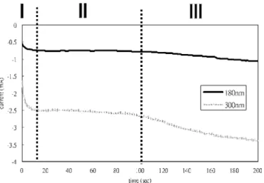
Figure 4 shows a typical current-time transient for nickel depo-sition into the colloidal crystal template with 180 and 300 nm col-loidal diameter, respectively, which is divided into three stages ac-cording to the filling behaviors. In the initial stage, from 0 to 15 s, nickel ions in the electroplating solution are induced to migrate by the applied potential and accumulate onto the working electrode, resulting in the increase of current values. Subsequently, nickel is formed at the bottom of the template not masked by the colloids by a reduction reaction, as depicted in Fig. 5a. In the second stage, from 15 to 100 s, nickel metal forms in the interstitial spaces of the tem-plate on the working electrode, as depicted in Fig. 5b. The current values are at a stable state. This phenomenon of smooth current curve with small oscillation has also been reported for nickel-filling the interstitial spaces of colloidal crystal template by Sumida et al.35 In the third stage, 100–200 s, the level of nickel-filling into the interstitial spaces almost reaches the surface of the colloidal crystal template, as depicted in Fig. 5c. The time-dependence variation of current differs from that in the second stage. In general, the behavior of electrodeposition can be described explicitly by employing Fara-day’s law.36Hence, it can be used to explain the behavior of nickel-filling the colloidal crystal template, which is expressed as
W = ItM
ZF 关1兴
where W is the weight of electroplated material, I and t are electro-plating current and time, respectively, M is the molecular weight of electroplated material, Z is the electric charge, and F is the Faraday constant. Then Eq. 1 can be modified, as shown in Eq. 2
A =
冉
1h
tM
ZF
冊
I 关2兴where is the density of electroplated material. The values of A and
h indicate the area and height of electroplated material, respectively.
According to the modified Faraday’s law共Eq. 2兲, the current value 共I兲 increases with increasing area 共A兲. When the height of nickel-filling is above the surface of the colloidal crystal template, the electrochemical reaction area changes from projected area of the interstitial spaces into planes. The growth of nickel metal extends from the spaces among the colloids toward the colloidal surface, until colloidal particles on the template surface are covered com-pletely. Hence, current value gradually elevates due to the gradual increase of electrochemical reaction area. The result differs from
that in the electrodeposition of polypyrrole into the voids of colloi-dal crystal,37where a rapid increase in current is suggested to be caused mainly by a rapid increase of the electrochemical reaction area when the growing surface reaches the membrane/bulk solution interface.
After removing the colloidal crystal template by calcinations at 450°C for 4 h, nickel 共with the presence of NiO on the surface兲 macroporous structures are obtained. Moreover, porous structure with different pore sizes can be formed by employing a colloidal crystal template having different colloid diameters. Figure 6a shows the SEM image of the surface of a 3D ordered nickel replica formed by depositing into a colloidal crystal template with 180 nm colloid particles. It is apparent that a close-packed arrangement on the top-most plane of the replica is exhibited. The inset of Fig. 6a shows a symmetry indicating that the spherical holes are located above three
Figure 4. Chronoamperometric response of nickel electrodeposition in the
interstitial spaces of colloidal crystal template with 180 and 300 nm colloid diameter.
Figure 5. Schematic diagrams of nickel infiltration into colloidal crystal
templates at different electrodeposition stages of共a兲 0–15, 共b兲 15–100, 共c兲 100–200 s.
neighboring hollow sites in the crystal. Each cavity formed by the polystyrene particles has three dark spots corresponding to the con-tact points with the three particles in the layer below. In the cross section of Fig. 6b, the well-ordered macroporous structures are dem-onstrated to be fabricated from bottom to top. These nickel nano-structures are also called inverse opals.
As shown in Fig. 7a, the nickel porous arrays in a large area, fabricated by depositing into colloidal crystal template with 300 nm colloid particles, appear well-ordered in the free surface of the共111兲 texture. The inset of Fig. 7a also shows that each air sphere rests on three neighboring spheres below along the关111兴 direction. More-over, the same 3D macroporous structure consisting of many spheri-cal void layers from the side view can be also observed, as shown in Fig. 7b. Hence, nickel inverse opals can be obtained via a template-mediated electrodeposition process.
In general, the height of nickel-filling into the colloidal crystal template increases with increasing electrodeposition time. Further, by using the colloidal crystal templates consisting of the different colloid diameters, different kinetics relationships for nickel-filling occurs with different electrodeposition current values. Hence, the height of nickel-filling in the colloidal crystal with 180 and 300 nm colloid diameter, respectively, can be discussed according to a modi-fied Faraday’s law共Eq. 3兲, as depicted by the following
Ah =
冉
1
tM
ZF
冊
I 关3兴The nickel-filling volume共Ah兲 increases with increasing the current values. As depicted in Fig. 4, the ratio between current values 共−2.6 mA兲 upon filling a colloidal crystal with 300 nm colloid di-ameter and 180 nm共−0.75 mA兲 is equal to 3.47 during the elec-trodeposition time of 100 s. This also results in a ratio of nickel-filling volume between 300 and 180 nm colloidal diameter being
almost equal to 3.47 at the same time. From the observation of Fig. 6b and 7b, the ratio between the height共4.1 m兲 for nickel-filling into colloidal crystal with 300 nm colloid diameter and 180 nm 共1.2 m兲 is equal to 3.42. It can be demonstrated that essentially both areas not occupied by the colloids are the same for the case using 180 and 300 nm colloid particles. These areas indicate the nickel-filling sites, shown as shaded areas of Fig. 8. First, both black trigonal regions for colloids with 180 and 300 nm diameter can be calculated to be 1317 and 3657 nm2, respectively. The numbers of
the trigonal region for colloids with 180 and 300 nm diameter in the
Figure 6. SEM surface images of 3D well-ordered nickel macroporous
structure prepared by using colloidal crystal templates with the colloid di-ameters of 180 nm at the共a兲 top view and 共b兲 side view.
Figure 7. SEM surface images of 3D well-ordered nickel macroporous
structure prepared using colloidal crystal templates with the 300 nm colloid diameters at the共a兲 top view and 共b兲 side view.
Figure 8. The illustration of projected area to be filled by Ni in the
for ionic fluid near the electrical double layers in the fluid system.38 Therefore, the migration speed of nickel ions in the interstitial spaces of colloidal crystal with 180 nm diameter is lower than for the case of 300 nm colloid particles. According to the semiquantita-tive analysis, the current values in the 300 nm colloidal system are larger than ones in the 180 nm colloidal system.
In the initial stage of the nickel electrodeposition process, from 0 to 15 s, the different morphologies of the porous array can be ob-tained by manipulating electrodeposition time. Figure 9a shows a monolayer of nickel porous array fabricated via an electrodeposition process for 10 s. Each air hole is surrounded by six spherical voids, which is a hcp network of circular holes. The pattern of pore array indicates a replica of colloidal crystal in 2D. The inset of Fig. 9a shows a cross-sectional image of a nickel macroporous monolayer in which only one layer of nickel macropore exists. When elec-trodeposition time is decreased to 5 s, nanodot arrays can be ob-tained as shown in Fig. 9b. This structural arrangement is similar to the porous arrays in a close-packed network. The inset figure with a high magnification shows that one hole is surrounded by six nickel nanodots. These sites occupied by nickel nanodots are the interstitial spaces among three neighboring colloidal particles. Because these sites covered by ITO thin film are exposed to the electrodeposition solution, they possess a large free surface area for the nickel reduc-tion and deposireduc-tion. Hence, the nickel ions in the electrodeposireduc-tion solution migrate toward these sites and more easily produce nan-odots of nickel metal via an electrochemical reduction reaction.
Further, the phenomenon of nickel-filling interstitial spaces at the template bottom can be described according to the chronoampero-metric response at short time, as shown in Fig. 9c. From 0 to 3 s, because of the supply of a negative potential, nickel ions with posi-tive charge near the electrode in the solution migrate fast and accu-mulate onto the ITO-glass substrate not covered by colloids. Subse-quently, nickel metal is reduced at these sites, resulting in a rapid increase of the electrodeposition current. Because of the presence of time-dependent diffusion regime,39 the increasing trend of elec-trodeposition current became smaller from 3 to 8 s. Nevertheless, these nickel ions are reduced to metal gradually at the same sites. Meanwhile, the nickel nanorod is fabricated in 5 s. Due to the ex-istence of a colloidal crystal template, the nickel metals cannot grow freely. The actual electrode area is the cross section of the interstitial spaces of the template, which leads to a stable current during the period of Ni filling into the template. Hence, current is about the same from 8 to 15 s. The nickel macroporous monolayer also forms in 10 s via the template-mediated process. Therefore, three types of current variation during electrodeposition respectively lead to the fabrication of nanorods and a macroporous monolayer.
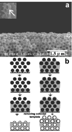
When the electrodeposition time proceeds for 120 s, a novel nickel nanostructure consisting of macroporous structure covered with a hollow sphere array can be formed after removing colloidal crystal template, as shown in Fig. 10a. From the figure inset, the nickel hollow spherical nanostructures can be observed clearly. The
behavior of nickel-filling interstitial spaces on the top surface of colloidal crystal template is different from the nanostructure at the bottom and middle. At the template bottom and middle, nickel metal is reduced and grows at the interstitial spaces among the colloidal
Figure 9. SEM images of a 2D nickel nanostructure based on 共a兲 a
macroporous monolayer and 共b兲 nanodot arrays prepared by a template-mediated electrodeposition process for 10 and 5 s, respectively.共c兲 Chrono-amperometric response of nickel electrodeposition in the interstitial spaces of colloidal crystal template with 300 nm colloid diameter for 15 s.
particles according to previous results, as depicted in the left of Fig. 10b. This mechanism results in a well-ordered, macroporous array on the top of the nanostructure, as shown in Fig. 7b. However, they are produced and coated around the colloidal surface, instead of interstitial spaces, on the template top. The mechanism of forming a hollow sphere array on the top is depicted in the right of Fig. 10b. This is attributed to an electrostatics-induced effect between colloi-dal particles and the nickel ions.40 The zeta-potential of P共St-co-MAA兲 colloidal particles is measured to be negative in our preliminary study 共not shown here兲. A great number of positive nickel ions in the solution are attracted toward the working electrode by the applied constant potential关−1 V to 共Ag/AgCl兲兴 during the electrodeposition process. This phenomenon results in many nickel ions being adsorbed onto the colloidal surface at the template top, until the colloidal crystal templates are filled with nickel metal and reach the top of the template. Finally, nickel metal is reduced pref-erentially at these sites, adsorbing nickel ions. Subsequently, nickel metal grows and completely coats the colloidal surface. After re-moving the template, the nickel hollow sphere nanostructures on the surface can be obtained if the process is terminated before the com-plete filling of the interstitial spaces. Therefore, at the bottom of colloidal crystal templates, well-ordered nanodot and nanoporous arrays form more easily due to the preferential filling of nickel into the interstitial spaces among the colloids. On the contrary, a
macroporous structure covered with hollow sphere arrays is fabri-cated, resulting from preferential adsorption coating of nickel around the colloids. Because this nanostructure possesses both a large surface area and a 3D well-ordered arrangement, it has poten-tial for many applications, including magnetic devices,41sensors,42 and surface-enhanced Raman spectroscopy.43
Conclusions
A high-quality, colloidal crystal template can be fabricated via the capillary-enhanced method. The mechanism for nickel-filling into the interstitial spaces of colloidal crystal templates can be di-vided into four stages according to the current values during the electroplating process. Further, the ratio of height of nickel-filling colloidal crystal with 300 and 180 nm diameter can be estimated using the ratio of their current values in the second stage of the electrodeposition process. In addition, well-ordered nanorods and nanoporous arrays can be formed in 5 and 10 s, respectively, and the nickel hollow spherical nanostructures can be obtained on the sur-face for an electrodeposition time of 120 s. Therefore, various kinds of nickel nanostructure can be produced via the one-step elec-trodeposition process. Moreover, their forming mechanism can be described according to the relation of current-time responses.
Acknowledgments
Financial support from the National Science Council, Taiwan, through contract no. NSC93-2216-E-006-039 is greatly appreciated. National Cheng Kung University assisted in meeting the publication costs of this article.
References
1. S. I. Matsushita, T. Miwa, D. A. Tryk, and A. Fujishima, Langmuir, 14, 6441 共1998兲.
2. J. H. Holtz and S. A. Asher, Nature (London), 389, 829共1997兲.
3. T. Yamada, H. S. Zhou, H. Uchida, M. Tomita, Y. Ueno, I. Honma, K. Asai, and T. Katsube, Microporous Mesoporous Mater., 54, 269共2002兲.
4. E. Yablonovitch, T. J. Gmitter, R. D. Meade, and A. M. Rappe, Phys. Rev. Lett., 67, 3380共1991兲.
5. M. Imada, S. Noda, A. Chutinan, and T. Tokuda, Appl. Phys. Lett., 75, 316共1999兲. 6. T. Bitzer, Honeycomb Technology, Chapman and Hall, London共1997兲. 7. R. E. Kesting, Synthetic Polymer Membranes, Wiley, New York共1985兲. 8. A. W. Castleman and K. H. Bowen, J. Phys. Chem., 100, 12911共1996兲. 9. A. P. Alivisators, Science, 271, 933共1996兲.
10. M. Sundaram, S. A. Chalmers, P. F. Hopkins, and A. C. Gossard, Science, 254, 1326共1991兲.
11. D. F. Sievenpiper, M. E. Sickmiller, and E. Yablonovitch, Phys. Rev. Lett., 76, 2480共1996兲.
12. V. Shapovalov, MRS Bull., 19, 24共1994兲.
13. Y. Zhou and M. Antonietti, Adv. Mater. (Weinheim, Ger.), 15, 1452共2003兲. 14. G. S. Attard, P. N. Bartlett, N. R. B. Coleman, J. M. Elliott, J. R. Owen, and J. H.
Wang, Science, 278, 838共1997兲.
15. Y. C. Wang, I. C. Leu, and M. H. Hon, J. Mater. Chem., 12, 2439共2002兲. 16. Y. A. Vlasov, X. Z. Bo, J. C. Sturm, and D. J. Norris, Nature (London), 414, 289
共2001兲.
17. A. A. Zakhidov, R. H. Baughman, Z. Iqbal, C. X. Cui, T. Khayrullin, S. O. Dantas, I. Marti, and V. G. Ralchenko, Science, 282, 897共1998兲.
18. Z. Z. Gu, A. Fujishima, and O. Sato, Chem. Mater., 14, 760共2002兲.
19. M. T. Wu, I. C. Leu, J. H. Yen, and M. H. Hon, Electrochem. Solid-State Lett., 7, C61共2004兲.
20. T. S. Eagleton and P. C. Searson, Chem. Mater., 16, 5027共2004兲.
21. Y. W. Chung, I. C. Leu, J. H. Lee, and M. H. Hon, J. Cryst. Growth, 275, e2389 共2005兲.
22. H. Yan, C. F. Blanford, J. C. Lytle, C. B. Carter, W. H. Smyrl, and A. Stein, Chem.
Mater., 13, 4314共2001兲.
23. C. T. Kresge, M. E. Leonowics, W. J. Roth, J. C. Vartuli, and J. S. Beck, Nature
(London), 359, 710共1992兲.
24. A. Imhof and D. J. Pine, Nature (London), 389, 948共1997兲.
25. D. Zhao, J. Feng, Q. Huo, N. Melosqh, G. H. Fredrickson, B. F. Chemelka, and G. D. Stucky, Science, 279, 548共1998兲.
26. M. D. Sacks and T. Y. Tseng, J. Am. Ceram. Soc., 67, 526共1984兲.
27. R. Mayoral, J. Requena, J. Moya, C. Lopez, A. Cintas, H. Miguez, F. Meseguer, L. Vazquez, M. Holgado, and A. Blanco, Adv. Mater. (Weinheim, Ger.), 9, 257共1997兲. 28. R. C. Salvarezza, L. Vázquez, H. Míguez, R. Mayoral, C. López, and F. Meseguer,
Phys. Rev. Lett., 77, 4572共1996兲.
29. P. Jiang, J. F. Bertone, K. S. Hwang, and V. L. Colvin, Chem. Mater., 11, 2132 共1999兲.
30. D. Mei, H. Liu, B. Cheng, Z. Li, D. Zhang, and P. Dong, Phys. Rev. B, 58, 35 共1998兲.
31. Y. W. Chung, I. C. Leu, J. H. Lee, and M. H. Hon, Appl. Phys. A, 79, 2089共2004兲.
Figure 10.共a兲 SEM surface image of a macroporous structure covered with
hollow sphere arrays prepared for 120 s by a template-mediated electrodepo-sition process.共b兲 The illustration for forming a nickel macroporous structure 共left figure兲 and a hollow-sphere-array-covered macroporous structure 共right figure兲.