國立臺灣大學生物資源暨農學院生物產業機電工程學系 博士論文
Department of Bio-Industrial Mechatronics Engineering College of Bio resources and Agriculture
National Taiwan University Doctoral Dissertation
抗 STAT3 圈套寡核苷酸應用於癌細胞診療之研究 On the Anti-STAT3 Decoy Oligonucleotides for
Cancer Cell Theranostics
王紹任 Shao-Jen Wang
指導教授:陳林祈 博士 Advisor: Lin-Chi Chen, Ph.D.
中華民國 104 年 7 月
July, 2015
摘要
本 研 究 旨 在 開 發 一 系 統 性 方 法 於 提 升 圈 套 寡 核 苷 酸 (decoy
oligodeoxynucleotide, dODN) 對於轉錄訊息傳遞活化子 3 (signal transducer and activator of transcription 3, STAT3) 的親和力,並將此系統性方法之產物運用於癌 細胞抑制,以及延伸發展成活化之轉錄因子的感測方法與靶向性診療系統。
過度活化的 STAT3 可導致癌細胞的抗凋亡表現與抗藥性,其過度表現的下
游蛋白質在此過程扮演了重要的角色。以 Bcl-xL 蛋白質為例,其已被證實會影響
細胞凋亡機制並降低多柔比星 (doxorubicin, DOX) 抑制癌細胞的功效。為了阻礙
過度活化的 STAT3 之作用,目前廣泛使用的圈套寡核苷酸模仿了核酸中與轉錄
因子結合的啟動子 (promoter) 序列,可抑制轉錄因子並進而促進癌細胞進入凋亡
程序。事實上圈套寡核苷酸可以被視為一種細胞內核酸抗體,其透過結構(雙股 螺旋)以及特定鹼基序列達到選擇性蛋白質結合。借鏡適體 (aptamer) 的發展概
念,本研究的目標為藉由系統性方法重新設計 STAT3 圈套寡核苷酸的鹼基序
列,讓其擁有更高的轉錄因子親合力,進而更有效地抑制異常活化的轉錄因子。
本研究所提出的系統性方法結合了網路伺服器運算 (PiDNA)、寡核苷酸親合 力試驗以及寡核苷酸細胞抑制試驗。該系統性方法的產物 (命名為 hpdODN-Pi) 較
目前廣泛使用的序列具備 (1) 10%~29%的 STAT3 親合力提升,(2) 1.7~2.3 倍的選
擇性提升 (STAT3-to-STAT1),(3) 約 20% 的抗凋亡蛋白質負調控能力提升,以及
(4) 約 20% 的癌細胞抑制效力提升。相較於 hpdODN-Pi 成功抑制癌細胞,其對纖
維母細胞(對照組,沒有 STAT3 過度活化)則不具顯著作用,顯示 hpdODN-Pi
可選擇性抑制STAT3 過度活化之癌細胞。
基於 hpdODN-Pi 的親合力特性,本研究提出一個運用 hpdODN-Pi 與螢光分
子形成之複合物 (complex of hpdODN-Pi and dye, 簡稱 PiD) 感測活化之轉錄因子
的方法(in situ)。當此複合物遭遇活化之轉錄因子時,嵌入於 hpdODN-Pi 雙股序
列中的螢光分子 (SYBR green, 或 Quinacrine) 便會因轉錄因子的結合競爭(轉錄
因子對序列的親合力高於螢光分子)而自圈套寡核苷酸脫離,此導致的螢光訊號
變化便可用以定量感測活化的轉錄因子,感測線性區間為5 nM 到 100 nM。直接
給予活細胞 PiD 時,因活化的轉錄因子觸發之螢光訊號變化亦可由螢光顯微鏡直
根 據 hpdODN-Pi 的 高 STAT3 親 合 力 與 PiD 的 成 果 , 本 研 究 提 出 以 hpdODN-Pi 和 DOX 形成之複合物 (DOX-intercalated hpdODN-Pi, 簡稱 DOXON)
對 STAT3 異常活化之癌細胞進行靶向性診療的概念與方法。靶向性診療系統需
具備三個必要機制,包含辨識目標、發出訊號、以及觸發藥物作用。DOXON 以 hpdODN-Pi 辨識活化之 STAT3 蛋白質並與其進行結合,嵌入的 DOX 因為結合競
爭而被釋放至細胞質中,在488 nm 光激發下可發出 590 nm 螢光訊號。因 STAT3
的核酸結合位被 hpdODN-Pi 佔據,導致細胞的抗凋亡基因表現下降,而且被釋放
的 DOX 進 入 細 胞 核 並 嵌 入 核 酸 中 , 促 使 細 胞 啟 動 凋 亡 機 制 。 其 中 , 由 於 hpdODN-Pi 的基因調節與 DOX 的機轉可產生協同作用,所以複合物的癌細胞抑 制效果大於各別的單獨抑制效果總合。此外,DOXON 只於 STAT3 過度活化之細
胞內產生作用(如 MCF-7 和 HepG2 細胞),反之則沒有效果(如心肌細胞和纖維
母細胞),證實了DOXON 為一有效的靶向性診療系統。
本研究的貢獻在於提出並驗證了圈套寡核苷酸可經由系統性方法重新設計以 提高其親合力與選擇性,進而提升圈套寡核苷酸抑制癌細胞的功效。此外,本研 究根據其親合力特性發展出的轉錄因子感測方法與靶向性診療系統,發掘出了圈 套寡核苷酸的多功能性,讓其不限縮於疾病治療研究而可更進一步運用於感測和 診療。上述的研究成果亦可望推廣至其它種類的轉錄因子,促進圈套寡核苷酸在 治療、診斷以及診療的研究發展。
Abstract
This study develops a systematic method that enhances the affinity of the decoy oligodeoxynucleotide (dODN) against the signal transducer and activator 3 (STAT3), which is a kind of oncogenic transcription factor (TF) in adult human cell. The product obtained through this systematic method is further applied in the cancer cell inhibition.
Besides, the product has also been used to develop an activated transcription factor sensing method and targeting theranostic system.
Over-activated STAT3 causes abnormal proliferation and drug resistance of the cancer cell. Behind, the over-expressed downstream proteins (e.g. cyclin D1 and Bcl-xL) play the very important role in inducing those phenomenons. For instance, the Bcl-xL has been proven that it disrupts the apoptotic process and decreases the cancer inhibitory potency of doxorubicin. To block the abnormal STAT3 activation, the dODN that mimics the c-fos promoter has been widely used. It inhibits the activity of the oncogenic transcription factor and triggers the apoptosis. In fact, the dODN is a kind of intracellular nucleotide antibody. It binds to the specific target protein with its double- strand structure and particular base arrangement. Refer to the aptamer evolution, this study aims to re-design the STAT3-dODN through a sytematic method to achieve the sequence with higher target binding affinity for equals to higher inhibitory potency.
We report an systematic selection (combines in silico and in vitro method) to quickly evolve a potent anti-STAT3 hairpin dODN (named as hpdODN-Pi). Binding assays proved that hpdODN-Pi featured the best STAT3-binding affinity and STAT3-to- STAT1 selectivity among the dODNs reported to date. In the MCF-7 cell model, RT- qPCR confirmed that hpdODN-Pi effectively suppressed the downstream mRNAs of STAT3 without affecting STAT1-related expression after transfection.
In the following, this study proposes a dye-intercalated dODN system for sensing the oncogenic STAT3. This system is constructed with the hpdODN-Pi and the intercalated fluorescent dye (name as Pid). For in vitro STAT3 detection and intracellular STAT3 imaging, the signal comes from the release of intercalated dye due to the binding competition from STAT3. The fluorescence signal outputs are designed as the “signal- off” type and the “signal-on” type with different intercalating molecule. Either system shows a detection range between 10-100 nM of protein.
In the last part of the research, the concept of tackling the STAT3-associated cancer cell lines with dox-intercalated decoy oligonucleotide complex (DOXON) is proved. Before internalization, DOXON remains non-dissociated and does not emit fluorescence. After internalization mainly through the pinocytosis and specific recognition of over-activated STAT3, DOXON dissociates into therapeutic dODN for STAT3 binding and releases DOX for taking anti-cancer effect. In addition, free DOX emits fluorescence indicating the presence of over-activated STAT3. Since the disassembly of DOXON is caused by STAT3 binding competition, the complex is selective and will not affect the normal cells without STAT3 over-activation. Moreover, the disassembled dODN and DOX exert dual pathway intervention and synergistic effect, which bring the precise and potent cell-inhibition.
Table of Contents
摘要 ... i
Abstract ... iii
Table of Contents ... v
List of Figures ... vii
List of Tables ... xiii
Frequent abbreviations ... xiv
1. Introduction ... 1
1.1. The “decoy” of the therapeutic oligonucleotides ... 2
1.2. Decoy oligodeoxynucleotide engineering ... 4
1.3. Decoy oligodeoxynucleotide in therapeutics ... 9
1.4. Oligodeoxynucleotide in diagnostics ... 16
1.5. Experimental considerations ... 20
1.6. Objectives ... 27
2. Literature review ... 28
2.1. Decoy ODN against the STAT3 and its therapeutic application ... 28
2.1.1. STAT3 activation in cancer ... 28
2.1.2. Therapeutic nucleotide for STAT3 regulation ... 32
2.1.3. STAT3 decoy oligodeoxynucleotide ... 35
2.2. dODN in sensing application ... 40
2.2.1. oligonucleotide-based sensing system ... 40
2.2.2. protein sensing system with engineered sequence ... 42
2.2.3. diagnosis application of dODN ... 51
2.3. DOX-dODN in synergistic anti-cancer and theranostic application .... 54
2.3.2. Dual pathway intervention ... 61
2.3.3. Intercalation drug loading ... 64
3. Enhancing the potency of STAT3-dODN and use it in cancer cell therapy ... 69
3.1. Brief introduction ... 69
3.2. Experimental ... 72
3.3. Results and Discussion ... 76
4. Potency enhanced dODN in protein sensing ... 112
4.1. Brief introduction ... 112
4.2. Experimental ... 115
4.3. Results and Discussion ... 118
5. Potency enhanced dODN in theranostics ... 162
5.1. Brief introduction ... 162
5.2. Experimental ... 164
5.3. Results and Discussion ... 167
Conclusion ... 199
References ... 201
List of Figures
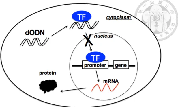
Figure 1.1 The principle of how dODN blocks the activity of the transcription factor in interest.
The dODN-bound TF is not able to cross the nucleus membrane to exert the down stream
functions. 4
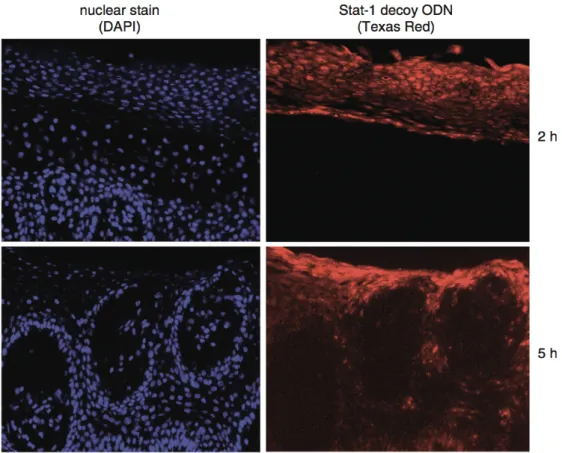
Figure 1.2 Fluorescence microscopic image that demonstrates the uptake of a Texas Red- labelled Stat-1 decoy ODN into the human skin cells (red). The nucleus was stained with
DAPI (blue). (Shangguan D, et al. 2008) 8
Figure 1.3 An illustration briefly describes the gene expression in cell. 18 Figure 1.4 Research framework of this study. The c-fos derived STAT3-hpdODN is potency-
enhanced with the proposed process. The enhanced hpdODN is further applied in the STAT3 sensing and anti-cancer theranostic application. 27 Figure 2.1 (A) Structure and function domains for STAT1-6 protein. (B) STAT3 activation and
signalling routes. (Bello et al., 2014) 29
Figure 2.2 Schematic illustration of (A) polymerase chain reaction and (B) rolling circle
amplification. 42
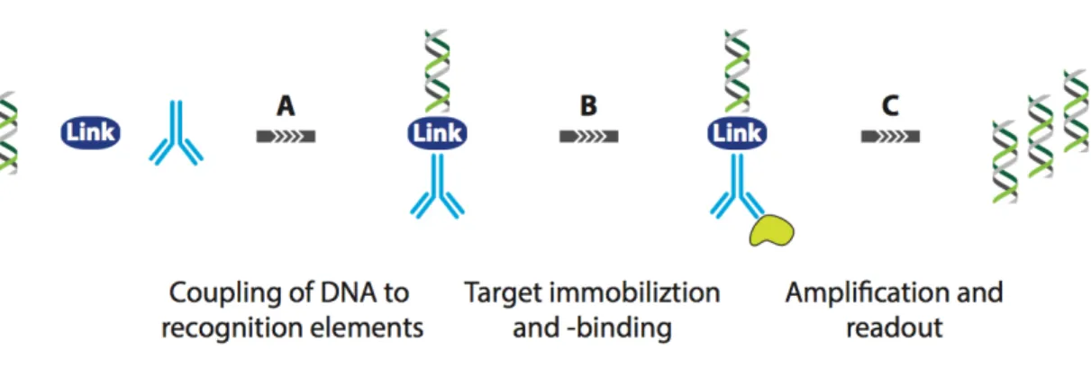
Figure 2.3 Schematic illustration of IPCR. (A)The dsDNA is firstly conjugated to the antibody with linkers. (B) Target protein binds to the anti-body and (C) the dsDNA is subsequently
amplified. 43
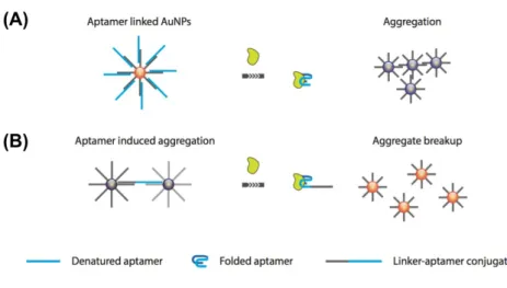
Figure 2.4 Gold nanoparticle-based protein sensing systems. Aptamer release-induced (A)
aggregation and (B) breakup. 46
Figure 2.5 Working principle of the CESA. 49
Figure 2.6 Working principle of the NEFSA. 50
Figure 2.7 The preparation of Apt-hybr-TCL-SPIONs and Dox@Apt-hybr-TCL-SPIONs
developed by Yu’s group. 60
Figure 2.8 (A)The complex of lipid-PEG and PLGA that contains the PTX (therapeutic purpose) and dye (diagnostic purpose), with the aptamer for targeting. (B) Self- assembled complex with aptamer as the targeting ligand. (C) A poly-aptamer-drug complex that carries the DOX through the intercalation mechanism. 65
Figure 3.1 The steps toward enhancing the potency (STEP) that used to generate a potent STAT3-inhibitory dODN comprises the following five steps. 77 Figure 3.2 The verification of the STAT1-dODN’s function in regulating the STAT1 down-
stream gene expression. 80
Figure 3.3 The cell mortality evaluation of STAT3-dODN, STAT3-dODN+IFN-γ, STAT3-
dODN+IFN-γ+STAT1-dODN. 81
Figure 3.4 Results of in silico selection (II) and counter selection (III). 83 Figure 3.5 Result of in vitro affinity-based selection (IV). 86 Figure 3.6 Result of in vitro function-based selection (V). 87 Figure 3.7 The gold standard for inhibiting the STAT3 pathway in cell. 89 Figure 3.8 The verification of the cell inhibition of hpdODN-Pi and the non-toxicity of the
scramble-dODN. 90
Figure 3.9 The verification of the gene regulation of hpdODN-Pi and the non-function of the
scramble-dODN. 92
Figure 3.10 The dose-dependence of hpdODN-Pi on the cell viability and mRNA of Bcl-2 and cyclin D1 (anti-apoptotic protein) in the MCF-7 cell model. 93 Figure 3.11 The prediction two-dimensional structure of the dODNs obtained form the mfold
prediction tool (The mfold web server: mfold.rit.albany.edu). 95 Figure 3.12 The CD spectra of the four hairpin dODNs (hpdODN-Pi, hpdODN-A, hpdODN-B,
and hpdODN-E) measured at 37℃. 96
Figure 3.13 ELONA experiments for STAT3 binding affinity and specificity assessment at room
temperature. 98
Figure 3.14 The STAT3-to-STAT1 selectivity coefficients of the dODNs. 99 Figure 3.15 Gene expression levels of Bcl-xL, cyclin D1 (STAT3 downstream) and IRF-1
(STAT1 downstream) of the MCF-7 cells treated with different dODNs. 103 Figure 3.16 Histograms of cell viability of 48-hrs dODN-treated MCF-7 cells obtained by MTT
assay. 104
Figure 3.17 Flow cytometry apoptosis analysis for the dODN-treated MCF-7 cells (24-hrs treatment) using the Annexin V-Alexa Fluor®488-PI double-stain method. 105
Figure 3.18 Apoptosis analysis of dODN-treated MCF-7 cells (24hrs) with the TUNEL assay. 106 Figure 3.19 The dose-dependence of hpdODN-Pi on the cell viability and mRNA cancer-relative mRNA level determination (with 2 µg/mL) in the PC-3 cell model. 109 Figure 3.20 Flow cytometry apoptosis analysis for the dODN-treated PC-3 cells (24-hrs
treatment) using the Annexin V-Alexa Fluor®488-PI double-stain method. 110 Figure 3.21 The cell viability and Bcl-xL expression level of the hpdODN-Pi treated CCD-966SK cell (fibroblast cell) and compared to the MCF-7 cell. 111 Figure 4.1 The schematic illustration of the SYBR green intercalated dODN system. 119 Figure 4.2 The preliminary experiment for verifying the concept of hpdODN-SYBR green
detection system. 122
Figure 4.3 The fluorescence intensity of hpdODN-SYBR complex under different pH and NaCl
concentration. 123
Figure 4.4 The UV-Vis spectra and circular dichroism spectra of the SYBR green-intercalated
hpdODN. 126
Figure 4.5 The fluorescence intensity recorded from the sample that composed of the hpdODN
and SYBR green with different dye/base pair ratio. 127
Figure 4.6 The basic detection feature of the hpdODN-SYBR system. 129 Figure 4.7 The three major parameters that determine the detection curve of the hpdODN-SYBR
system. 132
Figure 4.8 The modelled detection curve of the hpdODN-SYBR system. 133 Figure 4.9 The confocal fluorescence microscopy images obtained from the hpdODN-SYBR
complex treated cells. 136
Figure 4.10 The fluorescence images obtained from the MCF-7/CCD-966SK co-cultured 24-well
plate through a micro plate reader. 137
Figure 4.11 Potential alternatives for SYBR green, which provide the fluorescence signal when
being released. 139
Figure 4.12 The schematic illustration of the Quinacrine intercalated dODN system. 141 Figure 4.13 The preliminary experiment for verifying the concept of hpdODN-Quinacrine
detection system. 144
Figure 4.14 The fluorescence intensity of hpdODN-Quinacrine complex under different pH and
NaCl concentration. 145
Figure 4.15 The UV-Vis spectra and circular dichroism spectra of the Quinacrine-intercalated
hpdODN. 147
Figure 4.16 The basic detection feature of the hpdODN-Quinacrine system. 150 Figure 4.17 The three major parameters that determine the detection curve of the hpdODN-
Quinacrine system. 151
Figure 4.18 The modelled detection curve of the hpdODN-Quinacrine system. 152 Figure 4.19 The confocal fluorescence microscopy images obtained from the hpdODN-
Quinacrine complex treated cells. 155
Figure 4.20 The fluorescence images obtained from the MCF-7/CCD-966SK co-cultured 24-well
plate through a micro plate reader. 156
Figure 4.21 The STAT3-responsive Quinacrine release profile. 158 Figure 4.22 The specificity evaluation of hpdODN-Quinacrine complex. 159 Figure 4.23 The cell viability of the complex (either one or the other) treated MCF-7cell after 24
hours and 48 hours. 160
Figure 4.24 The cell viability of the different molecule treated MCF-7cell after 48 hours. 161 Figure 5.1 The schematic illustration of the DOX-intercalated hpdODN (DOXON) complex for
theranostic purpose. 168
Figure 5.2 The 2D and 3D structure prediction of the hpdODN generated by the mfold web
server and RNA composer web server. 171
Figure 5.3 CD spectra of the DOX-hpdODN mixtures. 172
Figure 5.4 UV-Vis absorption spectra of doxorubicin in the presence of hpdODN at the
concentrations = 0, 5.0, 10.0, 15.0, 20.0 and 25.0 µM. 173 Figure 5.5 Drug capacity of the DOXON (black line) and melting temperature of the hpdODN
(blue line) under different ion strength condition. 176
Figure 5.6 STAT3-responsive DOX release profile. DOXON was immobilised on 24-well plate wells, and 0.5 µM of protein was added to release DOX. 177
Figure 5.7 ELONAs for STAT3 binding affinity and specificity assessment for hpdODN and DOXON, carried out at room temperature with BSA as a non-specific control. 178 Figure 5.8 In the cell viability and gene expression determination, cells are treated with 15µg/mL
of each molecule for 16 hours. 181
Figure 5.9 Dose dependent cell death caused by the hpdODN, doxorubicin, and DOXON in the
MCF-7 cell model. 182
Figure 5.10 Using flow cytometry to determine the transfection rate. 183 Figure 5.11 The scatter plot of flow cytometry of DOXON-treated MCF-7 cell shows the high
quantity of apoptosis (50.3%, J4 region) with 5 µg/mL, 6 hours incubation. 184 Figure 5.12 Confocal fluorescence microscopy images of the DOXON-treated MCF-7 cell (left)
and the DOXON-treated CCD- 966SK cell (right). 186
Figure 5.13 Cell viability of the MCF-7cells treated with different agent. 187 Figure 5.14 Schematic representation of the major internalisation pathways of the macro-
molecule in the mammalian cells. 190
Figure 5.15 Fluorescence intensity of the uptake complex in the presence of different endocytic inhibitors in the MCF-7 cells, includes cytochalasin D (CytD), nocodazole, sodium azide
(NaN3), and wortmannin (WMN). 193
Figure 5.16 Fluorescence intensity of the uptake complex in the presence of different endocytic inhibitors in the MCF-7 cells, includes pertussis toxin (PTX), phospholipase C inhibitor (U-
73122), and poly I. 194
Figure 5.17 Fluorescence intensity of the uptake complex in the presence of different endocytic inhibitors in the MCF-7 cells, includes methyl-â-cyclodextrin (MBCD), chlorpromazine
(CPM), and dynasore. 195
Figure 5.18 Fluorescence intensity of the uptake complex in the presence of different endocytic inhibitors in the PC-3 cells, includes cytochalasin D (CytD), nocodazole, sodium azide
(NaN3), and wortmannin (WMN). 196
Figure 5.19 Fluorescence intensity of the uptake complex in the presence of different endocytic inhibitors in the PC-3 cells, includes pertussis toxin (PTX), phospholipase C inhibitor (U-
73122), and poly I. 197
Figure 5.20 Fluorescence intensity of the uptake complex in the presence of different endocytic inhibitors in the PC-3 cells, includes methyl-â-cyclodextrin (MBCD), chlorpromazine (CPM),
and dynasore. 198
List of Tables
Table 1.1 Methods for disrupting and homogenising cell for sample isolation. 23
Table 1.2 Common contaminants in the sample preparation. 25
Table 2.1 Transcription factor decoys oligonucleotides that have been developed. 37
Table 2.2 Techniques used in molecular diagnostics. 41
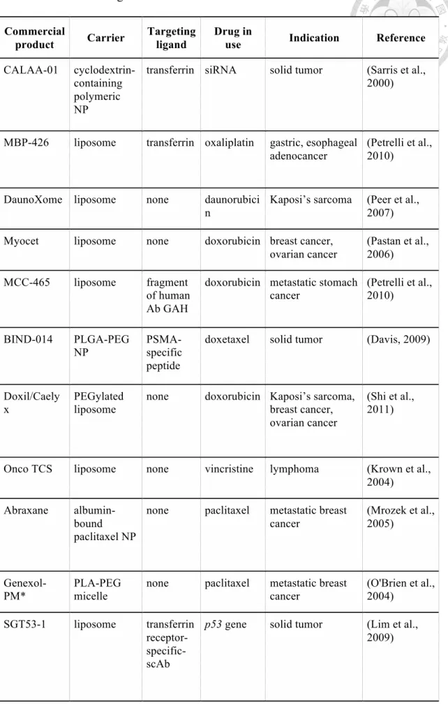
Table 2.3 NP-based drug that have been researched and delivered to clinical trial. 57 Table 2.4 Aptamer-drug complex/conjugate for cancer therapy in recent years. 67
Frequent abbreviations
STAT3 signal transducer and activator of transcription 3 STAT1 signal transducer and activator of transcription 1
dODN decoy oligodeoxynucleotide
hpdODN hairpin decoy oligodeoxynucleotide
DOX doxorubicin
TUNEL terminal deoxynucleotidyl transferase dUTP nick end labeling DOXON DOX-intercalated decoy oligodeoxynucleotide
RT-qPCR reverse transcription quantitative polymerase chain reaction
TF transcription factor
STEP steps toward enhancing the potency
1. Introduction
Using engineered oligonucleotide in therapeutics, diagnostics, and theranostics have attracted much attention in the recent years. Among the cancer researches, the oligonucleotides have shown great potential in effective cancer cell inhibition. More importantly, they show much less cytotoxicity compared to the conventional chemotherapeutic agents. Despite the promising safety the oligonucleotides drug have shown, they are facing the challenges about potency enhancement and multi-functional utility. For example, the oligonucleotide can be re-designed to have higher specificity for having higher potency. In addition, conjugation with other kinds of agents leads the oligonucleotide become multi-functional system.
Among so many oligonucleotides, we focus on a decoy oligodeoxynucleotide (dODN) that targets to signal transducer and activator of transcription 3 (STAT3), which is a kind of oncogenic transcription factor (TF) in adult human cell. By mimicking the favoured-promoter sequence of STAT3, the STAT3 is guided to bind to the dODN and be trapped. Followed by the activity blockage, the STAT3-dODN blocks the oncogenic gene expression in the down stream of STAT3.
In the following paragraphs, the origin of the dODN and how it can be engineered are introduced firstly. Followed by the therapeutics and diagnostics relative background introduction, the dODN applications in inflammatory diseased treatment and kinds of nucleotide-based diagnostic system are introduced. Among these, since the the dODN is rarely been used in diagnostics so far, the introduction involves most of other types of oligonucleotides (e.g. aptamer). Finally, the major experimental considerations are addressed to provide the concepts of how to appropriately operate the biological experiments.
1.1. The “decoy” of the therapeutic oligonucleotides
The first therapeutic oligonucleotide is reported by Zamecnik and Stephenson in 1978 (Zamecnik and Stephenson, 1978). A complimentary oligonucleotides targeting to the terminal sequences of the Rous sarcoma virus successfully inhibits the its replication.
Although it attracts little interest then, the following approach of automatic DNA synthesisers that Zamecnik reported attracts much more attention (Zamecnik et al., 1986). In a short time, many companies appeared to manufacture modified antisense oligonucleotides (e.g. phosphorothioation and methylphosphonation) as drugs. These oligonucleotides were complementary to the sequences of either viral or messenger RNAs. The antisense oligonucleotides block the expression of proteins by inhibiting the translation, instead of bind directly to the proteins like the conventional drugs.
However, the sequence that works stably in vivo has not been solved at that time.
Every oligonucleotide behaves differently, since the single-stranded nucleic acids has complex secondary and tertiary structures. Contributed from the structure, the oligonucleotide binds to the protein and performs like an antibody or enzyme. The variability of the oligonucleotide structures complicates the mechanism and leads to the un-expectable biological response. Besides, many synthetic oligonucleotides (especially the RNAs) exert the unknown function in vivo, which therefore upgrade the difficulty in making them as the medicine. In brief, the work presented by Zamecnik and Stephenson suggested the new ways of applying therapeutic oligonucleotides. Today, the translation of RNA can be inhibited by the small interfering RNAs, ribozymes, and DNAzymes, in addition to the aforementioned antisense. To inhibit the proteins, aptamers, decoys, and CpG oligonucleotides bind are widely researched and developed.
Epigenetic causes leads to the increased risk for diseases and it typically comes from the
into the corresponding messenger RNA (mRNA) or the incorrect translation of mRNA into an peptide or protein is occurred. To date, it remains a challenge of how to “fix” the defective gene. On the other hand, regulating the genetically caused changes in gene expression seems to be less challenging. To those disease-related abnormal gene expression, we might employ the inhibitory molecules that directly cause the protein block or decrease its activity. For example, the neutralising antibodies or small molecule inhibitors. Alternatively, the nucleic acid-based medicines, such as antisense oligodeoxynucleotides (ODNs) or small interfering RNA (siRNA), which blocks the translation of the malignant mRNA into protein.
Another possible solution to disrupt the abnormal expression of the malignant gene is to block the activity of the transcription factor that drives the malignant protein transcription. To this end, decoy ODNs (dODNs) have been researched and developed.
The first dODN is reported in 1990 for regulating the NF-κB (Bielinska et al., 1990), which is kind of TF controlling the inflammation in body. The dODNs are doubled- stranded DNA (dsDNA) which mimic the consensus binding site of a promoter that corresponding to the specific transcription factor. As a result, dODNs inhibit the function of the transcription factor by preventing their binding to the DNA. Thus, the modulation of gene expressions is approached. Notably, the dODNs block the function of activated transcription factor, instead of blocking the expression of the transcription factor itself (figure 1.1). Nowadays, the decoy oligonucleotides are not pursued as aggressively as other therapeutic oligonucleotides. However, in the emerging researches that successfully apply the dODNs in treating kinds of disease, the dawn of dODNs therapy is close.
Figure 1.1 The principle of how dODN blocks the activity of the transcription factor in interest. The dODN-bound TF is not able to cross the nucleus membrane to exert the down stream functions.
1.2. Decoy oligodeoxynucleotide engineering
The dODN is usually designed base on the promoter sequence in gene. Take the the dODN that targets to the STAT3 for example, it is based on the Serum Inducible Element (SIE) of the human c-fos promoter (Leong et al., 2003a). The oligonucleotide with 10-20 base pair was designed to bind the DNA binding domain of the STAT3. It abrogates the function of ability to bind to DNA response elements and induce transcription of target genes, STAT3 and elicits the anti-tumour effects through the inhibition of STAT3 down stream gene expressions. Into the cells, the introduced oligonucleotides faces the challenge of rapid degradation in serum. To conquer this
most common method is the phosphorothioation, which replaces the non-bridging oxygen with sulphur in the phosphate linkages (Brown et al., 1994). In addition to enhance the stability, researches have shown that the phosphorothioation also increases the cellular uptake efficiency and protein binding affinity. However, the modification also results in the increased nonspecific binding compared to the unmodified dODN (Brown et al., 1994).
• Means to determine the specificity
Generally, the consensus sequence of the dODN is derived as an integrated site from the transcription binding sites in the promoter of several genes. A tool for searching the appropriate sequence is, for example, the MatInspector (Cartharius et al., 2005). It is a software that involves the large library of matrix descriptions of transcription-factor binding sites to locate the matches in DNA sequences. This software is also suitable for designing an mutated ODN as control or to ensure that the scrambled ODN affects no other transcription factor (off-target effect). The consensus sequence typically comprises six to twenty bases and is targeting to the specific transcription factor. The exceptions is found when there is an overlapping binding sites between different transcription factors. For example, the cAMP response element (CRE) binds not only to the CRE binding protein (CREB), and also the members of the activator protein-1 (AP- 1) family. Another exception is the common bindings sites that interact with kinds of proteins and thus causes a competitive manner. For example, the STRE-binding transcription factors early growth response protein-1 (Egr-1) and Sp-1 are verified to compete for the similar sequence. To solve this problem, it is logical to derive the binding-sequence from the binding site in the promoter of the primary target gene, instead of adopting the consensus binding site. Another issue is observed in the large
transcription factors families. Like the AP-1 and the STAT family, each member may binds to the same or similar sequence. That is, the competition binding between each might occurred among the family. Therefore, the consensus sequence derived dODNs of transcription factor in the large family might have low discrimination.
Several experimental methods have been reported for verifying the specificity of the dODN. The most common used methods are electrophoretic mobility shift analyses (EMSA) and enzyme-linked immunosorbent assay (ELISA), and down stream gene expression. In a typical EMSA, the cell extracts are mixed with the radiation-labelled dsDNA probe. The probe comprise a binding sequence for the transcription factor. The tested dODN displaces the probe that binds to the transcription factor in the extract, which results in the reduction of the probe–protein complex. Followed by the antibody- based super-shift analyses, the identification of the targeted transcription factor can then be achieved by. ELISA, which also allows the researchers to estimate the binding affinity of the dODN toward transcription factor. In a typical ELISA, the activated transcription factor or the purified protein binds to the immobilised dsDNA probe comprising the consensus sequence. Similar to the EMSA, the dODN displaces the probe from the transcription factor. The result is visualised by using the antibody against the transcription factor. ELISA enables not only the definition of the decoy ODN specificity, but also suitable for selecting the sequence with higher affinity to provide higher efficacy. In addition, it can be used determine to clarify how chemical modifications affects the sODN. For example, we can use ELISA to evaluate the how phosphorothioation affect the affinity and specificity toward the transcription factor.
The third common method is to determine the variation of the gene expression that governed by the transcription factor. This method is feasible only in the cellular model and can be achieved by using reverse transcription quantitative polymerase chain
reaction (RT-qPCR). The extracted mRNAs are reverse transcript to the cDNA and amplified with the PCR approach to quantify the level of the certain gene expression.
This method provides the cellular evidence of dODN effect on the transcription factor and hence is widely used.
• Means to enhance the transfection efficiency
In the last section, the importance of sequence design on the efficacy is mentioned.
Besides that, the transfection efficiency of dODN also dominates the efficacy. That is, a dODN that is easily and fast uptake by the cell is expected to benefit the efficacy. The common way used to determine the transfection efficiency is counting the fluorescent dODN-transfected cell with the flow cytometry. The dODN is labelled with fluorescent dye, such as Texas Red or Alexa Fluor 594. Besides the flow cytometry, fluorescence microscopy or any fluorescence reader are also the possible instrument to carry out the determination. Furthermore, the tested sample is not limited in cultured cells. The tissues or organs ex vivo are also suitable for the fluorescence determination (Shangguan D, et al. 2008)(figure 1.2). Besides the fluorescent dye, radiation labelling is also widely employed. For example, the phosphorothioated dODNs can be labeled with the 35S to track its location in vitro. Notably, the 32P or 33P-labelling of the nucleic acids has been verified to be less suitable, due to the fast decay in the cell.
According to the past researches, four major parameters govern the transfection efficiency of dODN into the cell. They are concentration, length, nucleotide sequence, and nucleotide composition. The optimal condition for DNA transfection is 10 mM sequence with at least one hour of pre-incubation. In most cases, this condition provides a satisfactory outcome, which is a maximised inhibition of target gene expression. A 60- 80% higher inhibition can be obtained compared to the control. Secondly, the optimal
length of the dODN is less than 30 base pairs. On the other hand, the transfection efficiency of a 25 base pair dODN is higher than a 10 base pair one. That is why most of the dODNs reported recently is in the range of 15-25 base pair. Although the previous literatures have pointed out an optimal condition of concentration and length, the transection efficiency of each dODN is not consistent. This phenomenon is related to the involved transport system during crossing the cell membrane. The transport pathway is highly dependent on the sequence and composition of the nucleotide.
Therefore, the characteristics of these transport systems is worth to be explored. There are many method fro determining the transport system that is involved.
Figure 1.2 Fluorescence microscopic image that demonstrates the uptake of a Texas Red-labelled Stat-1 decoy ODN into the human skin cells (red). The nucleus was stained with DAPI (blue). (Shangguan D, et al. 2008)
• Stability of decoy oligodeoxynucleotide in cell
The dissolved dODN in aqueous solution of neutral pH and ambient temperature is stable. Many stability analysis reports have been published to support the use of the dODNs in clinical application. For the long-term storage, the freeze-dried dODNs under 5℃ is recommended. The stability of the transfected dODN in cell is usually estimated according to the duration of their efficacy. For example, continuously monitoring the mRNA expression of the cells to see whether the transcription factor is still regulated by the dODN or not. According to some animal experiments, the dODN (with phosphorothioation at the 3’- and 5’-terminal three bases) remains its function until 48 hours after transfection (Buchwald et al., 2002).
To precisely determine the stability in cell, the 35S-labelled dODN has been transfected into the human umbilical vein endothelial cells. With the aids of the native polyacrylamide gel electrophoresis and autoradiography, it is found that there is no degradation of the dODNN within 24 hours after the transfection. The results suggest that the dODNs that transfected into the cells remains stable and activity for quite a long period. The chemical modifications enhance the stability in a further extent. With the incubation of rat serum at 37℃, the calculated half-life of phosphorothioated dODNs is 16 hours, which is much longer than the unmodified one.
1.3. Decoy oligodeoxynucleotide in therapeutics
To date, the dODN has been used in transcription factor neutralisation to access its therapeutic value in a variety of the animal models and also in humans. Many studies have reported their therapeutic potential in the subacute and chronic inflammation, infectious diseases, and cancers. The researched dODNs mainly target to the
cancer therapy in the following chapter, here, the author would like to introduce the cases of using dODN in inflammatory diseases therapy, which plays an important role in the early stage of the dODN research and development.
• Inflammatory skin disease
T-Cell mediated skin diseases are the most common and severe chronic inflammatory skin diseases. For example, the psoriasis, which is believed an exaggerated Th1 or Th17-type immune response-induced disease. Besides, the allergic contact dermatitis (ACD), is also believed to be relevant to the Th1 immune response. Therefore, the crucial role of the Th1 cytokine IFN-γ is undisputed. Thus, it is reasonable that the transcription factor STAT1, as the main effector molecule of IFN-γ signalling, is considered as the major target in both the ACD and the psoriasis. In the typical therapy, the topical corticosteroids is widely used in treating the aforementioned disease. It provides the anti-inflammatory efficacy by inhibiting the transcription factors AP-1 and NF-κB. However, human that take the corticosteroids for a long time often suffers from the adverse side effects, like skin atrophy. Therefore, a new way for treating these kinds of disease is necessary.
The efficacy of the STAT1-dODN containing ointment on the ACD in guinea pigs and domestic pigs has been evaluated. In guinea pigs, single application of the STAT1- dODN in the ointment results in a dose-dependent improvement of decreasing the inflammation. The result is accompanied by a considerable decrease in leukocyte (polymorphonuclear neutrophils, monocytes, T-cells) infiltration into the dermis and reduced skin thickening. In addition, the STAT1-dODN ointment effectively decrease the expression of IL-1b, IL-8 and IL-12 as well as the TNF-α and IFN-γ. Compared to the topical steroids, the STAT1-dODN ointment appeared to be an effective therapeutic
agent. In the other case, the intra-articularly injected STAT1-dODN also successfully blocks the delayed-type hypersensitivity reaction of the antigen injection (Huckel et al., 2006).
Besides the STAT1, NF-κB is another worth-mentioned target for the treatment of inflammatory skin diseases therapy. In a past research, the NF-κB dODN ointment is successfully tested in a spontaneous atopic dermatitis model (NC/Nga mice). The test is carried out with (Dajee et al., 2006)or without (Nakamura et al., 2002)the additional dust-mite antigen challenge. Also, in a delayed-type hypersensitivity response model in mice (Dajee et al., 2006) .With the antigen, the NF-κB dODN ointment seems to be as effective as the corticosteroid. It down-regulates the expression of the pro-inflammatory gene products in the mouse skin.However, the use of the NF-κB dODN can also cause a remarkable number of the cell death. Therefore, the administration of NF-κB dODN is suggested to be limited in the topical use, since the systemic inhibition of NF-κB is harmful. In the case of given that NF-κB signalling knock-out mice, it suffers from the immune deficiency (e.g. lack lymphocyte activation) (Luque and Gelinas, 1997). Back to the skin disease issue, the use of the NF-κB dODN ointment still provide a less skin atrophy and other local adverse effects compared to the topical steroids.
Besides the NF-κB dODN, the STAT6-dODN is also an effective agent against the ACD in mice (Sumi et al., 2004). Notably, the STAT6-dODN has a relative long sequence (28-mer). Therefore, it has to be used with the aids of the haemagglutinating virus of Japan (HVJ)–liposome. The optimal effect is obtained by taking the subcutaneous injection with a concentrations of 5-20 mM. The topical route of the STAT6-dODN take shows a strain-specific effect, which suggests that it directly affect the Th2-immune response, instead of Th1.
• Rheumatoid arthritis
The rheumatoid arthritis is one of the prevalent and severe chronic inflammatory disease of the joints, which strongly influences the patient’s daily life. The rheumatoid arthritis is an immune-induced disease that enhance the inflammation and destruction of the joints. This kind of disease is believed to be associated with the imbalance of IFN-γ over IL-4 (Huckel et al., 2006). That is, the TNF-α and IL-1b signal via the transcription factor NF-κB, IL-6 and cause a IFN-γ-induced pro-inflammatory effects through activating the STAT1. The activated STAT1 hence up regulates the expression, such as the co-stimulatory molecule CD40. The CD40 induces the synthesis of the transcription factor IFN regulatory factor-1 (IRF-1) (Vince T. Nguyen, 2000, Wagner et al., 2002).Since the NF-κB or STAT1 are both pro-inflammatory molecule, using the dODNs to directly target these two transcription factors is reasonable. The previous literatures have tried to do so in two different animal models, one is the collagen- induced arthritis in the rat and the other is the antigen-induced arthritis in mice. In this case, the NF-κB dODN is carried by the HVJ–liposomes, while the STAT1-dODN is used as the naked DNA. Single treatment with the STAT1-dODN shows a dose- dependent (0.05–10 nmole) inhibitory effect for joint swelling and the histopathological signs of acute and chronic arthritis. In addition, the CD40 mRNA expression in stimulated macrophages is down regulated, which is a function that similar to the monoclonal antibody for the CD40 neutralising. In summary, the therapeutic effect of the STAT1-dODN mainly comes from affecting the CD40 signalling pathway in the antigen-presenting cells. In the other case, the collagen-induced arthritis model, injection of the liposomal NF-κB dODN provides a considerable moderation of the joint destruction, and also induced the cytokine release into the synovia (Tomita et al., 1999).
One thing worth to mention that the dODN affects not only a single joint, instead, the
systemic delivery method provides the benefit to cure all the joint. In a recent research, another dODN approach targeted to the transcription factor E2F has been verified to have protective effect in immunodeficient severe combined immune deficiency (SCID) mice that were transplanted with human cartilage and rheumatoid arthritis tissue. Such a protective effect is probably the result of the inhibition of synovial cell proliferation.
In the human body, Nitric oxide (NO) is a signalling molecule that plays a key role in the pathogenesis of inflammation, where it acts as a anti-inflammatory mediator. The endothelial NO synthesis is majorly up regulated by the cytokine IL-10, which is the result of the activation of the transcription factor STAT3 (Cattaruzza et al., 2003).The increased NO synthesis blocks the CD40-dominated expression of the pro-inflammatory cytokine IL-12 in the endothelial cells. In this case, the use of the STAT3-dODN blocks the function of the STAT3, hence decrease the production of the intracellular mediator of IL-10. The binding causes a weakened NO synthesis capacity, leading to an increased risk of inflammation in the joints. Refers to the coronary heart disease (CHD), which is a kinds of disease that highly associate with the NO. The aforementioned result provides the opportunity for employing the dODNs as a novel approach towards precision medicine (Warrington et al., 2005, Gazi et al., 2007).
• Acute and chronic transplant rejection
Alone the dODN research and development, moderating the transplant rejection is an meaningful therapeutic application for dODNs. The dODN is infused into the blood vessels of the donor organ and flushed out before re-establishing perfusion of the transplant to avoid the systemic exposure of the recipient. Notably, the brief period of warm ischaemia might hinder the endothelial cells of the donor organ from taking the naked DNA during the process. Transplant rejection is an important application of
dODN, because it is usually accompanied with not only the acute, but also chronic inflammation associated with the endothelial dysfunction and microcirculatory perfusion failure. To solve these situations, dODNs that directly target the NF-κB were first tested in the animal models of allograft rejection. The perfusion of rat renal allografts with naked DNA (Vos et al., 2000) results in a significantly increased survival of the transplants in the absence of immunosuppression. In a similar way, the administration of the STAT1-dODN in the rat small bowel allografts (Stojanovic et al., 2007)or that of a STAT1 or AP-1 dODN in rat cardiac allografts (Holschermann et al., 2006) both result in a significant improvement of the microcirculatory and histopathological signs of acute rejection. In those cases, cardiac allograft can actually be considered as an aggressive form of the CHD. The STAT1-dODN and the AP-1 dODN inhibit the cytokine-stimulated CD40 expression in the allograft endothelial cells. When it comes to the transplant rejection, the ischaemia/reperfusion (I/R) injury is one of the major concern. The use of the NK-kB dODN have shown benefits in therapeutic effects in the liver I/R injury (Xu et al., 2005).
• Others
Other indications in which animal experimental proof of principle studies have been performed include chronic inflammatory bowel disease, i.e. different mouse models of colitis in which NF-κB decoy ODNs were successfully administered intrarectally, either encapsulated in a viral envelope (Fichtner-Feigl et al., 2005) or as naked DNA, albeit fully phosphorothioate-modified (De Vry et al., 2007), osteoporosis in rats (NF-κB decoy ODN) (Shimizu et al., 2006),diabetic nephropathy in rats (AP-1 decoy ODN (Ahn et al., 2004) or ring-Sp-1 decoy ODN (Chae et al., 2006)), experimental autoimmune myocarditis in rats (NF-κB decoy ODN) (Liu et al., 2001)and carrageenin-
induced acute inflammation in the rat hind paw (NF-κB decoy ODN) (D'Acquisto et al., 2000). According to the preclinical and the clinical report to dat, the dODNs give a promising performance for being the highly effective anti-inflammatory medicines. In addition to its satisfactory efficacy, it also provides the fast-acting and extreme low side effects properties. Although the dODN may affect the immune response-related genes expression in some cases. For instance, the STAT1-dODN may weaken the subjacent physiological immune response agains the bacteria, viruses or parasites. If the effective and cell-specific drug delivery is achieved, the dODNs will become one of the therapeutic approaches with blockbuster potential.
• Nucleic Acid Based Drugs in Comparison
The dODN specifically binds to the targeted transcription factor, and inhibits the down stream genes expression. Notably, in most of the cases, dODN does not completely block the gene expression. The major advantage of the dODN is that is is able to be transported into their target cells rapidly without additives and thereafter regulate the targeted transcription factor. Inside the cell, especially the terminally phosphorothioated dODNs have a relative higher stability and hence provide the long-lasting effects. The long-term effect is believed to benefit the dosing and costs issues. When it comes to the side effect , it seems that the targeted cells (upon local delivery) and the whole organism (upon systemic administration) has a high tolerance towards these dODNs. In contrary, this kind of phenomenon is not normally seen in the application of the ssDNA, such as the antisense ODNs and the double-stranded siRNA. Compared to the other kinds of DNA drugs, dODN is a kind of medicine that regulates multiple pathway in the same time. It is because that many transcription factors (e.g. AP-1, STAT1, and NF-κB) govern the expression of many pro-inflammatory genes. Therefore, the inhibition of the
transcription factor will result in a decrease of the expression of several kinds of genes simultaneously. This feature may thus cause the dODN being considered as a pathway- non-specific medicine compared to the single-target gene knockdown by the antisense ODN or siRNA. However, this is also the advantage that make the dODN highly effective therapeutic agent. For example, the STAT1-dODN is such a powerful corticosteroid-like anti-inflammatory medicine without any appreciable side effects (Quarcoo et al., 2004, Huckel et al., 2006).This efficacious outcome is rarely seen in the case of anti-sense and siRNA, even though they have higher specificity.
1.4. Oligodeoxynucleotide in diagnostics
With the aids of the molecular biology, it is now possible to have an assay for imaging the specific molecular processes, such as the gene expression. Imaging gene expression enables us to determine the locations of particular gene expressing. Besides, we can even monitor the level and duration of that gene expression. Imaging the gene expression with the generic approaches to the gene expression of interest has been researched for years. The conventional imaging techniques use the radiolabeled substrate to interact with the protein of the gene to image the expression. To be convenient to the users, more and more general methods without the need for radiolabeled substrates are developed to image the gene expression. In this section, we would like to focus on the aptamer approach for imaging protein of the gene expression.
To date, there are only a few groups using the dODN to image the proteins that related to the specific gene expression. Among the diagnostic oligonucelotieds, aptamer has pave a way for the dODN to develop the protein imaging. Therefore, methods to introduce aptamer into target, principle, and applications of aptamer for imaging gene
• Fundamentals of gene expression
Even today, the process of gene expression (figure 1.3) and the components of gene regulation are only partially understood. Every cell has its own gene expression feature, which leads to the wide variety of functions or phenotypes. The development, maturation, proliferation, and even the oncogenesis of the cell is attributed to the kinds of gene expression. Promoter is a sequence motifs (~100 nucleotide bases) at the specific region of the gene that the transcription initiates. It is involved in the RNA polymerase binding and the transcription initiation. In the eukaryotic cell, the promoters do not always work alone. The other point that worth to be mentioned is that it can be influenced by the enhancers. The enhancer has no certain location, it exist in the either the upstream or the downstream from he promoter. Sometimes, the enhancer is even located within the transcription unit. The role of the enhancer is either increasing the initiation efficiency or participating in a specific regulation. After interacting with the promoter, the complex of the proteins and the RNA polymerase starts to join the transcription process. To image the gene expression, two main types of promoters are worth to be understood. They are constitutive and inducible promoters. The former can be used to produce continuous gene transcription, and the latter can be used to externally control the level of transcription. For example, constitutive promoters are cytomegalovirus, CMV (exogenous gene) and GAPDH (endogenous gene). Inducible promoters are tetracycline regulable (exogenous gene) and c-jun (endogenous gene).
Figure 1.3 An illustration briefly describes the gene expression in cell.
• Aptamer probes targets to the protein
Gene expression imaging with oligodeoxynucleotide can be achieved wither by the mRNA monitoring or down stream protein probing. In the case of using aptamer to probe the protein, the affinity between the sequence and the target protein is dominated not by the base pairing, but three-dimensional steric interactions between the molecules.
An aptamer possess a dissociation constants in a range between the micro-molar to pico-molar, which is comparable to that of the antibody. The systematic evolution of ligands by exponential enrichment (SELEX) has been used both to characterise the interaction of nucleic acids pool with proteins and to generate the new aptamers (Conrad et al., 1995, Buerger and Groner, 2003, Tavitian, 2003). The library contains the random sequence is used to screen for the high affinity probe targeting to a specific protein. The aptamers is able to recognise a single or multiple targets, including proteins, small molecules, or even the complex of the heterogeneous targets. The aptamers is able to be transported into the cell theoretically, thus the down stream of genes can potentially be targeted. Almost two decades ago, the first use of an aptamer to
target the neutrophil elastase (a surface protein) to achieve the in vivo inflammation imaging with a gamma camera is reported (Charlton et al., 1997). The used model is a rat with reverse passive Arthus reaction (local hypersensitive reaction on skin). In this case, the aptamer approached the maximum target-to-background ratio of 4 in 2 hours.
Afterward, the aptamer is fast removed with the blood clearance. From now on, more and more researches in this area are reported to facilitate other imaging applications in which the protein is targeted with the aptamers. These approaches allow the user to image the endogenous gene expression, by selecting the aptamers target toward the protein of that specific gene. In a following research, the aptamers are labeled with
99mTc for imaging purposes and to investigate aptamer pharmacokinetics toward the Tenascin C, which is an extracellular stroma protein present in tumour and neovasculature (Hicke and Stephens, 2000). Due to a long tumour half life and fast clearance from the body, 99mTc-labeled aptamers yielded a tumour-to-blood ratio of 50 within 3 hours. And the clear images of U251 and MDA-MB-435 are obtained with a gamma camera (Hicke et al., 2006). Like what has been mentioned above, since there are a few group doing the research of dODN imaging, the aptamer based system become a reliable reference. on the same basis of protein recognition, methods and knowledges from the aptamer imaging approach lead the dODN imaging research toward the right direction.
To date, the significant progress in the molecular imaging has been made, thus facilitate the developing of assays for in vivo gene expression imaging. Even though, there is still room for improvement to develop a specific, quantitative, highly sensitive, and practical assays for clinical applications. Generally, oligonucleotide-based imaging approaches will need to enhance the key features, including the stability of probes, prevention of the non-specific interaction, and minimising the background signal. Once the successful has
been developed, it will provide a general method to imaging gene expression. As well as to identify the early changes in gene expression signalling between the normal cellular function to an altered state of disease. Oligonucleotide-based imaging approaches have been proven in many preliminary animal applications and pilot human studies. Studies with transgenic animals, in vivo inducible expression, bicistronic vectors, and the monitoring of endogenous gene expression are all widely investigated and accompanied with the meaningful result. In the nest step, multi-modality reporters and the complex of reporters and probes will facilitate the the development process of in vitro and in vivo assays, which will allows the researchers to study the more complicated biological processes.
1.5. Experimental considerations
In this section, the experimental considerations for nucleic acids sample preparation is discussed. The following review aims to collect the information that helps in preparing reliable sample for intended applications and analysis.
To secure the quality of the analytical results, the first step is defining the parameters for sample preparation based on the complete workflow and the purpose. The following five issues should be clarified before starting the experiment.
1. Purpose of the intended application or analysis 2. The biomolecule that is of interest
3. Characteristics of the sample source used in the study 4. The detail of the analytical techniques used in the study
5. The capabilities of the analytical techniques and the criteria for optimal performance
• Sample collection
Starts from the sample collection stage, the quality of biomolecules start to decrease.
Therefore, the just prepared materials and samples are used in all our presented experiment. To those samples or materials that are not used immediately, they are stored in a -80°C refrigerator with the stabilising agent. When carrying out the mRNA isolation, the extreme cautiousness for minimising the introduction of exogenous RNases to the sample is needed. For the DNA isolation, the quality of the sample surely benefit the downstream analysis. Thus, all the DNA samples are processed in the well- cleaned biosafety cabinet (BSC).
To access the component inside the cell, the cultured cells are directly lysed and delivered to the following application or analysis. With some understanding of the basic characteristics of the cells of interest, the lysis protocol that efficiently disrupt the cell’s external barriers should preserve the integrity of the cell’s chemical components. This preservation is critical for molecules that will be studied in downstream applications.
Generally, the animal cells are easily disrupted due to the lack of the cell wall.
However, the variation between cell membrane compositions and the distribution of the molecules on it can make situation more complex. that is, an incomplete cell lysis or sample homogenisation leads to a reduced yield. In addition, it also increases the risk of problems in the following process. For example, the debris may clog the purification column or inhibit the enzymatic reactions like PCR. Besides, the nucleases and proteases may be liberated and activated while the cell disruption. The activated enzymes can extensively influence the eventual analysis of the target molecule.
Therefore, the sample should be protected from the action of such enzymes during the cell disruption and the subsequent purification. Particularly, it is crucial to inhibit the RNases and proteases to obtain the high-quality RNA and protein for the following
analysis. Table 1.1 summarises the most used methods and points out the corresponding appropriate sample type and target biomolecule. Generally, tender disruption are preferred when the cell of interest can be easily lysed (e.g. most of the cultured cells, blood cells, and some of the microorganisms). Tender disruption methods are also suitable when only one particular sub-cellular fraction is under consideration, like only cytoplasmic proteins or intact mitochondria are going to be analysed. Sometimes tender disruption methods are combined. For example, using the osmotic lysis then enzymatic treatment or freeze-thaw in the presence of detergent. When cells are less easily broken, the moderate disruption methods are employed. Usually, these methods involve the mechanical process to physically break the tissue.
Table 1.1 Methods for disrupting and homogenising cell for sample isolation.
Method Principle Starting
material Biomolecule Pros and cons osmotic shock
lysis
osmotic changing induced membrane disruption
gram(-) bacteria, erythrocytes, and cultured cells
protein isolation • simple, inexpensive
• inefficient for the cell with robust wall
• low yield
chaotropic salts lysis
causing a less hydrophilic environment and disrupting the hydrophobic interactions
all sample types (except Gram(+) bacteria)
nucleic acid
isolation • nucleases and proteases denature
• isolate nucleic acids and protein from the same sample
• high through out enzymatic
digestion
enzymatic cell wall digestion
plant tissue, bacteria, fungal cells,yeast
protein isolation, nucleic acid isolation
• gentle
• times costing
1.
low yielddetergent lysis cell membrane solubilisation
cultured cells, some tissue
protein isolation, nucleic acid isolation
• simple
• times costing
• low yield mechanical
homogenization
breaking the cell with blade
Most tissues (plant and animal), cultured cells, bacteria, yeast
protein isolation, nucleic acid isolation
• efficient
• biomolecule denature by heat
• residual debris
For most of the nano-medicine research, the cultured mammalian cells, which is easily disrupted, are widely used. In the case of cells grown in suspension, they are typically harvested by centrifugation, washed, and resuspended in the chosen lysis buffer. The buffer usually contains the relevant denaturants, inhibitors, and stabilisers. Overall, the use of vortex is enough to achieve the complete lysis. On there other hand, the adherent cells can be directly lysed in the culture plate with the same route mentioned above.
• Quality and purity considerations
The quality of the collected sample is important, since it directly represents the extent of the specific molecule in vivo. The poor quality shall lead to the misunderstanding of the cellular condition. In the RNA isolation, the degradation is the key concern. Thus, inhibition of the RNases is of leading importance in RNA isolation process. Together with the intact composition of the biomolecule, three-dimensional structure and the activity are also the important considerations. For example, the plasmid DNA with open circle or denaturation performs differently compared to the desired form. To ensure the quality of the isolated biomolecules, kinds of means can be used to determine the purity.
For example, the RNA integrity number (RIN) is a quality measurement for RNA. The output value 1 to 10 corresponding to the poorest to the highest quality (Schroeder et al., 2006). In the case of DNA, the quality can be measured by the Phred quality scoring (Ewing and Green, 1998, Ewing et al., 1998).
When preparing the sample, the requirement of the purity varies from the sample type, the target biomolecule, and the intended applications and analyses. In the case of the nucleic acids, the A260/A280 ratio is a basic but important index. Although the purity requirement differs from cases, thus there is no universal definition of purity. However, this ratio does provide the information about removal of the contaminants in the nucleic acid sample. This index is important because of many biological analyses are sensitive to the contaminants. In order to obtain the reliable analytical results, interfering compounds like salts, polysaccharides, and non-target molecules have to be removed.
Table 1.2 includes a list of common contaminants and means to deal with.