CASE REPORTS
Benefits of Endorectal Ultrasound for
Management of Smooth-Muscle Tumor
of the Rectum
Report of Three Cases
Jan-Sing Hsieh, M.D., Che-Jen Huang, M.D., Jaw-Yuan Wang, M.D.,
Tsung-Jen Huang, M.D.
.From the Department of Surgery~ Kaohsiung Medical College, Kaohsiung, Taiwan
Smooth-muscle tumor of the rectum is rare, and the thera- peutic strategy is still controversial. Endorectal ultrasound was used to evaluate three patients with smooth-muscle tumor of the rectum. Endorectal ultrasound demonstrated a homogenous hypoechoic tumor w-ithout invasion to the perirectal tissue in two patients. The tumor was 5 cm in diameter in one patient and 4 cm in diameter in the other patient, and they were excised locally. Their histologic types were leiomyoma and leiomyosarcoma. The third pa- tient had a recurrent leiomyosarcoma. Proctosigmoidos- copy found a linear lesion with ulcerated mucosa on the rectal wall. Endorect',d ultrasound observed a hypoechoic solid tumor of 3~5 cm X 1 cm, which involved the mucosal, submucosal, and muscle layers of the rectal wall. Disruption of the first hypoechoic layer was identified. Abdominoper- ineal resection was performed. Endorectal ultrasound fol- low-up revealed no evidence of recurrence in any of these patients. Endorectal ultrasound can help to define the ex- tent of disease and may be a useful adjunct in deciding about the appropriate surgical procedure in these diseases. [Key words: Smooth-muscle tumor; Rectum; Endorectal lfl- trasonnd]
Hsieh J-S, Huang C-J, Wang J-Y, Huang T-J. Benefits of en- dorectal ultrasound J{br management of smooth-muscle tu- mor of the rectum: report of three cases, Dis Colon Rectum
1999;42:1085-1088.
M
Yogenic t u m o r s o f t h e r e c t u m are r a r e l y e n - c o u n t e r e d a n d their m a n a g e m e n t is still c o n t r o - versial. T h e c o n v e n t i o n a l i m a g i n g t e c h n i q u e s , s u c h as b a r i u m e n e m a , p r o c t o s c o p y , o r c o m p u t e d t o m o g r a - p h y a n d digital rectal e x a m i n a t i o n h a v e b e e n u s e d for t h e p r e o p e r a t i v e i n v e s t i g a t i o n o f t h e s e d i s e a s e s . * H o w e v e r , t h e d i s t i n c t i o n b e t w e e n a b e n i g n a n d a m a l i g n a n t t u m o r is difficult w i t h t h e s e p r o c e d u r e s a n dAddress reprint requests to Dr. Hsieh: Department of Surgery, Kaohsiung Medical College, 100 Shih-Chuan 1st Road, Kaohsiung, Taiwan.
is m a d e o n l y b y h i s t o l o g i c e x a m i n a t i o n . 2, 3 Moreover, t h e r e is n o t a g e n e r a l l y a c c e p t e d o p t i m a l surgical a p p r o a c h , b e c a u s e t h e t u m o r s t e n d to r e c u r l o c a l l y e v e n after r a d i c a l r e s e c t i o n . Recently, e n d o r e c t a l ul- t r a s o u n d (EUS) h a s b e e n u s e d for p r e o p e r a t i v e as- s e s s m e n t o f rectal t u m o r s a n d h a s p r o v e d to b e a v e r y u s e f u l a n d a c c u r a t e m e t h o d . #~ W e p r e s e n t o u r e x p e - r i e n c e o f EUS in a i d i n g the d i a g n o s i s for m y o g e n i c t u m o r s o f the r e c t u m a n d t h e a p p l i c a t i o n o f EUS in a d j u s t i n g a d e q u a t e surgical m a n a g e m e n t . R E P O R T O F C A S E S
Case 1
A 7 3 - y e a r - o l d f e m a l e w a s h o s p i t a l i z e d w i t h a o n e - m o n t h histor T o f o c c a s i o n a l p a s s a g e o f b r i g h t b l o o dper rectum.
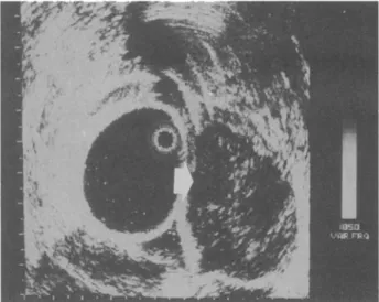
P h y s i c a l e x a m i n a t i o n s w e r e o t h e r w i s e n e g a t i v e . L a b o r a t o r y tests, i n c l u d i n g h e m o g r a m , liver f u n c t i o n tests, a n d b l o o d b i o c h e m i s t r y , w e r e all w i t h i n n o r m a l limits. P r o c t o s i g m o i d o s c o p y f o u n d a m a s s 5 c m a b o v e t h e anus, b u l g i n g p o s t e r i o r l y in t h e r e c t u m f r o m t h e 6 to 9 o ' c l o c k p o s i t i o n s , w i t h a n intact m u c o s a l surface. EUS (7.5 m Hz, BrOel a n d Kjaer, N a c r u m , S w e d e n ) w a s p e r f o r m e d w i t h t h e p a t i e n t l y i n g in left lateral p o s i t i o n . A latex s h e a t h w a s p l a c e d o v e r t h e u l t r a s o n i c t r a n s d u c e r at t h e distal tip o f t h e p r o b e . T h e p r o b e w a s i n s e r t e d 15 c m a n d t h e n w i t h - d r a w n after filling t h e s h e a t h w i t h d i s t i l l e d w a t e r . T h e t a r g e t a r e a w a s t h e n s c a n n e d b y m o v i n g t h e instru- m e n t b a c k w a r d a n d f o r w a r d . EUS e x a m i n a t i o n re- v e a l e d a t u m o r 4 c m in d i a m e t e r , w i t h a s m o o t h m a r g i n , arising f r o m t h e p r o p e r m u s c l e l a y e r o f t h e r e c t u m (Fig. 1). T h e t u m o r s h o w e d a h o m o g e n o u s 10851086 HSIEH E T A L Dis Colon Rectum, August 1999
h y p o e c h o i c pattern without invasion into the perirec- tal tissue. No evidence of lymph n o d e involvement could b e detected o n EUS. The tumor was locally excised b y a transsacral approach, Histologic exami- nation revealed a low-grade l e i o m y o s a r c o m a of the rectum. The patient r e m a i n e d well and asymptomatic two years later. EUS follow-up revealed no evidence of recurrence of this disease at that time.
Case 2
A 56-year-old male came to our hospital complain- ing of tenesmus a n d passing small-caliber stools for o n e year. He was in apparent g o o d health. Physical and laboratory examinations revealed n o abnormal- ity. Digital examination s h o w e d a rectal mass 3 cm from the anal verge. At p r o c t o s i g m o i d o s c o p y a bulg- ing mass was s e e n in the rectum extending into the l u m e n o n the right rectal wall. EUS disclosed a well- defined tumor, 5 cm in diameter, with a h o m o g e n o u s h y p o e c h o i c pattern (Fig. 2). On EUS the t u m o r ex- p a n d e d the s e c o n d h y p o e c h o i c layer, indicating mus- cularis propria. There w a s n o sign of invasion into the perirectal tissue nor involvement of regional l y m p h nodes. Local excision of the tumor was p e r f o r m e d transanally. Histologic study revealed a l e i o m y o m a of the rectum. T h e recovery period was uneventful a n d the patient was discharged in satisfactory condition o n e w e e k after the operation. Histologic study dem- onst.rated a benign l e i o m y o m a of the rectum. The patient underwent stria follow-up postoperatively. EUS revealed that he was free of tumor two years later.
Case 3
A 42-year-old male w a s admitted to our hospital for a recurrent rectal tumor. He u n d e r w e n t transanal ex-
Figure 2. Patient 2. Endorectal ultrasound image show- ing a hypoechoic well-delineated tumor that involves the muscularis propria (arrow).
cision of a rectal t u m o r at another hospital three years before this admission. The t u m o r was 6 cm in diam- eter a n d was histologically diagnosed as rectal leiomyosarcoma. Postoperatively, he r e m a i n e d asymptomatic for a further three years before devel- oping a recurrence, which w a s found on a follow-up proctoscopic examination. He was referred to our hospital for a detailed investigation of the recurrence. At physical examination he w a s found in g o o d gen- eral condition. Chest x-ray, abdominal ultrasound, a n d b l o o d biochemistry w e r e normal. A rectal exam- ination revealed a hard and r o b b e r y longitudinal mass measuring 3.5 cm × 1 cm. Proctosigmoidoscopy f o u n d a linear lesion with ulcerated m u c o s a on the posterior wall of the rectum and 1 cm adjacent to the anal verge. EUS o b s e r v e d a h y p o e c h o i c solid tumor of 3.5 cm x 1 cm, which involved the mucosa, s u b m u - cosat, and muscle layers of the rectal wall (Fig. 3). Disruption of tile first h y p o e c h o i c layer was identi- fied. Expansion of the m u c o s a with s o m e degree of irregularity related to the s u b m u c o s a and muscle lay- ers was observed. An a b d o m i n o p e r i n e a l excision of the rectum was performed. Histology again s h o w e d features consistent with leiomyosarcoma. He re- mained well and asymptomatic 1 year after the sec- o n d surgery.
Figure 1. Patient 1. Endorectal ultrasound image show- ing a hypoechoic tumor with well-defined border. The tumor grows within the muscularis propria (arrowhead).
D I S C U S S I O N
There is no standard treatment for rectal myogenic tumors at present, b e c a u s e of their rarity. A local excision is usually sufficient for the c o m p l e t e cure of a benign rectal leiomyoma, although malignant trans-
Vol. 42, No. 8 ENDORECTAL ULTRASOUND FOR MYOGENIC TUMOR 1087
Figure 3, Patient 3. Endorectal ultrasound showing the tumor growing within the muscularis propria (arrow) and protruding forward into the submucosal and muscle lay- ers. Expansion of the first hypoechoic layer with some degree of irregularity of the submucosal and muscle lay- ers was found.
formation of a b e n i g n s m o o t h - m u s c l e t u m o r has b e e n e m p h a s i z e d previouslyT, 8 On the other hand, the treatment of rectal l e i o m y o s a r c o m a is still controver- sial. Furthermore, an a d e q u a t e staging m e t h o d for gastrointestinal leiomyosarcomas has not b e e n eluci- dated. Several therapeutic modalities m a y b e involved in the treatment of l e i o m y o s a r c o m a s of the rectum, including l o w anterior resection, a b d o m i n o p e r i n e a l resection, local excision, and n o n o p e r a t i v e thera- py.8<3 The choice of surgical a p p r o a c h for a rectal l e i o m y o s a r c o m a d e p e n d s mainly on clinical a n d his- topathologic findings. Recently, EUS has p r o v e d to b e the preferred modality in preoperative assessment of rectal neoplasia with regard to d e p t h of invasion a n d nodal involvement. #-6 EUS is of substantial value be- cause it permits accurate demonstration of each layer structure in the rectal wall. Thus, the extent and d e p t h of t u m o r invasion are readily assessed, as they w e r e in our cases. The p r e s e n c e of metastatic spread to the regional l y m p h n o d e can also be detected.
Because the origin a n d invasiveness of the t u m o r could b e determined b y EUS preoperatively, the dif- ferentiation b e t w e e n a benign and malignant tumor b e c a m e possible. The extent of surgical resection can thus b e decided. T w o of our patients w e r e thought to have a t u m o r sufficiently localized, as assessed by clinical examinations a n d EUS, to be suitable for wide local excision; the other patient h a d a b d o m i n o p e r i - neal resection. As long as a d e q u a t e t u m o r clearance has b e e n obtained, there is p r o b a b l y no long-term difference in survival rates b e t w e e n those treated ini-
tially b y wide local excision v s . those treated by rad- ical resection. 10, 12 Some authors 14 r e c o m m e n d e d that features such as small tumor size, a b s e n c e of ulcer- ation, a n d fixation offer a g o o d chance o f survival with local excision. Usually m a c r o s c o p i c examination revealed that the rectal m u c o s a was relatively unin- v o l v e d b y the t u m o r tissue, despite the large size o f the neoplasm. In our third patient an ulceration on the mucosal surface could b e found o n proctoscopy. This finding of mucosal invasion could b e correlated with that of EUS and pathologic examinations. In such a situation radical surgery is r e c o m m e n d e d . Review of previous data concluded that site of the primary tu- m o r did not affect survival a n d that size was important only if adjacent structures w e r e involved a n d not completely excised. 15 However, local recurrence is also a c o m m o n p r o b l e m in the m a n a g e m e n t of rectal leiomyosarcoma. Previous findings indicated that lo- cal excision of those tumors arising from the muscu- laris propria of the rectum resulted in a high local recurrence rate regardless of the d e g r e e of differenti- ation of the tumor, but long-term survival figures w e r e similar w h e t h e r the tumors w e r e treated b y local or radical techniques.16 From o u r limited experience w e r e c o m m e n d that m o r e accurate staging m e t h o d s are m a n d a t o r y for the patient to select appropriate treat- m e n t and to c o m p a r e properly groups of patients treated with different modalities. Moreover, EUS m a y be a useful adjunct in deciding about the appropriate surgical p r o c e d u r e w h e n conservative surgery is in- tended, b e c a u s e it can help to define the extent of the disease.
REFERENCES
1. Tarasidis G, Brown BC, Skandalakis LJ, Mauer RC, Gray SW, Skandalakis JE. Smooth muscle cells tumors of the rectum and the anus: a collective review of the world literature. J Med Assoc Ga t991;80:685-99.
2. AsbunJ, Asbun HJ, Lang A, BlochJ. Leiomyosarcoma of the rectum. Ann Surg 1992;58:311-4.
3. Haque S, Dean PJ. Stromal neoplasms of the rectum and the anal canal. Hum Pathol 1992;23:762-7.
4. Orrom WJ, Wong WD, Rothenberger DA, Jensen LL, Goldberg SM. EndorectaI ultrasound in the preopera- tive staging of rectal tumors. Dis Colon Rectum 1990; 33:654-9.
5. Roubein LD, David C, Dubrow R. Endoscopic ultra- sonography in staging rectal cancer. Am J Gastroenterol 1990;85:1391-4.
6. Tanaka M, Fujishima H, Chijiwa Y, Nawata H, Eguchi T, Kinjo M. Endoscopic ultrasonographic findings in rectal leiomyoma. J Gastroenterol Hepatol 1995;10:103-5.
1088 HSIEH E T A L Dis Colon Rectum, August 1999 7. Newman Z. Leiomyosarcoma of the rectum developing
from benign leiomyoma. Ann Surg 1952;133:426-30. 8. Bores RR, Thorlakson RH. Leiomyosarcoma of the rec-
tum. Int Surg 1974;59:616-20.
9. Quan SH, BergJW. Leiomyoma and leiomyosarcoma of the rectum. Dis Colon Rectum 1962;5:415-25.
10. Randleman CD Jr, Wolff BG, Dozois RR, Spencer RJ, Weiland LH, Ilstrup DM. Leiomyosarcoma of the rectum and the anus. A series of 22 cases. Int J Colorectal Dis 1989;4:91-6.
11. Diamante M, Bacon HE. Leiomyosarcoma of the rectum: report of a case. Dis Colon Rectum 1967;10:347-51. 12. Khalifa AA, Bong WL, Ral VK, Williams MJ. Leiomyo-
sarcoma of the rectum: report of a case and review of the literature. Dis Colon Rectum 1986;29:427-32. 13. Minsky BD, Cohen AM, Hajdu SI. Conservative man-
agement of anal leiomyosarcoma. Cancer 1991;68: 1640-3.
14. Labow SB, Hoexter B. Leiomyosarcoma of the rectum: radical vs. conservative therapy and report of three cases. Dis Colon Rectum 1977;20:603-5.
15. Akwari OE, Dozois RR, Weiland LH, Beahrs OH. Leiomyosarcoma of the small and large bowel. Cancer 1978;42:1375-84.
16. Walsh TH, Mann CV. Smooth muscle neoplasms of the rectum and anal canal. Br J Surg 1984;71:597-9.