High-Energy Collision-Induced
Dissociation of Ceramide Ions from
Permethylated Glycosphingolipids
D. D. Ju, G. J. Wei, and G. R. HerDepartment of Chemistry, National Taiwan University, Taipei, Taiwan, Republic of China
Ceramide fragments from permethylated glycosphingolipids (GSLs) were studied by high- energy collision-induced dissociation (CID). In comparison with ceramlde fragments of underivatized GSLs, many more product ions including charge-remote fragment ions were observed. These ions provided detailed structural information on the ceramides. The relative intensity and the mass interval between the L and M ions were used to assign the position of the double bond. The position of the hydroxyl group was assigned with the Ln and K ions. Because the ceramide fragments and not the pseudomolecular ions were selected as the precursor ions, the size of GSLs had little effect on the quality of the product ion spectra. The sensitivity of this approach was in the range of picomoles. (1 Am Sot Muss Spectrom 1994, 5, 5588563)
G
lycosphingolipids (GSLs) are cell membrane components. There are two different structural moieties in GSLs: a ceramlde and a carbohy- drate residue. The ceramide consists of the long chain base (sphingoid) substituted at the amino group by a fatty acid. The growing interest in the diverse biologi- cal functions of GSLs as cell surface antigens, cell-cell recognition markers, and receptors for a variety of signal molecules has stimulated the search for new analytical techniques for their structural analysis.Mass spectrometty, a highly sensitive technique, has been a key tool for the structural elucidation of GSLs. The classical approach involved the conversion of GSLs to methyl, acetyl, or trimethylsilyl derivatives before electron ionization (EI) or chemical ionization (CI) mass spectrometric analysis [l-4]. This approach proved to be very useful to the structural analysis of glycans and ceramide. Since its development in the early 198Os, fast-atom bombardment (FAB) has quickly become the method of choice for the analysis of polar, nonvolatile compounds such as peptides, oligosaccha- rides, and GSLs. FAB, both in the positive and nega- tive ion mode, has been widely used in the structural analysis of derivatized and underivatized GSLs [5-91.
Mainly due to the many interfering matrix ions and the lack of structurally important fragment ions, recent works focus on the use of FAB in combination with collision-induced dissociation (CID) in the structural analysis of GSLs and ceramides [lo-171. Costello and Address reprint requests to G. R. Her, Department of Chemistry, National Taiwan University, Taipei, Taiwan, Republic of China.
co-workers have demonstrated the utility of using FAB in combination with four-sector tandem mass spec- trometry to elucidate the structure of GSLs [12-141. High-energy CID of the ceramide fragments from ur- derivatized GSLs provides information on the mass of the sphmgoid and N-acyl residue [14]. However, the location of other substituents such as double bonds and hydroxyl groups in the sphingoid and N-acyl chain cannot be determined. For intact ceramides, in addition to the much better sensitivity under FAB conditions, the procedure of amide reduction, hydro- boration and oxidation followed by high energy CID of the (M + H)+ ion provides the complete structural information of the ceramide including the position of the double bonds [13, 141. High energy CID of metal adduct ions of GSLs and ceramides has been studied extensively by Ann and Adams [15-171. For the cation- ized (especially lithium) ceramides and small GSLs, the CID spectra provide information on the length of the long chain base and fatty acid, as well as the location of the double bond and hydroxyl group in the N-acyl chain. However, this method has not been applied to GSLs with more than two sugar residues.
A low-energy CID study of ceramide ions from permethylated GSLs has been reported [18]. The prod- uct ions provide information about the length of the fatty acid and long chain base. However, ions related to the position of the double bonds and hydroxyl groups were not detected. In this article, we explore the potential of using high energy CID to study the structure of the permethylated ceramide fragments from GSLs with one-seven sugar residues.
0 1994 American Society for Mass Spechmmehy 104‘-0305/94/$7.00
Received July 26,1993 Revised January 5,1994 Accepted January 9,1994
J Am Sot Mass Spechom 19X,5,558-563 HIGH-ENERGY CID OF CERAMIDE IONS 559
Experimental
Materials and
Methods
Glucocerebrosides from human (Gaucher’s) spleen, type 1 and type 2 galactocerebrosides from bovine brain, N-palmitoyl-, N-stearoyl-, N-lignoceroyl-dihy- drolactocerebroside, N-palmitoyl-, N-oleoyl-, N- stearoyl-, and N-nervonoyl-cerebrosides, Gangliosides GMI, GDla, GTlb were all purchased from Sigma Inc. (St Louis, MO). Helium and xenon were purchased
from San-Fu Co. (Hsinchu, Taiwan). GSLs were per-
methylated as described by Ciucanu and Kerek 1191, and adapted for glycolipids by Larson et al. [20]. Briefly, a sample was put into a screw-to,p glass tube and subsequently dissolved in 200 PL of anhydrous dimethyl sulfoxide which contained finely powdered sodium hydroxyde. The solution was vortexed for 4 mm at room temperature. Ten microliters of methyl iodide (trideuteromethyl iodide for deuteromethyla- tion) was added and the solution was sonicated at room temperature for an additional hour. Dichloro- methane (1 mL) and water (1 mL) were added to the reaction mixture. After vortex and centrifugation, the dichloromethane phase was washed three times with 2 mL of water.
Mass Spectromet y
All spectra were recorded with a Jeol SX-102A (JEOL, Japan) double focusing mass spectrometer of reversed geometry. In FAB/MS, the FAB gun was operated at 6 kV using xenon as the ionizing gas. One microliter of a sample solution was mixed with 1 CCL of matrix (3- nitrobenzyl alcohol) on the FAB probe tip for FAB and FAB/CID analysis.
Mass spectra from constant B/E (product) linked scans and mass-analyzed ion kinetic energy mass spec-
tra were acquired ai a scan rate of 20 s/scan. Helium was used as the collision gas; the pressure of heiium was adjusted to reduce the ion beam to 50% of its initial value. The mass scale in the linked scan mode was calibrated with a mixture of alkali metal haldes bll.
Results and Discussion
The formation of permethylated derivatives for the analysis of glycolipids by positive ion FAB is a well- established strategy. Under the conditions of FAB, permethylated GSLs showed much higher sensitivity than their underivatized analogs [13]. Because of the chemical noise and/or the lack of structurally impor- tant fragment ions in FAB mass spectra, CID was chosen to elucidate the detail structure of the GSLs. When the FAB-generated MH+ ions were studied by high-energy CID, the spectra were primarily composed of fragment ions resulting from cleavage of the glyco- sidic linkages with minimal fragmentation in the ce-
ramide portion of the molecule. The lack of sphingoid- and N-acyl chain-related product ions suggested the need for acquiring product ion mass spectra of the ceramide fragments. Ceramide ions (Z type ion, using the nomenclature proposed by Costello et al. [14]) were often observed as one of the major fragments under the condition of positive ion FAB [22]. When the ceramide fragments of permethylated GSLs were stud- ied by high-energy CID, the spectra were very much different from the product ion spectra of underivatized ceramide fragments [14] in that many more product ions, in&ding the charge-remote losses of C,H, and Hz (or CnHzn+z > from the N-acyl chain, were ob- served. The nomendature and sites of cleavage of the major product ions are shown in Scheme I. The nomen-
clature used is not all the same as those proposed previously [14, 15-171 because some of the product ions have not been reported before and the compounds studied are permethylated GSLs rather than underiva- tized GSLs. En (n = 1,2,3.. . ) is used for ions contain- ing the N-a@ chain, whereas Ln is used for ions containing the long chain base. The nomenclature pro posed by Adams [15-171 was adopted for ions result- ing from the charge remote losses of alkyl groups from fatty acyl chains (K, L, and M ions). The product ion mass spectra of the ceramide fragments from the per- methylated N-palmitoyl-, N-stearoyl-, and N-ligno- ceroyl-dihydrolactocerebrosides are shown in Figure 1. The Ll (m/z 280) and Fl ions (m/z 270, 298, 382) indicated the chain length of the long chain base and fatty acid, respectively. These assignments were fur- ther supported by the L2 (m/z 310/312) and F2 (m/z 294,322,406) ions. The series of ions due to the loss of 14n + 2 were believed to be similar to those observed by Ann and Adams [15-171 in the CID of metal cation- ized ceramides. Most likely due to the conjugation (Scheme IIa), the K ion (m/z 366) has a higher abun- dance than the analogous higher mass ions [16]. The F3’ ions (m/z 324, 352, 436) were assigned tentatively as the loss of C,,H, and CH, from the sphinganine. In the study of trideuteromethylated N-stearoyl-dihy- drolactocerebroside, the F3’ ion was observed at m/z 355 (elimination of Ci,H,,, and CD,H) instead of 358
F6
M M+14
F”F3-C_G F6’ = F6 CH,OH Scheme I
560 JU ET AL.
n=l ,Fig(a)
Figure 1. The product ion spectra, obtained with linked scan- ning at constant B/E, of the ceramide fragments from the per- methylated (a) N-palmitoyl-dihydro-lactucerebmsides, m/z 550, (b) N-stearoyl-dihydro-lactocerebrosides, m/z 578, and Cc) N- lignoceroyl-dihydro-lactocerebrosides, m/z 662. ln all spectra, the Massey of the precursor and product ions are shown as norminal masses (e.g., m/z 550 is actually 550.7).
b
R’- +
6~’
+
C”*
Scheme II
J Am Sot Mass Spectrom 1994,5, 558-563
(elimination of C,,H,W and CH,). These data are con- sistent with the postulation of cleavage of the methyl group from the C(3) methoxy group of the sphinganine (Scheme IIbl.
GSLs containing an unsaturated long chain base, such as (4Ekphingenine, are more common. The prod uct ion mass spectra of the permethylated N-palmitoyl- and N-stearoyl-cerebrosides are shown in Figure 2. Unlike ceramides with a saturated long chain base, the Fl (acylamide) ion was not observed (Figure 2), which indicated the significant influence of the double bond in the long chain base on the formation of the Fl ion. The N-acyl chains in Figure la,b are the same as in Figure 2. Thus, the ions containing fatty acid (F2, F3’) had the same mass-to-charge ratio values. Because of the presence of a double bond in the long chain base, the ions containing the sphingoid (Ll, L2, K) were, as expected, two mass units less than the analogous ions observed in Figure 1. In addition to the F2, F3’, Ll, L2, and K ions, an ion (F6’1 indicating the position of the double bond (C,&,) in the sphingoid was also ob- served (Figure 2). This ion corresponds to the elimina- tion of C,H, and a methanol molecule from the ceramide ion to form a conjugated diene (Scheme III). The elimination of the C(3) methoxy group in the long
chain base was supported, in part, by the observation of the m/z 379 ion (elimination of C,,H,, and CD,OH) for the trideuteromethylated N-stearoylcerebroside (m/z 376 for permethylated N-stearoylcerebroside).
The observation of the [Z-(14n + 2)] ions presents the possibility to detect the location of the double bonds or other substituents in the sphingoid or N-acyl chains. The product ion mass spectra of ceramide frag- ments with a double bond in the fatty acyl chain (N-oleoyl- and N-nervonoyl-cerebrosidesl are shown in Figure 3. Similar to the CID data reported by Ann and Adams [16] for the (M + Li)+ ions, ions between the L and M ions are less abundant than the L and M ions. In addition to the reduction in ion abundance, the mass interval between the L and M ions is 54 u (C,H,) rather than 56 u (C,H,). The ion 14 u higher than the M ion (M + 14 ion> has higher abundance than the analogous higher mass ions, which can be rationalized due to the formation of a stable conjugated diene (Scheme IV). This approach for double bond assign- ment was tested with type 2 galactocerebroside. The Z ion from the permethylated type 2 galactocerebroside was observed at m/z 658. The product ion mass spec- trum of the 658 ion is very similar to Figure 3b; thus, the fatty acid in the type 2 galactocerebroside was identified to be a C24:l fatty acid with a double bond in the C(15)-C(16) position (XIeNOtiC acid). The hy-
droxyl group in the N-acyl chain could also be as- signed with this method. The Z ion from the permeth- ylated type 1 galactocerebroside was observed at m/z 690. In comparison with the product ion mass spec- trum of N-nervonoylkerebroside (Figure 3b), the same mass-to-charge ratio values for the Ln ions (m/z 278, 308/310) suggests that the hydroxyl group is in the
J Am Sot Mass Spectmm 1994,5,558-563 HIGH-ENERGY CID OF CERAMIDE IONS 561
I02
(a)50 -
n= 1, Fig(a) n=2, Fig(b)
Figute 2. The product ion spectra, obtained with linked scan- ning at constant B/E, of the ceramide fragments from the per- methylated (a) N-palmitoyl-cerebmside, m/z 548, (b) N- stearoylcerebrosides, m/z 576.
fatty acid rather than the long chain base. The hy- droxyl group was determined to be in the (r position of the lignoceric acid because the K ion was observed
at m/z 394 which is 30 mass units (-0CH3 versus -H) higher than the K ion observed in Figures 2 and 3.
A product ion spectrum obtained using a linked scan of constant B/E shows good product ion resolu- tion; however, the resolution in the selection of the precursor ion is not adequate for ceramides with very similar mass-to-charge ratio values. In the analysis of
r
MeN;L
0 F6’ ion =FGcH,OH .._. _. n=l. Fiela) n=l, FiiibjFigure 3. The product ion spectra, obtained with linked scan- ning at constant B/E, of the ceramide fragments from the per- methylated (a) N-oleoyl-cerebroside, m/z 574, (b) N-rmvonoyl- cerebmside, m/z 658.
glucocerebrosides, a mixture with microheterogeneity in the ceramide, four different ceramide ions (m/z 660,658,632,548) were observed. Based on their linked scan at constant B/E mass spectra (not presented), the fatty acids in the m/z 632 and 548 ions were assigned
Cer=z+ +
+
OMe + - + If2 The M+14 ion Scheme IV Scheme III562 JU ET AL.
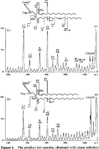
as behenic acid and palmitic acid, respectively. Be- cause liked scan at constant B/E does not provide adequate resolution for the differentiation of m/z 660 and 658 ions, mass selection followed by ion kinetic energy analysis was used to acquire the product ion spectra of the m/z 660 and 658 ions (Figure 4). The Ln ions (m/z 278, 308) and K ion (m/z 364) in Figure 4a and b were the same as those observed in Figure 3b; therefore, the long chain base was assigned as a sphing-4-ene residue. The F2, F3’, and F6’ ions at m/z 406, 436, 460 suggested that the N-acyl chain of the m/z 660 ion was a lignoceric acid residue (Figure 4b). The fatty acid of the m/z 658 ion (Figure 4a) was determined to be a C,,., acid because the Fn ions (m/z 404,434,458) were two mass units less than the analo- gous ions observed in Figure 4b. Based on the L and M ions (Figure 4a), the double bond in this C24:l acid was determined to be in the C(15)-C(16) position (nervonic acid). In linked scan at constant B/E mass
4Ea
Figure 4. The product ion spectra, obtained with mass selection followed by ion kinetic energy analysis of the ceramide frag- ments from the permethyleted glucocerebrosides. (a) The m/z 658 precursor ion. (b) The m/z 660 precursor ion. Glucocerebro- sides were isolated from human (Gaucher’s) spleen. Four differ- ent GSLs were detected in this mixture; two of the four product ion spectra are shown in this figure.
J Am Sot Mass Spectrom 1994,5,558-563
spectra, the position of the double bond is determined according to the abundance and the mass separation (54 u) of the L and M ions. Because of the poor product ion resolution in the MIKES, there is some uncertainty in the mass assignment of the L and M ions. Therefore, the position of the double bond was assigned mainly based on the abundance of the L and M ions. In samples where two or more ceramide fragments have very similar mass and the difference in relative inten- sity is less obvious, a four-sector tandem mass spec- trometer would be needed to make a clear assignment.
It is in the analysis of GSLs, rather than intact ceramides, that this approach showed a clear advan- tage over the published methods. Due to the size of the glycan, many GSLs have molecular weights much larger than ccramides and cerebrosides. Because the ceramide fragments rather than the MHC ions were selected as the precursor ions in this approach, the masses of the precursor ions remain in the range of 500-700 u. This represents a much more favorable situation than the methods with the selection of the (M + H)+, (M - H)-, or (M + Li)’ as the precursor ions because the quality of the product ion spectra often becomes rapidly worse with increasing precursor ion mass. For example, the method of metal ion adduc- tion provides an excellent approach for ceramides and small GSLs [15-171. To our knowledge, this method has not been applied to GSLs with more than two sugar residues. This is, at least in part, due to the fact that the product ion spectra of large GSLs do not have the quality of those of ceramides and small GSLs.
Sensitivity is important in the analysis of GSLs because GSLs are often isolated in small amounts. The detection limit of this method compares favorably with published methods. In the analysis of a GSL with two sugar residues, N-palmitoyl-dihydrolactocerebroside, 5 pmol of the permethylated sample (under the assump- tion of 100% yield in permethylation) produced the product ion mass spectrum shown in Figure 5. For a much larger GSL, GTlb, the FAB mass spectrum was
characterized with ceramide ions (m/z 576, 604) and carbohydrate containing ions as shown in Figure 6a. The product ion mass spectrum of the m/z 604 ion
from 46 pmol of the permethylated GTlb is shown in Figure 6b. Since there are two different ceramide frag- ments (m/z 576, 604) with relative intensity of 2:3 (Figure 6a), the spectrum in Figure 6b represents ap- proximately 28 pmol of GTlb molecules with eicosa- sphiigosine as the long chain base. Since the molecular weight of permethylated GTlb is in the range of 2500 u and a 6-keV Xe gun is used in this analysis, the sensitivity might be further improved with a higher energy Cs+ ion gun.
Conclusions
High-energy CID of the ceramide fragments from un-
J Am %C Mass Spectrom 1994, 5, 558-563 HIGH-ENERGY CID OF CE RAMIDE IONS 563
550
Figure 5. The product ion spectra, obtained with linked scan- ning at constant B/E, of 5 pm01 of permethylated N-palmitoyl- dihydro-lactocerebroside.
-CH3OI,=WI
376
Figure 6. (a) The FAB mass spectrum of permethylated GTlb. The peaks marked with * are matrix peaks of 3-nitrobenzyl alcohol. (b) The producf ioh ‘mass specbxqt of the ceramide fragment at m/z 604 from 46 pmol of the pemwthylatti GTlb. The spectlum corresponds to approximately 28 pm01 of GTlb molecules with eicosasphingosine as the long chaii base.
location of substituents such as double bonds and hydroxyl groups in the sphingoid and N-acyl chain
[ 141. We have shown that permethylation, a very sim- ple procedure, can overcome this problem. Upon high-energy CID, the product ion mass spectra of the ceramide fragments provide detailed information re- garding the structure of the ceramides. Since the ce- ramide fragments instead of MH’ ions are chosen as the precursor ion, the molecular weight of GSLs has no effect on the mass of the precursor ions and the quality of the product ion spectrum can be preserved. The sensitivity of this approach makes possible the analysis of large GSLs in the range of picomoles.
Acknowledgment
Financial support from the National Science Council of the Re- public of China is greatly appreciated.
References
1. 2. 3. 4. 5. 6. 7. 8. 9. 10. 11. 12. 13. 14. 15. 16. 17. 18. 19. 20. 21. 22.Karlsson, K.-A. FEBS Left. 1973,32,317-320.
Ten&erg, S.; PimIoot, W.; Karlsson, K.-A. In Biological Mass Specfromefry; A. L. Burlingame and J. A. McCloskey, Ed.; John Wiley: New York, 1990; pp. 477-490.
Ariga, T.; Yu, R. K.; Suzuki, M.; Ando, S.; Miyatake, T. J. Lipid Res. 1982, 23, 437-442.
Can; S. A.; Reinhold, V. N. Biomed. Mass Spectrom. 1984, II,
633-641.
Kushi, Y.; Handa, S. 1. Biochem. 1982, 91, 923-931.
Hemling, M. E.; Yu, R. K.; Sedgwick, R. D.; Rinehart, K. L., Jr. Biochemistry. 1984, 23, 5706-5713.
Arita, M.; Iwamori, M.; Higuchi, T.; Nagai, Y. J. Biofhem. 1984, 95, 971-981.
Iwamori, M.; Ohashi, Y.; Nagai, Y. In Mass Spectmmetry in the Health and Life Sciences; A. L. Burlingame and N. Castagholi, Jr., Eds.; 1985, pp. 379-398.
Pahlsson, I’.; Niison, B. Anal. Biochem. 1988, X8,115-120. Kwhi, Y.; Rokukawa, C.; Handa, S. Anal. Biochem. 1988, ‘175, 167-176.
Ohashi, Y.; Iwamori, M.; Ogawa, T.; Nagai, Y. Biocheinisfly 1987, 26, 3990-3995.
Damon, B.; Costello, C. E. Biochemisfy 1988, 27, 1534-1543.
Damon, B.; Vath, J. E.; Costello, C. E. Anal. Biochem. 1990,
184,151-x4.
Costello, C. E.; Vath, J. E. In Methods in Enzymology; Mc- Closkey, J. A., Ed.; Academic Press: San Diego, CA, 1990; Vol. 193, pp. 738-768.
Ann, Q.; Adams, J. J. Am. Sot. Mass Specfrom. 1992, 3, 260-263.
Ann, Q.; Adams, J. Anal. Chem. 1993, 65, 7-13. Adams, J.; Arm, Q. Mass Spectrom. Reu. 1993, 12, 51-85. Dub, J. S.; Her, G. R Biologiuzl Mass Spectromfry 1992, 21, 391-396.
Ciucanu, I.; Kerek, F. Carbohydr. Res. 1984, 132, 209-217. Larson, G.; Karlsson, H.; Hansson, G. C.; Pimlott, W. Carbo- hydr. Res. 1987, 161, 281-290.
Sate, K.; Asada, T.; Ishihara, M.; Kunihiro, F.; Kammei,.Y.; Kubota, E.; Costello, C. E.; Martin, S. A.; Scoble, H. A.; Biemann, K. Anal. Chem. 1987, 59, 1652-1659.
Guo, N.; Her, G. R.; Reinhold, V.; Brennan, M. J.; Siraganian, R. P.; Ginsburg, V. J. Biol. Chem. 1989, 264, 13267-13272.