行政院國家科學委員會專題研究計畫 成果報告
利用斑馬魚建立心臟衰竭研究之動物模式:專一心臟
troponin-C 蛋白表現受抑制之斑馬魚心肌功能研究
計畫類別: 個別型計畫 計畫編號: NSC93-2314-B-002-219- 執行期間: 93 年 08 月 01 日至 94 年 07 月 31 日 執行單位: 國立臺灣大學醫學院內科 計畫主持人: 何奕倫 共同主持人: 謝豐舟,蔡懷楨,陳志榮 報告類型: 精簡報告 處理方式: 本計畫可公開查詢中 華 民 國 94 年 10 月 13 日
Topic
Conditional Antisense-Knockdown of Zebrafish Cardiac Troponin C as a New Model for Dilated Cardiomyopathy
Yi-Lwun Ho, MD, PhD1*; Wei-Yuan Tsai2*, Gar-Ling Wang, 2 Yen Hung Lin,
MD1; Fong-Jou Hsieh, MD3,# and Huai-Jen Tsai, PhD2,#
Division of Cardiology1, Department of Internal Medicine and Department of
Obstetrics and Gynecology3, National Taiwan University Hospital and National
Taiwan University College of Medicine, No.7 Chung-Shan S. Road, Taipei 100,
Taiwan; Institute of Molecular and Cellular Biology2, National Taiwan University,
1 Roosevelt Road, Section 4, Taipei 106, Taiwan.
Addresses for Correspondence#: Huai-Jen Tsai, Institute of Molecular and
Cellular Biology, National Taiwan University, 1 Roosevelt Road, Section
4,Taipei 106, Taiwan. Fong-Jou Hsieh, Department of Obstetrics and
Gynecology, National Taiwan University Hospital and National Taiwan
University College of Medicine, No.7 Chung-Shan S. Road, Taipei 100, Taiwan
Abstract
Background – Mutations of cardiac troponin C (cTnC) can cause dilated
cardiomyopathy in humans. We attempted to generate a zebrafish transgenic
line that enables us to produce the antinsense RNA strand of cTnC after
induction as a new animal model for studying dilated cardiomyopathy.
Methods and Results – We constructed plasmids that the reverse
tetracycline–controlled transactivator (rtTA) was driven by the cardiac myosin
light chain 2 promoter. This heart-specific rtTA bound another bidirectional
promoter to express the green fluorescence protein (GFP) reporter gene and
to transcribe the antisense RNA of cTnC in the presence of doxycycline (Dox).
After microinjection of these constructs into one-cell fertilized eggs, we
cultured, screened, and generated a transgenic line of zebrafish (CA17) which
enabled us to express GFP and to conditionally produce the antisense RNA of
cTnC in heart after induction. When 12-h postfertilization (hpf) embryos of F2
derived from CA17 were given Dox (10 µg/ml), the expression of endogenous
cTnC mRNA was not affected until 6 days postfertilization (dpf). The heart
rates of the embryos in CA17 were significantly slower than those of embryos
in the control T03 transgenic line (only expressing the heart-specific GFP) at 6
beats/min, p < 0.01). Moreover, the various values of cardiac chambers
measured in the induced F2 embryos from CA17 were significantly greater
than those of T03 embryos; while the ventricular ejection fraction of CA17 was
lower than that of the T03 both at 6 dpf (44%±4% vs. 50%±3%, p < 0.01) and
at 12 dpf (39%±5% vs. 52%±4%, p <0.01). In addition, asynchronized atrial
and ventricular contractions were noted in a few (1.3%) F2 embryos from the
induced CA17 transgenic fish. The mortality rate of F2 adult fish of the CA17
group was significantly higher (30% vs. 0%, p < 0.001) than that of F2 adults of
the T03 group after Dox induction.
Conclusions – Using conditional expression of antisense RNA of zebrafish
cTnC, we have created a new animal model with phenotypes simulating
dilated cardiomyopathy.
Key Words: Zebrafish; Cardiac troponin C; Antisense RNA; Dilated
Contraction of the myocardium is regulated by the cardiac troponin complex.
Calcium ions bind to the regulatory sites of cardiac troponin C (cTnC) and
facilitate interactions between the actin and tropomyosin. Mutation of cTnC
can cause myocardial dysfunction.1 A cTnC mutation has been reported to
cause human familial dilated cardiomyopathy.2 Mutation analysis of the
troponin complex in dilated cardiomyopathy patients may prove valuable in the
early identification of individuals with an adverse prognosis and a high risk of
premature death. 2 However, no animal model of dilated cardiomyopathy
related to cTnC dysfunction has been reported.
The zebrafish (Danio rerio) is an excellent animal model for cardiac
research.3-5 It has a 2-chambered heart that is similar in many genetic ways to
the 3- or 4-chambered hearts of higher vertebrates.6-7 Transparent embryos
make it possible to observe the dynamic expression of heart genes in a
noninvasive manner. Embryos of zebrafish receive oxygen through diffusion.
This kind of oxygenation system allows embryos to survive for up to (at least)
1 week even with cardiac defects.8 A 1.6-kb regulatory region of the zebrafish
cardiac myosin light chain 2 gene (cmlc2) has been demonstrated to
specifically drive green fluorescent protein (GFP) expression in the heart of
myocardium-specific expression of GFP under a tetracycline (Tet)-on
conditional expression system.10 To establish a zebrafish model that is feasible
for studying dilated cardiomyopathy, we designed 2 constructs in this study.
One was an upstream plasmid which contained a cmlc2 promoter to
specifically drive the expression of a reverse Tet–controlled transactivator
(rtTA) in the heart. The other was a downstream plasmid which contained a
Tet-binding domain (TetO) and possessed a bidirectional promoter to express
the GFP reporter and to transcribe cTnC-antisense RNA. Antisense
knockdown has been widely applied both in vitro and in vivo studies to block
the translation of endogenous mRNA.11,12 After gene transfer, a germ
line-transmitted zebrafish carrying a heart-specific Tet-On system was
generated to conditionally produce the antisense RNA of the cTnC gene.
Under the impediment of the translation of the endogenous cTnC gene,
cardiac functional assays were carried out in this transgenic zebrafish after
induction.
Methods Experimental Fish
Zebrafish of the AB strain were cultured and maintained according to the
determined according to criteria described by Kimmel et al.14
Molecular Cloning of Zebrafish cTnC (Figure 1)
The primers used to clone the cDNA sequence of zebrafish cTnC were
designed on the basis of the homologous analysis of cTnC from other known
vertebrates. The complete coding region sequence of zebrafish cTnC was
amplified by polymerase chain reaction (PCR) using cTnC-SpeI-F
(TGACTAGTATGAACGACATCTACAAAGCAGC) and cTnC-PstI-R
(TGCTGCAGCTATTCCACCCCTTCATG). The PCR product was used to
construct the expression plasmid after it was cut with SpeI and PstI. cTnC
polypeptides from various vertebrates were aligned using the BCM Research
Laucher (available at http://searchlauncher.bcm.tmc.edu/) and a phylogenetic
tree was established from the Gene Data Bank of Japan website (available at
http://www.ddbj.nig.ac.jp/). The complete coding region sequence was
deposited in GenBank (ascension no.: AF434188, maintained by the NCBI).
Whole-mount in Situ Hybridization (Figure 2)
In order to determine the tissue-specific expression pattern of the cTnC we
cloned, we carried out whole-mount in situ hybridization using a
digoxigenin-labeled RNA probe as described previously.15
pICML1-rT16-1b is an upstream plasmid used to produce cardiac-specific rtTA. It was
derived from plasmid pICMLE-(-870/787),9 except that the GFP cDNA was replaced
by rtTA-M2.10 The resulting pICML1-rT16-1b consisted of the regulatory region of
cmlc2 (the -870 to –1 nt region, exon 1, intron 1, and a portion of exon 2), rtTA-M2,
and inverted terminal repeats of an adeno-associated virus. Plasmid
pBIEK-cTnC-antisense is a downstream plasmid used to produce antisense RNA of
cTnC and to express the GFP reporter gene. It was derived from plasmid pBILE,16 in
which SpeI and PstI were used to remove the luciferase gene, which was replaced by
ligation with the opposite-direction cTnC cDNA to the rtTA-dependent bidirectional
promoter, tetO (Clontech).
Gene Transfer
Fertilized eggs at the 1-cell stage were collected and microinjected with linearized
constructs of pICML1-rt16-1b and pBIEK-cTnC-antisense following a previous
study17 except that a total of 15 µg/ml of each plasmid was used. This was the
cTnC-antisense group (CA17). Meanwhile, we also microinjected linearized plasmids
of pICML1-rt16-1b and pIBIE, in which the cTnC segment was not included, to serve
as a control group (T03 group).
Transgenic Line Screening
mature fish were kept together in tanks. The heart-specific GFP expression in F1
embryos was examined under a fluorescent microscope after induction with 10 µg/ml
of Dox (Sigma) for 3 days. GFP-positive progeny were selectively bred and mated
with wild-type (wt) fish when they reached adulthood.
Induction in Embryos
Heterozygotic F2 embryos derived from the transgenic germ lines were kept in
water until 12 h post fertilization (hpf), at which time they were treated with 10
µg/ml of Dox. After induction, GFP expression was observed under a
fluorescent microscope. Fish embryos that had heart-specific GFP expression
without leaks were selected, orientated ventrally, and tilted at 45o when taking
photos using an S2Pro digital camera (FinePix) with a 10-s exposure time at
ISO 400 for image analysis of heart functions.Morphological changes of the
heart and heart rate were also examined.
Induction in Adult Fish
The 6-month-old adult fish derived from the homozygote of transgenic lines of
the cTnC-antisense line (CA17) and the control line (T03) were selected to
carry out Dox induction. Ten fish from each group were raised in a tank and
treated with 10 µg/ml Dox. In order to prevent Dox from being photodegraded,
water in the tank was replaced daily. After the induction, we observed the
fluorescent signal in fish under fluorescent microscope for 3 weeks. Extraction of Zebrafish Embryonic mRNA
The heterozygous F2 embryos derived from the CA17 and T03 groups were
treated with Dox (10 µg/ml) when they were at the 12-hpf stage. Another
negative control group consisted of those embryos derived from the CA17
transgenic line without Dox treatment. Five embryos at 6 dpf from each of
these 3 groups were selected, and their mRNAs were extracted for further
analysis.
Reverse-Transcription Polymerase Chain Reaction (RT-PCR)
In order to detect the endogenous cTnC transcription and the antisense RNA
of cTnC transcription after induction, we collected 6-dpf F2 embryos derived
from the CA17 and T03 lines. Their total RNA was extracted. All of the RT-PCR
procedures followed those of a previous study18 except that (1) in the
first-strand cDNA synthesis, we used the Oligo-dT primer to detect the
endogenous the cTnC transcription and used TnCA-RT1F primer
(CGACATCTACAAAGCAGCGGC- AG; Fig. 1C) to detect the cTnC-antisense
strand transcription; and (2) in the following PCR amplification, we used
TnCA-RT2F (GTCTCCGTCCCTCATCAGTTCCTC) (Figure 1C).
Video File Capture and Processing
The lens adaptor of a stereomicroscope was connected to a DV camera. The
LCD monitor of the camera was turned on and a connected to the computer
with Ulead video studio 7.0 system. The image was captured for 10 s and
recorded as an AVI file in the computer. Then the AVI file was converted to
bmp files sequentially according to a previously described method.19 One bmp
file contained 1 frame of cardiac motion. The cardiac motion was viewed frame
by frame to define the systolic and diastolic phases. An area with the
most-even fluorescent distribution was selected, and the region of the
fluorescent heart to be analyzed was marked. The ventricular volume was
calculated using the formula for a prolate spheroid:20 Volume = 4/3 × π × a ×
b2 where a and b are the long and short radii of the ventricle, respectively. The
cardiac ejection fraction was calculated by the following formula: (cardiac
diastolic volume – cardiac systolic volume) ÷ cardiac diastolic volume.
Statistical Analysis
All data are expressed as the mean ± standard deviation (SD). Statistical
differences among groups were obtained using the Student's t-test. It was
Results Primary Structure of Zebrafish cTnC
The complete coding sequence of zebrafish cTnC we cloned was 486 bp
(Figure 1c), which encoded a 161-amino acid polypeptide (Figure 1a). The
zebrafish cTnC polypeptide shared 90% identities with those of a mammal,
chicken, and Xenopus (Figure 1), in which 4 E-F hand calcium-binding
domains were highly conserved. On the basis of the phylogenetic tree, the
zebrafish cTnC was clustered into the cardiac/slow skeletal muscle group.
Expression of cTnC is Heart-Specific
As shown in the in situ hybridization, the transcripts of cTnC we cloned were
heart-specific (Figure 2). The cTnC transcripts were first detectable in 17-hpf
embryos, and cTnC was expressed bilaterally in heart precursory cells behind
the notochord (Figure 2a). Transcription of the cTnC gene was detected only in
the heart-tube of 24-hpf embryos (Figure 2b), in the whole heart of 48-hpf
embryos (Figure 2c), and in the mature heart of 72-hpf embryos (Figure 2d).
The intensity of the fluorescent signal expressed in the ventricle was much
stronger than that in the atrium. The reason was there is only 1 layer of
cardiomyocytes in the atrium whereas there are 2 to 3 layers of
Inducible GFP Expression in the Germ Line-Transmitting Zebrafish The 12-hpf embryos derived from F1 of the transgenic cTnC-antisense line
(CA17) and control T03 were collected and treated with 10 ug/ml Dox. The
CA17 line expressed both the GFP and the antisense mRNA of cTnC, while
the T03 line expressed the GFP only. Without Dox treatment, there was no
green fluorescent signal shown in the fish. However, after induction, the green
fluorescent signal began to appear specifically in the heart about 12 h after
treatment (Fig 4a).
Transcription of cTnC Antisense RNA in Transgenic Fish
As shown in Figure 4b, cTnC mRNA was produced by the transgenic lines of
both CA17 and T03 after induction by Dox; whereas the antisense RNA
transcribed from the cTnC gene was clearly shown in embryos derived from
CA17 transgenic fish after Dox induction. Although a very weak signal of
antisense RNA was shown prior to induction, the green fluorescent signal was
not detected. On the contrary, the antisense RNA of cTnC was not produced in
embryos derived from the T03 control transgenic line. As long as cTnC
antisense RNA was expressed in CA17 embryos, the expression of
endogenous cTnC mRNA remained the same.
At 3 dpf, heart rates of the CA17 group were slightly faster than those of the
T03 group (Figure 5). With further growth, the situation gradually reversed. The
heart rates of CA17 were significantly slower than those of the T03 at 6
(150±10 vs. 194±11 beats/min, p < 0.01) and 12 dpf (128±12 vs. 168±8
beats/min, p <0.01).
Ventricular Assessments of CA17 and T03 Zebrafish at 6 and 12 dpf End-diastolic and end-systolic diameters were significantly different between
the T03 and CA17 groups at both 6 and 12 dpf (Figure 6, Tables 1 and 2). The
ventricular ejection fraction of CA17 was lower than that of T03 transgenic fish
(44%±4% vs. 50%±3%; p < 0.01). At 12 dpf, the ventricular ejection fraction of
CA17 was even lower than that of T03 (39%±5% vs. 52%±4%; p <0.01).
Measurements from the hearts of the T03 transgenic fish were calculated to
obtain the arithmetic mean and standard deviation. The range of mean ± 2
standard deviations was defined as the normal range. Percentages of the
upper outlier for cardiac chambers and lower outlier for the ejection fraction
and heart rates in the CA17 group are presented in Table 3.
Atrial and Ventricular Asynchrony in CA17 Zebrafish
At 6 dpf, atrial and ventricular contractions were synchronized in F2 embryos
transgenic fish developed asynchronized atrial and ventricular contractions at
12 dpf (Figure 7 and supplemental video file).
Slurred Movement and Excess Mortality in cTnC-Antisense Transgenic Adult Fish after Dox Induction
After the Dox has been continuously administrated to 6-month-old fish derived
from homozygotic parents, we found that the movement of fish from CA17 was
slower than that of T03 group. Furthermore, 30% (3/10) of fish from the CA17
group had died by day 13; whereas no mortality had occurred in the T03
group.
Discussion
cTnC and cardiac troponin T mutations have been reported to cause human
familial dilated cardiomyopathy.2 The clinical manifestations include heart
failure and sudden death.2 Cardiac troponin T variants produced by aberrant
splicing of multiple exons in animals have been related to high instances of
dilated cardiomyopathy. 21 However, there has been no report of a transgenic
animal model for cTnC dysfunction. On the other hand, cTnC is the target
protein of a novel calcium sensitizer (levosimendan).22 This drug has proven to
be a well-tolerated and effective treatment for patients with severe
heart failure is worth studying.
The zebrafish heart contains 4 components (sinus venosus, atrium,
ventricle, and bulbus arteriosus). Although it has a prototypic vertebrate heart,
the zebrafish has been used to study bradycardia, heart valve formation and
cardiac regeneration.23-29 By fusing the Tet repressor and activating the
transcription domain of virion protein 16 of the herpes simplex virus,
Tet-controlled transactivator (tTA) is generated.30 A Tet-On system is further
developed by mutagenesis, substituting 4 amino acids in tTA to generate rtTA
which requires Tet for binding to the Tet operon and subsequent activation of
the downstream gene.31 For pharmacokinetic reasons, controlling genes by
means of rtTA is particularly advantageous in the study of transgenic animals.
We have successfully developed such a Tet-On system in zebrafish that
enables the transgene to specifically be expressed in the heart after
induction.10 This technique also makes it feasible for later onset of abnormal
gene expression in adult transgenic fish. Therefore, this methodology extends
the utilization of zebrafish as a model organism from congenital cardiac
disorders to acquired heart diseases.
In this study, cTnC antisense mRNA was only noted in CA17 fish induced
ventricular size and the prevalence of ventricular enlargement had increased
significantly in the CA 17 group at 6 and 12 dpf. The ventricular ejection
fraction of the CA17 group was also significantly lower than that of the T03
group at 6 and 12 dpf. On the other hand, relative bradycardia was noted
despite the impaired contractility in the CA17 group. Chronotropic
incompetence was another major finding in the cTnC antisense zebrafish.
Even 1.3% of CA17 transgenic fish manifested asynchronized atrial and
ventricular contractions. Atrioventricular conduction block can lead to sudden
death which has been reported for troponin related dilated cardiomyopathy in
humans.2 According to Sedmera et al., trabecular bands form direct myocardial
continuity between the atrioventricular canal and the apex of the ventricle in
zebrafish.32 Therefore, cTnC may play a role in these trabecular bands.
It is interesting to observe the activities of adult fish derived from the
homozygotes of the CA17 transgenic line after continuous treatment with Dox.
The movement of cardiomyopathy-induced fish was slower, and 30% of them
experienced early death; whereas fish from the T03 control group lived
normally. It seemed that the low cardiac output failed to supply adequate
perfusion to skeletal muscles, resulting in a slurring of body movements. The
either a power failure or conduction abnormality of the hearts. These
phenomena are frequently observed in human patients with heart failure.
Recently, the zebrafish has become an important vertebrate model
organism, because this species can be used to study human diseases,
physiology, and pharmacology.33 The cTnC antisense zebrafish in this study
offers a new investigative tool for human heart failure.
Acknowledgments
This study was supported by the National Science Council, Taipei, Taiwan
References
1. Gomes AV, Potter JD. Molecular and cellular aspects of troponin
cardiomyopathies. Ann NY Acad Sci 2004; 1015: 214-224.
2. Mogensen J, Murphy RT, Shaw T, Bahl A, Redwood C, Watkins H,
Burke M, Elliott PM, McKenna WJ. Severe disease expression of
cardiac troponin C and T mutations in patients with idiopathic dilated
cardiomyopathy. J Am Coll Cardiol 2004; 44: 2033-2040.
3. Poss KD, Wilson LG, Keating MT. Heart regeneration in zebrafish.
Science 2002; 298: 2188-2190.
4. Raya AKC, Koth CM, Buscher D, Kawakami Y, Itoh T, Raya RM, Sternik
G, Tsai HJ, Rodriguez-Esteban C, Izpisua-Belmonte JC. Activation of
notch signaling pathway precedes heart regeneration in zebrafish. Proc
Natl Acad Sci USA 2003; 100: 11889-11895.
5. Shu X, Cheng K, Patel N, Chen F, Joseph E, Tsai HJ, Chen JN.
Na,K-ATPase is essential for embryonic heart development in the
zebrafish. Development 2003; 130: 6165-6173.
6. Fishman MC, Stainier DY. Cardiovascular development. Prospects for a
genetic approach. Circ Res 1994; 74: 757-763.
embryonic decisions. Development 1997; 124: 2099-2117.
8. Sehnert AJ, Huq A, Weinstein BM, Walker C, Fishman M, Stainier DY.
Cardiac troponin T is essential in sarcomere assembly and cardiac
contractility. Nat Genet 2002; 31: 106-110.
9. Huang CJ, Tu CT, Hsiao CD, Hsieh FJ, Tsai HJ. Germ-line transmission
of a myocardium-specific GFP transgene reveals critical regulatory
elements in the cardiac myosin light chain 2 promoter of zebrafish. Dev
Dyn 2003; 228: 30-40.
10. Huang CJ, Jou TS, Ho YL, Lee WH,Jeng YT, Hsieh FJ, Tsai HJ.
Conditional expression of a myocardium-specific transgene in zebrafish
transgenic lines. Dev Dyn 2005; (in press).
11. Yokoyama K, Imanoto F. Transcriptional control of the endogenous
MYC potooncogene by antisense RNA. Proc Natl Acad Sci USA 1987;
84: 7363-7367
12. McGarry J, Lindquist S. Inhibition of heat shock protein synthesis by
heat-inducible antisense RNA. Proc Natl Acad Sci USA 1986; 83:
399-403.
13. Westerfield M. The zebrafish book, Eugene, or University of Oregon
14. Kimmel CB, Ballard WW, Kimmel SR, Ullmann B, Schilling TF. Stages
of embryonic development of the zebrafish. Dev Dyn 1995; 203:
253-310.
15. Jowett T. Double in situ hybridization techniques in zebrafish. Methods
2001; 23: 345-358.
16. Chou CY, Hong LS, Tsai HJ. Uniform GFP-expression in transgenic
medaka (Oryzias latipes) at the F0 generation. Transgen Res 2001; 10:
303-315.
17. Wang TM, Chen YH, Liu CF, Tsai HJ. Functional analysis of the
proximal promoter regions of fish rhodopsin and myf-5 genes using
transgenesis. Mar Biotechnol 2002; 4: 247-255.
18. Huang CJ, Lin JY, Tsai HJ. Two distinct c-ski cDNAs of fish, tilapia
(Oreochromis aurea). Mol Reprod Dev 1999; 54: 223-231.
19. Ho YL, Shau YW, Tsai HJ, Lin LC, Huang PJ, Hsieh FJ. Assessment of
zebrafish cardiac performance using Doppler echocardiography and
power angiography. Ultrasound Med Biol 2002; 28: 1137-1143
20. Hou PC, Burggren WW. Cardiac output and peripheral resistance
during larval development in the anuran amphibian Xenopus laevis. Am
21. Biesiadecki BJ, Elder BD, Yu ZB, Jin JP. Cardiac troponin T variants
produced by aberrant splicing of multiple exons in animals with high
instances of dilated cardiomyopathy. J Biol Chem 2002; 277:
50275-50285.
22. Sorsa T, Pollesello P, Solaro RJ. The contractile apparatus as a target
for drugs against heart failure: interaction of levosimendan, a calcium
sensitiser, with cardiac troponin c. Mol Cell Biochem 2004; 266: 87-107.
23. Poss KD, Wilson LG, Keating MT. Heart regeneration in zebrafish.
Science 2002; 298: 2188-2190.
24. Raya A, Consiglio A, Kawakami Y, Rodriguez-Esteban C,
Izpisua-Belmonte JC. The zebrafish as a model of heart regeneration.
Cloning Stem Cells 2004; 6: 345-351.
25. Raya A, Koth CM, Buscher D, Kawakami Y, Itoh T, Raya RM, Sternik G,
Tsai HJ, Rodriguez-Esteban C, Izpisua-Belmonte JC. Activation of
Notch signaling pathway precedes heart regeneration in zebrafish. Proc
Natl Acad Sci USA 2003; 100: 11889-11895.
26. Armstrong EJ, Bischoff J. Heart valve development: endothelial cell
signaling and differentiation. Circ Res 2004; 95: 459-470.
Graef IA, Crabtree GR. A field of myocardial-endocardial NFAT
signaling underlies heart valve morphogenesis. Cell 2004; 118:
649-663.
28. Milan DJ, Peterson TA, Ruskin JN, Peterson RT, MacRae CA. Drugs
that induce repolarization abnormalities cause bradycardia in zebrafish.
Circulation 2003; 107: 1355-1358.
29. Hsieh DJ, Liao CF. Zebrafish M2 muscarinic acetylcholine receptor:
cloning, pharmacological characterization, expression patterns and
roles in embryonic bradycardia. Br J Pharmacol 2002; 137: 782-792.
30. Gossen M, Bujard H. Tight control of gene expression in mammalian
cells by tetracycline-responsive promoters. Proc Natl Acad Sci USA
1992; 89: 5547-5551.
31. Gossen M, Freundlieb S., Bender G., Muller G., Hillen W., Bujard H.
Transcriptional activation by tetracycline in mammalian cells. Science
1995; 268: 1766-1769.
32. Sedmera D, Reckova M, deAlmeida A, Sedmerova M, Biermann M,
Volejnik J, Sarre A, Raddatz E, McCarthy RA, Gourdie RG, Thompson
RP. Functional and morphological evidence for a ventricular conduction
Physiol 2003; 284: H1152-H1160.
33. Langheinrich U. Zebrafish: a new model on the pharmaceutical catwalk.
Table 1. Cardiac measurements of F2 embryos at 6 dpf from transgenic fish
after continuously Dox induction from 12 hpf.
T03 (N = 15) CA17 (N = 26) p value
Diastolic
Long diameter (um) 159±5 164±14 0.106
Short diameter (um) 99±5 109±6 < 0.01
Systolic
Long diameter (um) 130±6 132±13 0.24
Short diameter (um) 87±4 90±4 0.02
End-diastolic volume (um3) 870,226±122,525 1,027,030±157,561 0.13
End-systolic volume (um3) 491,800±45,258 568,976±837,191 < 0.01
Ventricular ejection fraction (%) 50±3 44±4 < 0.01
Heart rate (beats/min) 194±11 150±10 < 0.01
T03, control transgenic line which expressed the GFP after induction; CA17,
transgenic line which expressed the GFP and transcribed the antisense strand
Table 2. Cardiac measurements of F2 embryos at 12 dpf from transgenic fish
after continuous Dox induction from 12 hpf.
T03 (N = 15) CA17 (N = 20) p value
Diastolic
Long diameter (um) 225±8 252±16 < 0.01
Short diameter (um) 125±9 146±18 < 0.01
Systolic
Long diameter (um) 196±8 211±19 < 0.01
Short diameter (um) 110±4 124±14 < 0.01
End-diastolic volume (um3) 1,831,044±245,255 2,870,476±846,137 < 0.01
End-systolic volume (um3) 1,303,185±155,464 1,738,336±472,814 < 0.01
Ventricular ejection fraction (%) 52±4 39±5 < 0.01
Heart rate (beats/min) 168±8 128±12 < 0.01
T03, control transgenic line which expressed the GFP after induction; CA17,
transgenic line which expressed the GFP and transcribed the antisense strand
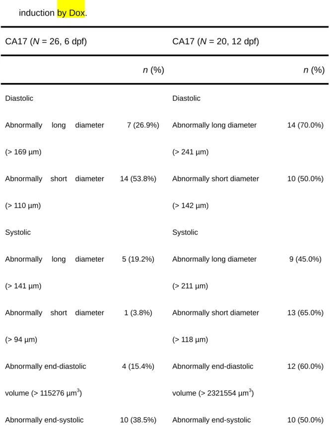
Table 3. Prevalence of abnormal cardiac measurements in F2 embryos
derived from the cTnC-antisense transgenic line (CA17) of zebrafish after
induction by Dox.
CA17 (N = 26, 6 dpf) CA17 (N = 20, 12 dpf)
n (%) n (%)
Diastolic Diastolic Abnormally long diameter
(> 169 µm)
7 (26.9%) Abnormally long diameter (> 241 µm)
14 (70.0%)
Abnormally short diameter (> 110 µm)
14 (53.8%) Abnormally short diameter (> 142 µm)
10 (50.0%)
Systolic Systolic Abnormally long diameter
(> 141 µm)
5 (19.2%) Abnormally long diameter (> 211 µm)
9 (45.0%)
Abnormally short diameter (> 94 µm)
1 (3.8%) Abnormally short diameter (> 118 µm) 13 (65.0%) Abnormally end-diastolic volume (> 115276 µm3) 4 (15.4%) Abnormally end-diastolic volume (> 2321554 µm3) 12 (60.0%)
volume (> 581424 µm3) volume (> 1614112 µm3) Abnormally ventricular
ejection fraction (< 44%)
12 (46.2%) Abnormally ventricular ejection fraction (< 45%)
18 (90%)
Abnormally heart rate (< 173 beats/min)
26 (100%) Abnormally heart rate (< 153 beats/min)
Legends
Fig. 1. Molecular cloning of cardiac troponin C (cTnC). In vertebrates, the amino acid sequences of cTnC are highly conserved from fish to mammal. The
overall identities are exceed 90% not only in the 4 calcium-binding domains
but also in the entire fragment. The nucleotide sequences of others species
were obtained form the NCBI EST database (a). In the phylogenic tree,
zebrafish troponin C was clustered in the ss/cTnC group (b). The full coding
region sequence is shown in (c).
Fig. 2. Expression pattern of the zebrafish cardiac troponin C (cTnC) gene. Images of 18 hpf (A) and 24 hpf (B) are in the dorsal view; the image at 48 hpf (C) is in the ventral view; and image at 72 hpf (D) is left-side up. The
expression of cTnC was restricted to the heart field from fusion of the 2
bilateral cells (at 18 hpf) to the mature heart (at 72 hpf).
Fig. 3. Construction map of antisense knockdown of cardiac troponin C (cTnC). After adding Dox, the transactivator, rtTA-M2, was driven by the cmlc2 promoter combined with Dox after 16 hpf. This complex was capable of
expression of the reporter green fluorescent protein (GFP) and the
transcription of cTnC antisense RNA.
Fig. 4. Detection of the expression of green fluorescent protein (GFP) and the transcription of cardiac troponin C (cTnC) antisense RNA. The 12-hpf embryos derived form the cTnC-antisense transgenic line CA17 were collected
and incubated in 10 µg/ml Dox. Green fluorescence was observed in the heart
at 48 hpf (A). The reverse-transcription polymerase chain reaction was used to
detect the transcription of endogenous cTnC mRNA and the transcription of
cTnC antisense RNA (B). Embryos were treated with Dox after 12 hpf, and the
total RNA was extracted when these embryos reached 6 dpf. Antisense cTnC
was strongly detected in the CA17 group after induction. Although the
antisense RNA of the cTnC gene was slightly detected in the non-induced
group, the green fluorescent signal was not detected. Expression of the
endogenous cTnC gene was almost the same in the T03 and CA17 groups.
This suggests that the antisense of cTnC did not interfere in the expression of
the endogenous cTnC gene. Arrow indicates the position of the heart.
Fig. 5. Heart rates at different stages in the T03 and CA 17 groups.
heart rates of embryos derived from the CA17 group were significant slower
than those of embryos from theT03 group at 6 and 12 dpf. * p < 0.01.
Fig. 6. Changes in ventricular morphology. The end-systolic and
end-diastolic ventricular dimensions of the F2 embryos of the CA17 group
were larger than those of the T03 group.
Fig. 7. Asynchronized contractions of the atrium and ventricle were observed in some F2 embryos derived from the CA17 group.