Magnetic Resonance Imaging for Prenatal Diagnosis: An Adjunct Diagnostic Tool?
5
0
0
全文
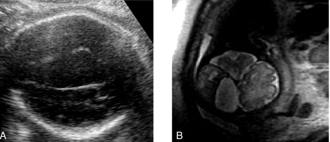
(2) Li-Chia Huang, et al.. Continuation or termination of pregnancy was dependent on the severity of fetal anomalies. RESULTS. Fetal anomalies involving the central nervous and skeletal systems were initially found by prenatal ultrasound. The majority of the fetuses had nervous system abnormalities. All of the prenatal ultrasound findings were compared with the findings from prenatal or postnatal magnetic resonance imaging. The descriptions of the seven fetuses are summarized in the Table. Prenatal ultrasound precisely diagnosed the structural anomalies but prenatal magnetic resonance imaging provided the exact location and the extent of disease. Arachnoid cyst was diagnosed in fetus 1 and fetus 2 by prenatal ultrasound (Table, Fig. 1). Prenatal magnetic resonance imaging revealed the exact anatomical sites and extent of severity. Postnatal and prenatal magnetic resonance images were compatible. Both of the fetuses were delivered at term and required neonatal shunts. In fetus 3, microcephaly and ventriculomegaly were diagnosed by prenatal ultrasound; however, prenatal magnetic resonance imaging revealed severe frontal lobe hypoplasia of the brain. The possibility of neonatal brain sequelae was considered and the parents were informed of the prognosis. In fetus 4, sacroccygeal teratoma with intra-pelvic mass was antenatally diagnosed. Magnetic resonance imaging showed the extent of tumor location, tumor size and the anatomical. 199. association with vital organs (Fig. 2). In this case, termination of pregnancy was suggested at 22 weeks’ gestational age because the extent of disease was severe. Ventriculomegaly was initially diagnosed by prenatal ultrasound in fetuses 5 and 6. Prenatal magnetic resonance imaging confirmed the diagnosis of colpocephaly in both of the patients. The mother with fetus 5 continued pregnancy and the baby was delivered in our hospital. However, neonatal information was not retrieved for fetus 6 because the patient did not receive further management at our hospital. Leukomalacia was detected on magnetic resonance imaging in fetus 7 while prenatal ultrasound only showed hydrocephalus. The fetus was delivered and a ventricular-peritoneal shunt was inserted. DISCUSSION. Congenital abnormalities account for 20% to 25% of perinatal deaths. Various kinds of prenatal diagnostic modalities are used to predict fetal disorders and genetic disease, and to determine the health of the unborn fetus. Ultrasound is the most commonly used noninvasive diagnostic screening tool for imaging fetal anatomy. It is harmless to both the fetus and the mother. Ultrasound can evaluate gestational age, amniotic fluid volume, fetal position, placental location, fetal growth and structural birth defects, and identify multiple pregnancies. However, maternal habitus, gestational age, oligohydramnios and fetal malpresentation limit. Table. Comparison between prenatal ultrasound, prenatal and postnatal MRI Case Prenatal ultrasound Postnatal MRI Prenatal MRI Management and Outcome Arachnoid cyst 1 Compatible Neonatal C-P shunt Supratentorial Arachnoid cyst Arachnoid cyst 2 Compatible Neonatal C-P shunt Interhemispheric Arachnoid cyst Microcephaly 3 NA Lost to follow up Ventriculomegaly Ventriculomegaly Frontal lobe hypoplasia 4 Sacroccygeal teratoma NA Termination of pregnancy Sacroccygeal teratoma 5 Ventriculomegaly Compatible Continued pregnancy Agenesis of corpus callosum with colpocephaly 6 Ventriculomegaly NA Lost to follow-up Unilateral colpocephaly 7 Hydrocephalus Hydrocephalus Neonatal V-P shunt NA Leukomalacia NA = postnatal MRI not performed; C-P shunt = cyst-peritoneal shunt; V-P shunt = ventriculo-peritoneal shunt..
(3) 200. A. Magnetic Resonance Imaging for Prenatal Diagnosis. B. Fig. 1. A: Ultrasound showing supratentorial arachnoid cyst. B: MRI precisely locates the cyst and its involvment with peripheral brain structures.. A. B. Fig. 2. A: Ultrasound revealing sacroccygeal teratoma. B: MRI shows the tumor involvement and the components of the tumor.. the diagnosis of structural anomalies. MRI can solve the limitations found in prenatal ultrasound. Prospective studies have revealed some advantages of prenatal magnetic resonance imaging [2]. Magnetic resonance imaging provides detailed and reproducible diagnosis of fetal structural anomalies. MRI is most useful in evaluation of abnormalities of the fetal brain, neck, chest, and abdomen. Evaluation of anomalies in the fetal brain and spine with ultrasound is limited. MRI has been shown to augment ultrasound diagnosis of CNS abnormalities. The normal and abnormal. appearance of the brain on ultrasound is based on the ability to obtain specific images of the cerebrum, cerebellum, and spine. Ventriculomegaly is the most common abnormal structure found during the antenatal ultrasound in fetal brain scanning. Mild to moderate enlargement of the ventricles is frequently associated with other anomalies [3]. Compared with ultrasound, MRI more accurately shows the cause of ventriculomegaly and identifies associated brain anomalies. Abnormalities of the corpus callosum, such as partial agenesis may be difficult to diagnose on ultrasound. MRI can differentiate between partial.
(4) Li-Chia Huang, et al.. agenesis and thinning of the corpus callosum. Other abnormalities including arachnoid cyst and holoprosencephaly can be easily diagnosed by prenatal ultrasound, although MRI can provide additional information, such as anatomical location. Sacrococcygeal tumors are usually diagnosed prenatally by ultrasound and are classified according to the amount of extra-or intra-pelvic components. Degree of increased vascularity detected on the tumors by ultrasonography is related to the severity of the disease and inversely to postnatal outcome. However, MRI better defines intra-spinal and intra-pelvic extension of the tumor. MRI differentiates tumors that are predominantly cystic from sacral myelomeningocele tumors [4]. Prenatal magnetic resonance imaging also provides further information regarding lesions of the fetal neck, thorax and abdomen. MRI can help define the relationship between the lesion and the airway, major neck vessels, the degree of liver herniation into the chest in diaphragmatic hernia, and assess the intra-abdominal location and degree of tumor involvement. However, MRI does not provide sufficient information for fetal renal anomalies. Magnetic resonance imaging provides additional information and aides in further obstetric counseling and management. It provides vital information that can be utilized for better antepartum management. Prenatal diagnosis also allows the obstetrician and neonatologist to be. 201. adequately prepared for a potentially ill neonate at delivery. It is unlikely that MRI will replace ultrasound as the primary obstetric imaging modality in the near future, but MRI is helpful in evaluating gross fetal anomalies and disturbances of fetal growth and development when ultrasound is limited by oligohydramnios, fetal position or maternal obesity. In this article, we described our experience in assessing fetal abnormalities based on MRI. Indications for MRI during pregnancy are limited but well defined. It is generally useful as an additional tool for further evaluation of problems that are initially detected by ultrasound. Improved prenatal diagnosis by magnetic resonance imaging can lead to changes in perinatal management, including the timing of delivery, reduction in parental anxiety, and contribute more data to aid in obstetric counseling and postnatal care. REFERENCES 1. Bekker MN, van Vugt JM. The role of magnetic resonance imaging in prenatal diagnosis of fetal anomalies. [Review] Eur J Obstet Gynecol Reprod Biol 2001;96:173-8. 2. Casele H, Meyer J. The selective use of magnetic res onance im a gi ng in p re nat al di agnosis. Ultrasound Obstet Gynecol 2004;23:105-10. 3. Hubbard AM. Ultrafast fetal MRI and prenatal diagnosis. [Review] Semin Pediatr Surg 2003;12:14353..
(5) 202. 2004. 1. 1. 2005. 12. 31. sacrococcygeal teratoma. 2007;12:198-202. 404. 2. 2007. 4. 9. 2007. 9. 10. 2007. 7. 13.
(6)
數據
相關文件
This retrospective magnetic resonance imaging study of patients under 21 years of age found that these young patients are susceptible to all stages of disk displacement and that
Thus, our study revealed that the fre- quency of clinical symptoms of TN was closely related to the site of NVC of the trigeminal nerve, that is, the distance from the trigeminal
The magnetic resonance imaging study of brain revealed a large extra-axial mass involving right cerebellopontine angle region causing moderate pressure effect on trigeminal nerve
Taking sequential images at fixed intervals while inject- ing the contrast agent and then graphing the contrast effect along the time axis produces a time–signal intensity curve,
The merits of diffusion-weighted MR imaging of the sub- mandibular gland are that the ADC has an important auxil- iary role in MR diagnosis in cases that lack specific findings
A prospective study of magnetic resonance and radiographic imaging in relation to symptoms and clinical findings of the temporomandibular joint in children with juvenile
Watt-Smith, “Angioleiomyoma of the hard palate: report of a case and review of the literature and magnetic resonance imaging findings of this rare entity,” Oral Surgery, Oral Med-
Carcinoma ex pleomorphic adenoma (CXPA) is a rare malignant salivary gland tumor, mostly involving the parotid and submandibular glands.. Minor salivary gland involvement is even
