Med Oncol (2013) 30:513 DOI 10.1007/s12032-013-0513-z
ORIGINAL PAPER
High expressions of histone methylation- and
phosphorylation-related proteins are associated with prognosis of oral squamous cell
carcinoma in male population of Taiwan
Jen-Hao Chen
•Kun-Tu Yeh
•Yu-Min Yang
•Jan-Gowth Chang
•Huey-Er Lee
•Shih-Ya Hung
Received: 26 January 2013 / Accepted: 15 February 2013 ₃ Springer Science+Business Media New York 2013
Abstract Since 2008, oral squamous cell carcinoma
(OSCC) has climbed to the fourth place in cancer mortality
in the male population of Taiwan. Epigenetic regulations
including DNA methylation and histone modification
control gene expression and play important roles during
cancer progression. Since the relationship between histone
modification and prognosis of OSCC is inconclusive, we
collected 215 formalin-fixed and paraffin-embedded
tis-sues from male patients having OSCC and surveyed them
by tissue microarray-based immunohistochemical staining.
The association between five histone modification-related
genes, clinicopathological parameters, and prognosis of
OSCC was examined. From tissue microarray
immuno-histochemistry staining results, we found that the nuclear
staining intensity of ARK2 (Aurora kinase B-a serine/
threonine-protein kinase of H3S10) was associated with
poor clinical outcomes (B3-year survival, p = 0.005). The
cytosolic staining intensity of the ARK2 protein was
associated with tumor stage (p = 0.006) and tumor size (T) of
TNM staging system (p = 0.026). Cytoplasmic staining
intensity of G9a (H3K9 methyltransferase) was associated
with histological grade of differentiation (p = 0.026). EZH2
(H3K27 methyltransferase) and SUV39H1 (H3K9
methyl-transferase) overexpressions in nuclei were, respectively,
associated with lymph node metastasis (N, p = 0.016) and
stage (p = 0.009). Our result suggests that overexpressions of
histone modification-related proteins-ARK2, G9a, EZH2, and
SUV39H1 but not SUV39H2 are associated with prognosis of
OSCC in the male population of Taiwan. These proteins,
especially ARK2, may serve as effective prognostic factors
and can also be used as biomarkers for predicting various
clinical outcomes of OSCCs in the Taiwanese population.
J.-H. Chen ₃ H.-E. Lee
Faculty of Dentistry, College of Dental Medicine, Kaohsiung Medical University, Kaohsiung 80756, Taiwan
J.-H. Chen ₃ H.-E. Lee (&)
Department of Prosthodontics, Kaohsiung Medical University Hospital, Kaohsiung 80756, Taiwan
e-mail: [email protected] J.-H. Chen
Dental Department, Kaohsiung Municipal Hsiaokang Hospital, Kaohsiung 81267, Taiwan
K.-T. Yeh
Departmant of Pathology, Changhua Christian Hospital, Changhua 50006, Taiwan
Y.-M. Yang
Department of Laboratory Medicine, E-DA Hospital, Kaohsiung County 84001, Taiwan
J.-G. Chang ₃ S.-Y. Hung
Epigenome Research Center, China Medical University Hospital, Taichung 40447, Taiwan
J.-G. Chang
Department of Laboratory Medicine, China Medical University Hospital, Taichung 40447, Taiwan
J.-G. Chang
School of Medicine, China Medical University, Taichung 40447, Taiwan
J.-G. Chang ₃ S.-Y. Hung (&)
Graduate Institute of Integrated Medicine, College of Chinese Medicine, China Medical University, Taichung 40447, Taiwan e-mail: [email protected]
Page 2 of 9 Med Oncol (2013) 30:513
Keywords OSCC ₃ Histone methylation ₃
Histone phosphorylation ₃ Histone modification
Background
Oral squamous cell cancer (OSCC) exhibits high morbidity
and mortality rates across the world. According to the Cancer
Registry Annual Report in Taiwan from 2009 to 2011, oral
cancer climbed to the fourth most common cancer in the male
population of Taiwan and increased every year at an amazing
growth rate. A recently published study of the National
Taiwan University Hospital revealed that Taiwan’s rate of
incidence of oral cancer is the highest in the world [
1]. Risk
factors in the Taiwanese population included betel-quid
chewing, cigarette smoking, and alco-hol consumption [
2].
Malignant neoplasms contributed to about 3.5 % of cancer
around the oral cavity, whereas -OSCCs accounted for more
than 95 % of all malignant neoplasms around the oral cavity [
3]. The primary treatment for OSCCs is surgical intervention
with or without radio-therapy and chemotherapy [
4].
However, for patients with tumors at an advanced stage, their
prognoses were usually discouraging, and the overall 5-year
survival rate was 46 % [
5]. This is because OSCCs often
exhibit extensive local invasion and frequent regional lymph
node metastasis [
6].
Human genome is highly compacted by the nucleopro-tein
complex called ‘‘chromatin’’. The basic unit of chro-matin is
the nucleosome; DNA is tightly packed in nuclei with the help
of packaging proteins called ‘‘histones’’ (H2A, H2B, H3, and
H4) [
7]. Histone modification is one of the epigenetic
mechanisms that is the post-translational modification of
N-terminal tails of histone proteins by acetylation, methylation,
phosphorylation, ubiquitylation, sumoylation, ADP
ribosylation, biotinylation, and poten-tially other
modifications [
8]. These modifications play essential roles in
generating the dynamic state of chromatin [
7]. Several
families of enzymes catalyze post-translational modifications
of histones, including acetyltransferases and deacetylases,
methyltransferases and demethylases, and others [
9]. Histone
deacetylases (HDACs) have been reported to promote
transcriptional repression and gene silencing [
1
0]. Recent
reports show histone modification-related proteins to be
associated with the prognosis of OSCCs. HDAC6
overexpression is found to be correlated with OSCCs’
aggressiveness; HDAC2 overexpression is associated with
advanced-stage, larger tumor size, and lymph node metastasis,
and both of them are related to diminish overall survival [
1
1,
12].
In addition to histone acetylation-related proteins, the
epigenetic aberrations caused by histone phosphorylation
(such as ARK2) and methylation (such as G9a, EZH2,
SUV39H1, and SUV39H2) are found referable to cancer
Fig. 1 ARK2 overexpression was found mainly in the nuclei with a poor 3-year survival rate in 215 OSCC male patients. a Tissue microarray-based immunohistochemistry staining patterns of ARK2 in tissue from oral squamous cell carcinoma (OSCC) patients. The image of magnification 4009 is from the red square of the respective 1009 image. b Kaplan-Meier curves of disease-free survival of ARK2 protein overexpression in the nuclei. ARK2 intensity was significantly associated with disease-free survival in an 8-year follow-up study
progression in many tumors [ 1 3– 1 5]. ARK2 (Aurora-B kinase,
phosphorylates histone H3 at serine 10) is a chro-mosomal passenger protein and that is essential for chro-mosome segregation and cytokinesis; the overexpression of ARK2 is concerned with metastasis in OSCCs in the Japa-nese population [ 1 6]. G9a, a histone methyltransferase for lysine 9 of histone 3 (H3K9), is also a major player in gene silencing and is essential for early embryogenesis to regulate developmental gene
expression [ 1 7, 1 8]. EZH2, a methyl-transferase for lysine 27 of
histone 3 (H3K27), is a member of the polycomb group of genes, which are important for transcriptional regulation through nucleosome modification, chromatin remodeling, and interaction
with other transcrip-tion factors [ 1 9]. In a recent study, EZH2 is
Med Oncol (2013) 30:513 Page 3 of 9
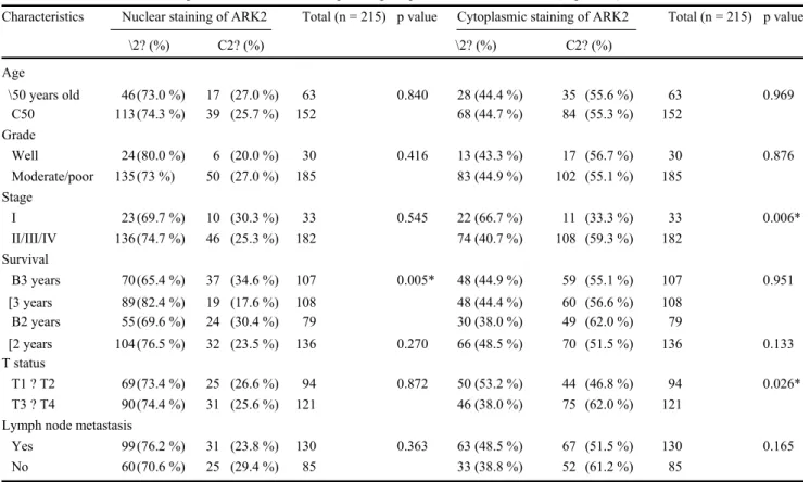
Table 1 Association of ARK2 expression levels with clinicopathological parameters in 215 OSCCs (*, p \ 0.05)
Characteristics Nuclear staining of ARK2 Total (n = 215) p value Cytoplasmic staining of ARK2 Total (n = 215) p value
\2? (%) C2? (%) \2? (%) C2? (%) Age \50 years old 46 (73.0 %) 17 (27.0 %) 63 0.840 28 (44.4 %) 35 (55.6 %) 63 0.969 C50 113 (74.3 %) 39 (25.7 %) 152 68 (44.7 %) 84 (55.3 %) 152 Grade Well 24 (80.0 %) 6 (20.0 %) 30 0.416 13 (43.3 %) 17 (56.7 %) 30 0.876 Moderate/poor 135 (73 %) 50 (27.0 %) 185 83 (44.9 %) 102 (55.1 %) 185 Stage I 23 (69.7 %) 10 (30.3 %) 33 0.545 22 (66.7 %) 11 (33.3 %) 33 0.006* II/III/IV 136 (74.7 %) 46 (25.3 %) 182 74 (40.7 %) 108 (59.3 %) 182 Survival B3 years 70 (65.4 %) 37 (34.6 %) 107 0.005* 48 (44.9 %) 59 (55.1 %) 107 0.951 [3 years 89 (82.4 %) 19 (17.6 %) 108 48 (44.4 %) 60 (56.6 %) 108 B2 years 55 (69.6 %) 24 (30.4 %) 79 30 (38.0 %) 49 (62.0 %) 79 [2 years 104 (76.5 %) 32 (23.5 %) 136 0.270 66 (48.5 %) 70 (51.5 %) 136 0.133 T status T1 ? T2 69 (73.4 %) 25 (26.6 %) 94 0.872 50 (53.2 %) 44 (46.8 %) 94 0.026* T3 ? T4 90 (74.4 %) 31 (25.6 %) 121 46 (38.0 %) 75 (62.0 %) 121
Lymph node metastasis
Yes 99 (76.2 %) 31 (23.8 %) 130 0.363 63 (48.5 %) 67 (51.5 %) 130 0.165
No 60 (70.6 %) 25 (29.4 %) 85 33 (38.8 %) 52 (61.2 %) 85
correlated with the prognosis of OSCCs in the Japanese
population [
2
0]. SUV39H1 is the first SET
domain-con-taining histone lysine methyltransferase (HKMT) that was
discovered in 2000; SUV39H1 and SUV39H2 knockout mice
have shown genomic instability [
2
1]. However, the
expression of these histone phosphorylation and methylation
proteins in OSCCs in the Taiwanese population is still
undetermined. The aim of this study is to investigate the
relationship of ARK2, G9a, EZH2, SUV39H1, and SUV39H2
with clinicopathological parameters from 215 OSCC patients
of the male population in Taiwan. Here, we examined the
expression of these five proteins in OSCCs to elucidate the
relationship between their protein expression and
clinicopathological findings by the immunohistochem-istry
method. If we can better understand the characteristics about
the clinical relevance of those histone modification proteins in
OSCCs, it may help clinicians provide more appropriate
cancer treatments for OSCC patients.
Materials and methods
Patients and clinical methods
This study was approved by the Institutional Review Board
of the Changhua Christian Hospital (Changhua, Taiwan).
Two hundred and fifteen male Taiwanese patients with oral
cancer who had been diagnosed at the Changhua Christian
Hospital (Chang, Taiwan) from 2000 to 2007 were included in
this study. The medical records of these investigated patients
were reviewed. All these patients received surgical
intervention with or without adjuvant therapies (radiother-apy,
chemotherapy). All tumors were classified according to the
TNM classification system [
2
2].
Tissue microarray-based immunohistochemical
staining analysis and antibodies
Tissue microarray-based immunohistochemical staining
for ARK2, G9a, EZH2, SUV39H1, and SUV39H2
pro-teins was performed as follows. Briefly, the 5-lm
par-affin-embedded tumor sections (5 9 8 = 40 cores normal and
cancer array from OSCC patients) were deparaffi-nized,
retrieved with heat in 10 mM citrate buffer (pH 6.0) at 121
LC for 10 min, and treated with 3 % hydro-gen peroxide to
remove endogenous peroxidase activity. The primary
antibodies for the study were ARK2 (Santa Cruz;
sc-14327), G9a (Santa Cruz; sc-22877), EZH2 (Invitrogen;
49-1043), SUV39H1 (Abgent; AP1190a), and SUV39H2
(Abgent; AP1281a). Primary antibodies were used as
suggested by the manufacturers and were used at a dilution
of 1:50 after optimization. Tissue
Page 4 of 9 Med Oncol (2013) 30:513
Table 2 Association of G9a expression levels of cytoplasm with clinicopathological parameters in 215 OSCCs (*, p \ 0.05)
Characteristics Cytoplasmic staining of Total p value
G9a (n = 215) \2? (%) C2? (%) Age \50 years old 41 (65.1 %) 22 (34.9 %) 63 0.065 C50 78 (51.3 %) 74 (48.7 %) 152 Grade Well 11 (36.7 %) 19 (63.3 %) 30 0.026* Moderate/poor 108 (58.4 %) 77 (41.6 %) 185 Stage I 19 (57.6 %) 14 (42.2 %) 33 0.780 II/III/IV 100 (54.9 %) 82 (45.1 %) 182 Survival B3 years 61 (57.0 %) 46 (43.0 %) 107 0.626 [3 years 58 (53.7 %) 50 (46.3 %) 108 0.518 B2 years 46 (58.2 %) 33 (41.8 %) 79 [2 years 73 (53.7 %) 63 (46.3 %) 136 T status T1 ? T2 56 (59.6 %) 38 (40.4 %) 94 0.272 T3 ? T4 63 (52.1 %) 58 (47.9 %) 121 Lymph node metastasis
Yes 69 (53.1 %) 61 (46.9 %) 130 0.407
No 50 (58.8 %) 35 (41.2 %) 85
sections were treated with secondary antibodies and with
biotin–streptavidin complex for 30 min each at 37 LC.
Diaminobenzidine was used as the chromogene for the
immunoperoxidase reaction. The slides were
counter-stained with hematoxylin and examined for the intensity of
nuclear and cytoplasmic staining in tumor cells by the
pathologist in a blind manner. In this study, when more
than 50 % of cells displayed nuclear staining and intensity
scores of moderate and strong, then it was considered to be
protein overexpression. Patient tissue specimens were
broadly distributed by immunohistochemical staining
cat-egory (0, 1?, 2?, or 3?) according to the guidelines based
on Hofmann et al. [
2
3
].
Statistical analysis
Statistical analyses were performed using SPSS statistical
package (SPSS, Chicago, IL, USA). The corrections
between protein expression and clinicopathological
parameters in OSCCs were examined by Pearson’s chi
squared test. The survival curve was constructed by the
Kaplan-Meier method and the different survival curves
were compared using the log-rank test. A difference of p \
0.05 was considered statistically significant.
Results
Expression of ARK2 in OSCCs
Qi et al. [
1
6
] demonstrate that ARK2 is expressed in both
normal oral squamous epithelia and OSCC cases; however,
ARK2 is significantly higher in OSCC cases (especially
poorly differentiated cases). The expression of ARK2
protein in OSCC was examined using an antibody to
ARK2 in paraffin-embedded sections. Typical staining
patterns of the ARK2 protein are shown in Fig.
1
a. In the
OSCC specimens, positive ARK2 protein staining was
found in both the nucleus and the cytosol (Fig.
1
a).
Association of ARK2 expression levels
with clinicopathological parameters in 215 OSCC
patients
The relationships between ARK2 intensity in the biopsy
specimens and the clinicopathological parameters of the
215 OSCC male patients are shown in Table
1
. As shown
in Table
1
, ARK2 nuclear intensity was associ-ated with
3-year survival (p = 0.005). The nuclear inten-sity of the
ARK2 protein was not associated with age, grade, stage, T
status, and lymph node metastasis. On the other hand,
ARK2 cytosolic intensity was associated with stage (p =
0.006) and T status (p = 0.026) but not with age, grade,
survival, and lymph node metastasis (Table
1
).
Survival analysis of ARK2 nuclear intensity
In this study, when more than 50 % of the cells displayed
nuclear staining and intensity scores that were moderate (?)
and strong (??), it was considered ARK2 overex-pression.
Nuclear overexpression of ARK2 was found in 162
OSCCs (? and ?? groups, 75.3 %; Fig.
1
b). In the 8-year
survival rate analysis, disease-free survival was
significantly worse in patients who were ARK2
overex-pressed (? and ??) in nuclei when compared with patients
who were not ARK2 overexpressed (p = 0.042 by log-rank
test; Fig.
1
b), suggesting that patients with higher ARK2
expression in the nucleus showed poor prognosis than
those with no ARK2 expression, and ARK2 might serve as
a novel biomarker for predicting prognosis in patients with
OSCC.
Med Oncol (2013) 30:513 Page 5 of 9
Fig. 2 Tissue microarray-based immunohistochemistry staining pat-terns of four histone modification-related proteins. Immunohisto-chemistry staining results of G9a (a), EZH2 (b), SUV39H1 (c), and
SUV39H2 (d) in tissues from oral squamous cell carcinoma (OSCC) patients. The image of magnification 4009 is from the red square of the respective 1009 image
Association of G9a expression levels
with clinicopathological parameters in 215 OSCC
patients
On the other hand, as shown in Table
2
, G9a cytosolic
intensity was only associated with grade (p = 0.026). On
the other hand, G9a protein cytosolic intensity was not
associated with age, stage, survival, T status, and lymph
node metastasis. The nuclear expression of G9a was not
related to any clinicopathological parameters (data not
shown). Typical staining patterns for G9a protein are
shown in Fig.
2
a.
Association of EZH2 expression levels
with clinicopathological parameters in 215 OSCC
patients
On the other hand, as shown in Table
3
, EZH2 nuclear
intensity was only associated with lymph node metastasis
(p = 0.016). On the other hand, EZH2 protein nuclear
intensity was not associated with age, grade, stage,
sur-vival, and T status. The cytosolic intensity of EZH2 was
not related to any clinicopathological parameters (data not
shown). Typical staining patterns for EZH2 protein are
shown in Fig.
2
b.
Association of SUV39H1 and SUV39H2 expression
levels with clinicopathological parameters in 215
OSCC patients
Typical staining patterns for SUV39H1 and SUV39H2
pro-tein are shown in Fig.
2
c, d. As shown in Table
4
,
SUV39H1 nuclear intensity was only associated with stage
(p = 0.009) but not with age, grade, survival, T status, and
lymph node metastasis. No significant association was
found between SUV39H1 cytosolic intensity and
SUV39H2 intensity (data not shown).
Page 6 of 9 Med Oncol (2013) 30:513
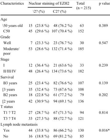
Table 3 Association of EZH2 expression levels of nuclei with clin-icopathological parameters in 215 OSCCs (*, p \ 0.05)
Characteristics Nuclear staining of EZH2 Total p value (n = 215) \2? (%) C2? (%) Age \50 years old 15 (23.8 %) 48 (76.2 %) 63 0.389 C50 45 (29.6 %) 107 (70.4 %) 152 Grade Well 7 (23.3 %) 23 (76.7 %) 30 0.547 Moderate/ 53 (28.6 %) 132 (71.4 %) 185 poor Stage I 12 (36.4 %) 21 (63.6 %) 33 0.239 II/III/IV 48 (26.4 %) 134 (73.6 %) 182 Survival B3 years 25 (23.4 %) 82 (76.6 %) 107 0.139 [3 years 35 (32.4 %) 73 (67.6 %) 108 B2 years 18 (22.8 %) 61 (77.2 %) 79 0.202 [2 years 42 (30.9 %) 94 (69.1 %) 136 T status T1 ? T2 27 (28.7 %) 67 (71.3 %) 94 0.814 T3 ? T4 33 (27.3 %) 88 (72.7 %) 121 Lymph node metastasis
Yes 44 (33.8 %) 86 (66.2 %) 130 0.016*
No 16 (18.8 %) 69 (81.2 %) 85
Discussion
Immunohistochemical staining is an essential method for
diagnostic pathology but it has inconsistency of quality
assurance in data interpretation form reported results.
Despite the widespread use of immunohistochemistry,
significant problems remain with regard to variability in
tissue fixation, processing, staining methodologies, and
reagents, and interpretation of staining results persists
between different slides [
2
4
]. In this study, we used tissue
microarray-based immunohistochemistry staining, which
allowed us to generate staining profiles for the various
antibodies that are specific to the same fixation and
staining procedures for 40 cores normal and cancer array
from OSCC patients on the same slide. It has consistency
of quality assurance and a cost-effective assessment of
inter-laboratory variation.
OSCCs represent a significant problem because of their
high incidence and unsatisfactory survival rate in Taiwan.
In OSCCs, tumor size, histological differentia-tion, and
mode of carcinoma invasion are known to be correlated
with tumor metastasis and patient prognosis [
6
,
2
5
].
Detecting the relationship between genetic
abnormalities and clinicopathological characteristics in
OSCCs might provide important prognostic indicators of
patient prognosis and survival. For example, P53 and
Ki-67 overexpressions have been identified as markers of
malignancy with an aggressive clinical course in OSCCs [
26
,
2
7
].
Besides the well-known alteration of histone acetylation
patterns, cancer cells also display widespread changes in
his-tone phosphorylation and methylation patterns [
2
1,
2
8–
3
1].
So, the histone phosphorylation protein (-ARK2), and
meth-ylation proteins (-G9a, EZH2, SUV39H1, and SUV39H2)
were investigated in this study. ARK2 (a kinase for H3S10)
overexpression is found to be associated with the stages of
malignant progression in thyroid carcinomas and with poor
prognosis in endometrial carcinomas [
1
3,
3
2]. Qi et al. [
1
6]
showed that ARK2 overexpression is correlated with lymph
node metastasis and with histological grade of differentiation
in 40 OSCC patients in Japan (26 men and 14 women). The
intracellular localization of ARK2 in tumor cells was mainly
in nuclei, especially in proliferative areas, and significant
over-expression is found in peritumoral areas of
well-differentiated cases and through the nest of poorly
differentiated cases [
1
6]. Furthermore, Pannone et al. [
3
3]
also have shown similar results in intracellular findings,
indicating a significant association between ARK2
overexpression and advanced tumor stage, larger tumor size,
poorer survival, shorter time to progression, and the worst
prognosis in OSCCs. In our result, ARK2 overexpression was
found mainly in nuclei with a poor 3-year survival rate,
diversely associated with advanced tumor stage, and more
severe in T status of those 215 OSCC male patients in
cytoplasmic staining. The pos-sible reason of this variation
may be due to differences such as the selected antibody,
genetic background, and risk factors between selected
populations. ARK2 protein may work as an independent
prognostic factor and can be used as a bio-marker for the
aggressiveness of OSCCs in the Taiwanese population.
G9a is a methyltransferase for H3K9, which promotes
lung cancer cell invasion and may play an early role in
metastasis cascade [
3
4
]. The nuclear intensity of the G9a
protein is correlated with reduced overall survival and
disease-free interval in lung cancer [
3
4
]. In our result, G9a
cytoplasmic intensity but not nuclear intensity in 215
OSCCs was associated with histological grade of
differ-entiation (tumor grade), which determines whether the
OSCC case is benign or malignant. This is the first report
that shows G9a expression to be associated with poorer
tumor grade in OSCC patients.
The polycomb group protein-EZH2 functions as a
methyltransferase for H3K27 as the catalytic subunit of
polycomb repressive complex 2; EZH2 is frequently
overexpressed in a more biological malignancy state of
solid tumors including glioblastoma multiforme, prostate,
Med Oncol (2013) 30:513 Page 7 of 9
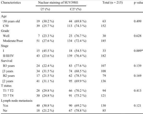
Table 4 Association of Characteristics Nuclear staining of SUV39H1 Total (n = 215) p value SUV39H1 expression levels of
nuclei with clinicopathological \2? (%) C2? (%)
parameters in 215 OSCCs (*, p \ 0.05) Age \50 years old 19 (30.2 %) 44 (69.8 %) 63 0.499 C50 39 (25.7 %) 113 (74.3 %) 152 Grade Well 7 (23.3 %) 23 (76.7 %) 30 0.628 Moderate/Poor 51 (27.6 %) 134 (72.4 %) 185 Stage I 15 (45.5 %) 18 (54.5 %) 33 0.009* II/III/IV 43 (23.6 %) 139 (76.4 %) 182 Survival B3 years 24 (22.4 %) 83 (77.6 %) 107 0.139 [3 years 34 (31.5 %) 74 (68.5 %) 108 B2 years 17 (21.5 %) 62 (78.5 %) 79 0.169 [2 years 41 (31.1 %) 95 (69.9 %) 136 T status T1 ? T2 28 (29.8 %) 66 (70.2 %) 94 0.413 T3 ? T4 30 (24.8 %) 91 (75.2 %) 121
Lymph node metastasis
Yes 40 (30.8 %) 90 (69.2 %) 130 0.121
No 18 (21.2 %) 67 (78.8 %) 85
and breast cancers [
3
5
–
3
7
]. Furthermore, high EZH2
expression has revealed poor prognostic relevance in many
head- and neck-related tumors, including leukoplakia
malignant transformation, salivary gland myoepithelial
tumor, and adenoid cystic tumor [
3
8
–
4
1
]. In our result,
EZH2 overexpression was observed mainly in nuclei and
was associated with lymph node metastasis in the 215
OSCC patients which is consistent with the results of
Ki-dani et al. [
2
0
]. Their report also indicates that EZH2
expression is associated with histological differentiation,
clinical stage, and tumor size in the 102 OSCC patients in
Japan [
2
0
]. Therefore, EZH2 might also serve as an
effi-cient biomarker for discriminating between the aggressive
state and indolent state of OSCCs.
SUV39H1 and SUV39H2 are also histone
methyltrans-ferase for H3K9. SUV39H1-mediated methylation has
been linked to the tumor-suppressive function in T cell
lymphoma and AML [
4
2
,
4
3
]. Patani et al. [
4
4
] have
shown that the expression profile of SUV39H1 is
significantly associated with tumor grade, TMN stage, and
disease-free survival in breast cancer. Our result showed
that nuclear intensity of SUV39H1 but not SUV39H2 was
associated with tumor stage, and it also implied that
SUV39H1 is related to OSCC cancer progression. This is
the first report showing SUV39H1 overexpression to be
associated with advanced tumor stage in OSCC patients.
To decipher the relationship between clinical outcome
and histopathological behavior of OSCCs, and to identify
patients at risk of developing malignancy, further
metas-tases, and poorer prognosis, this study was conducted.
Combined multiple markers are perhaps needed for a more
accurate judgment of the clinical outcome of OSCC
patients; so, a further analysis between multiple
biomarkers and clinicopathological behaviors should be
made later, and this may lead to the finding of more
successful treat-ment targets in OSCC patients.
Conclusion
In this article, we focused on five markers of histone
methylation and phosphorylation proteins-ARK2, G9a,
EZH2, SUV39H1, and SUV39H2. Our results showed that
ARK2, G9a, EZH2, SUV39H1, and especially ARK2, but
not SUV39H2 are highly correlated with
clinicopatholog-ical significances, and this may lead to the discovery of
important biomarkers which can help to predict patient
survival and prognosis in the male population with OSCC
in Taiwan.
Acknowledgments This work was supported by grants from the National Science of Council (NSC-99-2320-B-037-006-MY3) and CMU Hospital (DMR-102-XXX).
Page 8 of 9 Med Oncol (2013) 30:513
Conflict of interest The authors have declared that no conflict of interest exists.
References
Chen TC, Wang CP, Ko JY, Yang TL, Lou PJ. The impact of pathologic close margin on the survival of patients with early stage oral squamous cell carcinoma. Oral Oncol. 2012;48(7): 623–8. doi: 10.1016/j.oraloncology.2012.01.015.
1. Ko YC, Huang YL, Lee CH, Chen MJ, Lin LM, Tsai CC. Betel quid chewing, cigarette smoking and alcohol consumption related to oral cancer in Taiwan. J Oral Pathol Med. 1995;24(10):450–3.
2. Muir C, Weiland L. Upper aerodigestive tract cancers. Cancer. 1995;75(1 Suppl):147–53.
Liao CT, Wang HM, Ng SH, Yen TC, Lee LY, Hsueh C, et al. Good tumor control and survivals of squamous cell carcinoma of buccal mucosa treated with radical surgery with or without neck dissection in Taiwan. Oral Oncol. 2006;42(8):800–9. doi:
10.1016 /j.oraloncology.2005.11.020.
3. Pugliano FA, Piccirillo JF, Zequeira MR, Fredrickson JM, Perez CA, Simpson JR. Clinical-severity staging system for oral cavity cancer: five-year survival rates. Otolaryngology–head and neck surgery : official journal of American Academy of Otolaryngol-ogy-Head and Neck. Surgery. 1999;120(1):38–45.
4. Yamamoto E, Miyakawa A, Kohama G. Mode of invasion and lymph node metastasis in squamous cell carcinoma of the oral cavity. Head Neck Surg. 1984;6(5):938–47.
5. Kornberg RD, Lorch Y. Twenty-five years of the nucleosome, fundamental particle of the eukaryote chromosome. Cell. 1999; 98(3):285–94.
Dawson MA, Kouzarides T. Cancer epigenetics: from mechanism to therapy. Cell. 2012;150(1):12–27. doi: 10.1016/j.cell.2012.06.013. Arrowsmith CH, Bountra C, Fish PV, Lee K, Schapira M.
Epi-genetic protein families: a new frontier for drug discovery. Nat Rev Drug Discov. 2012;11(5):384–400. doi: 10.1038/nrd3674. 6. Ng HH, Bird A. Histone deacetylases: silencers for hire. Trends
Biochem Sci. 2000;25(3):121–6.
7. Sakuma T, Uzawa K, Onda T, Shiiba M, Yokoe H, Shibahara T, et al. Aberrant expression of histone deacetylase 6 in oral squa-mous cell carcinoma. Int J Oncol. 2006;29(1):117–24.
Chang HH, Chiang CP, Hung HC, Lin CY, Deng YT, Kuo MY. Histone deacetylase 2 expression predicts poorer prognosis in oral cancer patients. Oral Oncol. 2009;45(7):610–4. doi: 10.1016/j.oraloncology.2008.08.011.
Sorrentino R, Libertini S, Pallante PL, Troncone G, Palombini L, Bavetsias V, et al. Aurora B overexpression associates with the thyroid carcinoma undifferentiated phenotype and is required for thyroid carcinoma cell proliferation. J Clin Endocrinol Metab. 2005;90(2):928–35. doi: 10.1210/jc.2004-1518.
Taby R, Issa JP. Cancer epigenetics. CA Cancer J Clin. 2010; 60(6):376–92. doi: 10.3322/caac.20085.
Sharma S, Kelly TK, Jones PA. Epigenetics in cancer. Carcino-genesis. 2010;31(1):27–36. doi: 10.1093/carcin/bgp220. Qi G, Ogawa I, Kudo Y, Miyauchi M, Siriwardena BS, Shi-mamoto
F, et al. Aurora-B expression and its correlation with cell proliferation and metastasis in oral cancer. Virchows Archiv. 2007;450(3):297–302. doi: 10.1007/s00428-006-0360-9. 8. Rice JC, Briggs SD, Ueberheide B, Barber CM, Shabanowitz J,
Hunt DF, et al. Histone methyltransferases direct different degrees of methylation to define distinct chromatin domains. Mol Cell. 2003;12(6):1591–8.
9. Tachibana M, Sugimoto K, Nozaki M, Ueda J, Ohta T, Ohki M, et al. G9a histone methyltransferase plays a dominant role in euchromatic histone H3 lysine 9 methylation and is essential for
early embryogenesis. Genes Dev. 2002;16(14):1779–91. doi: 10.1101/gad.989402.
19. Simon JA, Tamkun JW. Programming off and on states in chromatin: mechanisms of Polycomb and trithorax group com-plexes. Curr Opin Genet Dev. 2002;12(2):210–8.
Kidani K, Osaki M, Tamura T, Yamaga K, Shomori K, Ryoke K, et al. High expression of EZH2 is associated with tumor prolif-eration and prognosis in human oral squamous cell carcinomas.
Oral Oncol. 2009;45(1):39–46. doi:
10.1016/j.oraloncology.2008. 03.016.
20. Peters AH, O’Carroll D, Scherthan H, Mechtler K, Sauer S, Schofer C, et al. Loss of the Suv39h histone methyltransferases impairs mammalian heterochromatin and genome stability. Cell. 2001;107(3):323–37.
21. Sobin LH, Fleming ID. TNM Classification of Malignant Tumors, fifth edition (1997). Union Internationale Contre le Cancer and the American Joint Committee on Cancer. Cancer. 1997;80(9):1803–4.
Hofmann M, Stoss O, Shi D, Buttner R, van de Vijver M, Kim W, et al. Assessment of a HER2 scoring system for gastric cancer: results from a validation study. Histopathology. 2008;52(7): 797–805. doi: 10.1111/j.1365-2559.2008.03028.x.
Hsu FD, Nielsen TO, Alkushi A, Dupuis B, Huntsman D, Liu CL, et al. Tissue microarrays are an effective quality assurance tool for diagnostic immunohistochemistry. Mod Pathol. 2002;15(12): 1374– 80. doi: 10.1097/01.MP.0000039571.02827.CE.
Jerjes W, Upile T, Petrie A, Riskalla A, Hamdoon Z, Vourvachis M, et al. Clinicopathological parameters, recurrence, locoregional and distant metastasis in 115 T1–T2 oral squamous cell carci-noma patients. Head Neck Oncol. 2010;2:9. doi: 10.1186/1758
-3284-2-9.
22. Kudo Y, Takata T, Ogawa I, Sato S, Nikai H. Expression of p53 and p21CIP1/WAF1 proteins in oral epithelial dysplasias and squamous cell carcinomas. Oncol Rep. 1999;6(3):539–45. Myoung H, Kim MJ, Lee JH, Ok YJ, Paeng JY, Yun PY.
Cor-relation of proliferative markers (Ki-67 and PCNA) with survival and lymph node metastasis in oral squamous cell carcinoma: a clinical and histopathological analysis of 113 patients. Int J Oral Maxillofac Surg. 2006;35(11):1005–10. doi:
10.1016/j.ijom.2006. 07.016.
23. Nguyen CT, Weisenberger DJ, Velicescu M, Gonzales FA, Lin JC, Liang G, et al. Histone H3-lysine 9 methylation is associated with aberrant gene silencing in cancer cells and is rapidly reversed by 5-aza-2’-deoxycytidine. Cancer Res. 2002;62(22): 6456–61.
Kondo Y, Shen L, Ahmed S, Boumber Y, Sekido Y, Haddad BR, et al. Downregulation of histone H3 lysine 9 methyltransferase G9a induces centrosome disruption and chromosome instability in cancer cells. PLoS One. 2008;3(4):e2037. doi: 10.1371/journal.
pone.0002037.
Valk-Lingbeek ME, Bruggeman SW, van Lohuizen M. Stem cells and cancer; the polycomb connection. Cell. 2004;118(4): 409– 18. doi: 10.1016/j.cell.2004.08.005.
Kondo Y, Shen L, Cheng AS, Ahmed S, Boumber Y, Charo C, et al. Gene silencing in cancer by histone H3 lysine 27 trime-thylation independent of promoter DNA methylation. Nat Genet. 2008;40(6):741–50. doi: 10.1038/ng.159.
Kurai M, Shiozawa T, Shih HC, Miyamoto T, Feng YZ, Kashima H, et al. Expression of Aurora kinases A and B in normal, hyperplastic, and malignant human endometrium: Aurora B as a predictor for poor prognosis in endometrial carcinoma. Hum Pathol. 2005;36(12):1281–8. doi: 10.1016/j.humpath.2005.09.014. 24. Pannone G, Hindi SA, Santoro A, Sanguedolce F, Rubini C,
Cincione RI, et al. Aurora B expression as a prognostic indicator and possible therapeutic target in oral squamous cell carcinoma. Int J Immunopathol Pharmacol. 2011;24(1):79–88.
Med Oncol (2013) 30:513 Page 9 of 9
Chen MW, Hua KT, Kao HJ, Chi CC, Wei LH, Johansson G, et al. H3K9 histone methyltransferase G9a promotes lung cancer invasion and metastasis by silencing the cell adhesion molecule Ep-CAM. Cancer Res. 2010;70(20):7830–40. doi: 10.1158/0008
-5472.CAN-10-0833.
Suva ML, Riggi N, Janiszewska M, Radovanovic I, Provero P, Stehle JC, et al. EZH2 is essential for glioblastoma cancer stem cell maintenance. Cancer Res. 2009;69(24):9211–8. doi: 10.1158/
0008-5472.CAN-09-1622.
Varambally S, Dhanasekaran SM, Zhou M, Barrette TR, Kumar-Sinha C, Sanda MG, et al. The polycomb group protein EZH2 is involved in progression of prostate cancer. Nature. 2002; 419(6907):624–9. doi: 10.1038/nature01075.
Kleer CG, Cao Q, Varambally S, Shen R, Ota I, Tomlins SA, et al. EZH2 is a marker of aggressive breast cancer and promotes neoplastic transformation of breast epithelial cells. Proc Natl Acad Sci USA. 2003;100(20):11606–11. doi:
10.1073/pnas.19337 44100.
Cao W, Feng Z, Cui Z, Zhang C, Sun Z, Mao L, et al. Up-regulation of enhancer of zeste homolog 2 is associated positively with cyclin D1 overexpression and poor clinical outcome in head and neck squamous cell carcinoma. Cancer. 2012;118(11): 2858–71. doi: 10.1002/cncr.26575.
34. Cao W, Younis RH, Li J, Chen H, Xia R, Mao L, et al. EZH2 promotes malignant phenotypes and is a predictor of oral cancer
development in patients with oral leukoplakia. Cancer Prevent Res. 2011;4(11):1816–24. doi: 10.1158/1940-6207.CAPR-11
-0130.
Vekony H, Raaphorst FM, Otte AP, van Lohuizen M, Leemans CR, van der Waal I, et al. High expression of Polycomb group protein EZH2 predicts poor survival in salivary gland adenoid cystic carcinoma. J Clin Pathol. 2008;61(6):744–9. doi: 10.1136/
jcp.2007.054262.
Vekony H, Roser K, Loning T, Raaphorst FM, Leemans CR, Van der Waal I, et al. Deregulated expression of p16INK4a and p53 pathway members in benign and malignant myoepithelial tumours of the salivary glands. Histopathology. 2008;53(6):658– 66. doi: 10.1111/j.1365-2559.2008.03184.x.
Braig M, Lee S, Loddenkemper C, Rudolph C, Peters AH, Schlegelberger B, et al. Oncogene-induced senescence as an initial barrier in lymphoma development. Nature. 2005;436(7051): 660–5. doi: 10.1038/nature03841.
Lakshmikuttyamma A, Scott SA, DeCoteau JF, Geyer CR. Re-expression of epigenetically silenced AML tumor suppressor genes by SUV39H1 inhibition. Oncogene. 2010;29(4):576–88. doi: 10.1038/onc.2009.361.
40. Patani N, Jiang WG, Newbold RF, Mokbel K. Histone-modifier gene expression profiles are associated with pathological and clinical outcomes in human breast cancer. Anticancer Res. 2011; 31(12):4115–25.