Research Express@NCKU - Articles Digest
Research Express@NCKU Volume 7 Issue 2 - December 26, 2008 [ http://research.ncku.edu.tw/re/articles/e/20081226/2.html ]
Dynamics of femtosecond laser photo-
modification of collagen fibers
Shean-Jen Chen
1,*and Chen-Yuan Dong
21Department of Engineering Science, College of Engineering, National Cheng Kung University
2Department of Physics, National Taiwan University [email protected]
The article is extracted from: V. Hovhannisyan, W. Lo, C. Hu, S.-J. Chen, and C. Y.
Dong, “Dynamics of femtosecond laser photo-modification of collagen fibers,”
Optics Express, vol. 16, no. 11, pp. 7958-7968, May 2008.
S
ince the introduction of nonlinear optical microscopy (NLOM) from 1990, microscopic examination and manipulation in biology and medicine entered a new age. Unlike traditional one-photon techniques, NLOM with femtosecond pulse laser depends on the nonlinearexcitation of fluorescent molecules or induction of polarization effects such as harmonic generation for imaging biological specimens. A number of significant advantages are associated with this approach:
1. The high incident photon flux needed in inducing the nonlinear optical response in biological materials limit specimen response to the focal volume. For imaging purposes, the excellent axial
depth discrimination achieved allows confocal-like image quality to be achieved without the use of confocal apertures.
2. Localization of photodamage to the focal volume results in a significant reduction of the overall specimen photodamage. As a result, prolonged sample longevity can be achieved.
3. Since the near-infrared (NIR) photons needed for NLOM are absorbed and scattered less by biological specimens, deeper sample penetration depths can be achieved.
4. The wide spectral separation between the multiphoton excitation (MPE) and emission wavelengths allows complete fluorescence spectra to be studied.
These advantages render NLOM to be the preferred imaging modality in numerous biomedical applications.
In addition to imaging, nonlinear interaction between a femtosecond pulse laser source and specimens allows 3D specimen photo-modification to be achieved. Femtosecond laser assisted micromachining has been demonstrated in inorganic materials such as silicon and glass. For biological specimens,
femtosecond laser techniques enabled gene transfection, 3D tissue engineering scaffolds, the study of neuron regeneration, and so on.
1 of 4
Research Express@NCKU - Articles Digest
NLOM depends on the induction of the nonlinear response in the specimens of interest for imaging and manipulation purposes. In the lowest order of two-photon excitation (TPE), a fluorescent molecule is excited by the absorption of two near-infrared photons of the appropriate wavelength in reaching the excited state. Quantum mechanically, transition probability of the TPE process has been solved and it is well known that the TPE transition probability is proportional to the square of incident intensity.
Contrary to TPE, the process of harmonic generation utilizes the non-linear polarization response in achieving specimen imaging. In general, the polarization of a material may be expanded as a power of E the incident electric field:
, (1)
where the second order term contributes to the second harmonic generation (SHG) process in which two photons are converted to one photon at precisely half the wavelength or twice the energy.
Non-centrosymmetric biological structures such as collagen and muscle fibers have been demonstrated to be effective generators of the second harmonic signal. Since the TPE fluorescence and SHG signals are spectrally separated, the combination of these two imaging modalities allows spectral imaging with high contrast. In tissue imaging, this approach has been successfully applied for imaging cellular and
extracellular matrix elements.
The NLOM addresses a wide variety of biomedical issues by multiphoton imaging of the specimens of interests. Furthermore, combined with femtosecond-laser assisted microprocessing (FLAM), we can investigate into unprecedented manipulation and imaging. For example, one can imagine that FLAM can be implemented to carve out the necessary 3D matrix for tissue engineering optimization. Combined with multiphoton imaging, we can both manipulate and monitor the biological structures we design.
This approach has tremendous applications in a wide array of biomedical applications. To investigate the feasibility of applying FLAM, we have also tested and applied FLAM in the microprocessing of collagen fibers, a critical component of biological tissues.
1. Femtosecond laser assisted microsurgery: We demonstrated that with repeated scanning, rat tail collagen fiber can be severed. The cutting site is indicated by an increase of autofluorescence (green pseudo-color in Fig. 1). We also noticed that associated with extended femtosecond- illumination is a decrease of SHG and increase in the autofluorescence signals. These phenomena are demonstrated in Fig. 2.
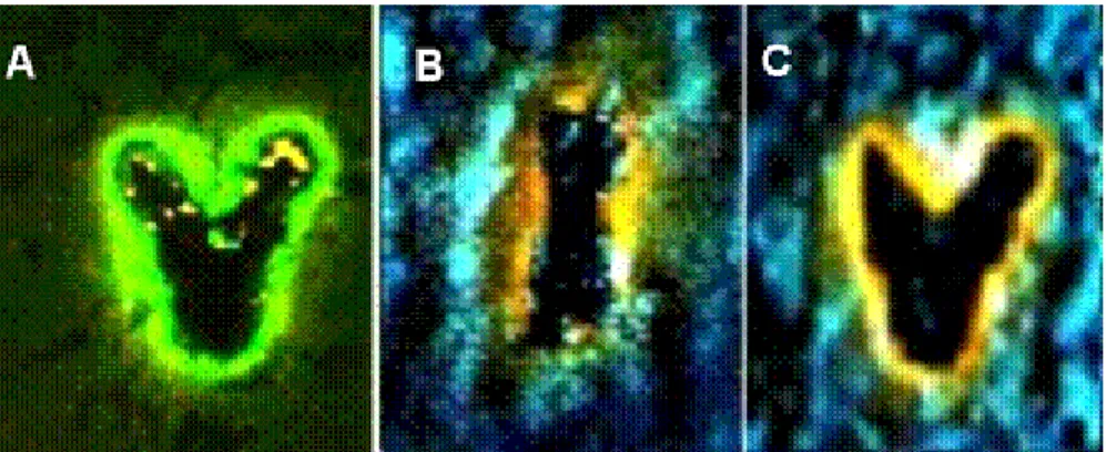
2. Microprocessing: With the desired scanning patterns, FLAM may be used for microlithographic applications. Demonstrated in Fig. 11 are the carvings of the letters “V” and “I” in bovine cornea.
2 of 4
Research Express@NCKU - Articles Digest
Fig. 1. Femtosecond-assisted laser microprocessing of rat tail collagen fiber. Left: before, and right: after femtosecond-laser incision along the blue line. Red: SHG; Green: autofluorescence.
Fig. 2. Photodamage on rat tail collagen. Note the decrease of SHG and increase in autofluorescence signals at the two regions of interests. Red: SHG; green: autofluorescence.
Fig. 3. Femtosecond-assisted laser microprocessing induced microlithography in bovine cornea. Note
3 of 4
Research Express@NCKU - Articles Digest
the photo-lithographic letters of “V” in A, C, and the letter “I” in B.
In the summary, we investigate the non-ablative, non-thermal photo-modification of collagen fibers by femtosecond Ti: Sapphire laser. The effect was induced and simultaneously registered during the repetitive laser scanning of type I collagen (rat tail and bovine Achilles’ tendon), and bovine cornea. An irreversible increase in two-photon autofluorescence and a decrease in second harmonic generation intensities were associated with the collagen femtosecond laser photo-modification. Confocal spectral imaging revealed the formation of new fluorescent species. Controllable nonlinear photo-modification of collagen fibers and bovine cornea with ~2 μm spatial resolution was demonstrated.
4 of 4