Research Express@NCKU - Articles Digest
1 of 2
Research Express@NCKU Volume 28 Issue 7 - February 26, 2015
[ http://research.ncku.edu.tw/re/articles/e/20150226/3.html ]High resolution acoustic radiation force imaging for
detecting the elastic properties of cornea
Cho-Chiang Shih
1, Chih-Chung Huang
1,*, Qifa Zhou
2, K. Kirk Shung
21 Department of Biomedical Engineering, National Cheng Kung University, Tainan City, Taiwan 2 Department of Biomedical Engineering, University of Southern California, Los Angeles, CA, USA cchuang@mail.ncku.edu.tw
IEEE TRANSACTIONS ON MEDICAL IMAGING, VOL. 32, NO. 7, JULY 2013
S
oft tissue hardening usually has a direct relationship with the occurrences of diseases, such as breast tumor, hepatic fibrosis, and ophthalmic pathologies. However, it is difficult to identify the regions of hardening in tissues by using conventional ultrasound gray-level image. Therefore, several ultrasonic elastographies have been proposed to provide the elastic information of tissue in clinical applications. Since the ophthalmic tissues are composed by microstructures, the current ultrasonic elastographies cannot provide sufficient resolution for imaging the cornea. Therefore, The purpose of this study was todevelop a high resolution acoustic radiation force imaging (ARFI) for detecting the localized hardness changes in cornea.
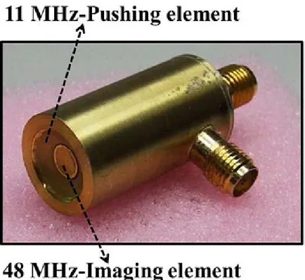
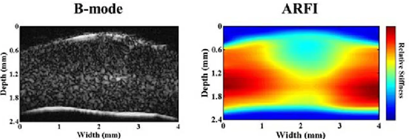
A dual confocal ultrasound transducer was designed in this study for ARFI (Figure 1). The low frequency element was used to generate the acoustic radiation force to create the tissue displacement. The high frequency element was utilized to detect the dynamic displacements of localized tissue in order to reconstruct a high resolution ARFI. The cornea experiments were carried out using fresh porcine eyeballs. Figure 2 shows that the region of localized sclerosis in cornea cannot be distinguished by ultrasound gray-level image, however, it was identified clearly by using high resolution ARFI. All the results demonstrated that the high resolution ARFI imaging has a great potential for clinical diagnosis in ophthalmology.
Research Express@NCKU - Articles Digest
2 of 2