Excimer-laser-induced activation of Mg-doped GaN layers
Yow-Jon Lin
a)and Wen-Fung Liu
Department of Electrical Engineering, Feng Chia University, Taichung 407, Taiwan, Republic of China Ching-Ting Lee
Institute of Microelectronics, Department of Electrical Engineering, National Cheng-Kung University, Tainan, Taiwan, Republic of China
共Received 14 October 2003; accepted 10 February 2004兲
In this study, we investigated the 248 nm excimer-laser-induced activation of the Mg-doped GaN layers. According to the observed photoluminescence results and the x-ray photoelectron spectroscopy measurements, we found that the dissociation of the Mg–H complexes and the formation of hydrogenated Ga vacancies 共i.e., VGaH
2) and/or the Ga vacancies occupied by interstitial Mg during the laser irradiation process, led to an increase in the hole concentration.
© 2004 American Institute of Physics. 关DOI: 10.1063/1.1695436兴
Applications of wide bandgap GaN-based III–V nitride compounds in blue light-emitting diodes, laser diodes, and high-power electronic devices have been reported. One of the problems in GaN-based optoelectronic devices is the low hole concentration of Mg-doped GaN, which makes it diffi- cult to form excellent ohmic contacts in metal/Mg-doped GaN samples. There have been a few recent studies on the effects of oxygen in the p-type activation process.
1,2In ad- dition, a low-energy electron-beam irradiation 共LEEBI兲 treatment,
3,4a microwave treatment,
5a thermal annealing treatment,
6and thermal annealing with a minority-carrier in- jection method
7have been also employed for the effective activation and generation of holes. Jang et al.
8have indicated that the acceptor concentration would increase by a factor of
⬃2 after KrF excimer laser irradiation. They suggested that the activation efficiency of Mg dopants was enhanced by the photon-assisted breaking of Mg—H bonds and/or the re- moval of hydrogen atoms in the presence of oxygen, which produced p-type GaN with an increased hole concentration.8
However, they did not investigate the excimer-laser-induced activation mechanism further.
8 Therefore, to date, the excimer-laser-induced activation of Mg-doped GaN has not yet been well understood. For this study, we demonstrate that the activation of Mg-doped GaN could be achieved by re- petitive laser beam irradiation with a pulsed KrF excimer laser under an air atmosphere. After laser irradiation, Ga ox- ides formed on the Mg-doped GaN surface, and hydroge- nated Ga vacancies 共i.e., V
GaH
2) and/or Ga vacancies, occu- pied by the interstitial Mg (Mg
Ga), formed in the Mg-doped GaN film, due to Ga outdiffusion. Therefore, after laser irra- diation, a hole concentration of 4.1 ⫻1017 cm
⫺3 could be achieved in the Mg-doped GaN film.
The epitaxial layers used in the experiments were grown on c-plane sapphire substrates using a metalorganic chemical vapor deposition system. Trimethylgallium, ammonia, and bis-cyclopentadienylmagnesium, were used as the Ga, N, and Mg sources, respectively. An undoped GaN buffer layer with a thickness of 650 nm was grown on the sapphire sub- strate at 520 °C, followed by the growth of an Mg-doped
GaN layer 共762 nm兲 at 1100 °C. The grown samples 共as- grown samples 兲 were annealed for the purpose of generating holes at 750 °C for 30 min in an ambient N
2(N
2-activated samples 兲, then irradiated in air, by a single pulse from a KrF excimer laser 共laser-irradiated samples兲. The laser was operated at 248 nm with a pulse duration of approximately 50 ns. The incident laser fluence was 250 mJ/cm
2( ⬍600 mJ/cm2). It is known that the threshold fluence for the decomposition or damaged of GaN is above 600 mJ/cm
2, under which the GaN surface is heated to its decomposition temperature of ⬃900 to 1000 °C.9,10 Next, all the samples were cleaned in chemical cleaning solutions of trichloroeth- ylene, acetone, and methanol. Prior to the making of the Hall and photoluminescence 共PL兲 measurements, the as-grown, N
2-activated, and laser-irradiated samples were treated in an aqua regia solution for 10 min. Hall and PL measurements were performed at room temperature in order to evaluate the electrical and optical properties of the Mg-doped GaN layers, for each activation method. When an He–Cd laser was used as an excitation source, only the 2.8 eV PL band was ob- served, in the all samples. Two Gaussian functions were used to fit the PL peak, neglecting interference effects. The van der Pauw–Hall measurements were used to determine the hole concentration of the Mg-doped GaN epilayer. From the observed Hall measurements, the hole concentration of the N
2-activated and laser-irradiated samples was calculated to be 3.6 ⫻1017 cm
⫺3 and 4.1 ⫻1017 cm
⫺3, respectively. The hole mobility of the N
2-activated and laser-irradiated samples was calculated to be 11.1 cm
2/V s and 10.9 cm
2/V s, respectively. For this work, the Ga 2 p
3/2core-level spectrum was measured via x-ray photoelectron spectroscopy 共XPS兲.
Next, all the samples were cleaned in chemical cleaning solutions of trichloroeth- ylene, acetone, and methanol. Prior to the making of the Hall and photoluminescence 共PL兲 measurements, the as-grown, N
2-activated, and laser-irradiated samples were treated in an aqua regia solution for 10 min. Hall and PL measurements were performed at room temperature in order to evaluate the electrical and optical properties of the Mg-doped GaN layers, for each activation method. When an He–Cd laser was used as an excitation source, only the 2.8 eV PL band was ob- served, in the all samples. Two Gaussian functions were used to fit the PL peak, neglecting interference effects. The van der Pauw–Hall measurements were used to determine the hole concentration of the Mg-doped GaN epilayer. From the observed Hall measurements, the hole concentration of the N
2-activated and laser-irradiated samples was calculated to be 3.6 ⫻1017 cm
⫺3 and 4.1 ⫻1017 cm
⫺3, respectively. The hole mobility of the N
2-activated and laser-irradiated samples was calculated to be 11.1 cm
2/V s and 10.9 cm
2/V s, respectively. For this work, the Ga 2 p
3/2core-level spectrum was measured via x-ray photoelectron spectroscopy 共XPS兲.
cm
⫺3, respectively. The hole mobility of the N
2-activated and laser-irradiated samples was calculated to be 11.1 cm
2/V s and 10.9 cm
2/V s, respectively. For this work, the Ga 2 p
3/2core-level spectrum was measured via x-ray photoelectron spectroscopy 共XPS兲.
The XPS measurements were performed using a monochro- matic Mg K ␣ x-ray source. For energy reference purposes, we took a Au 4 f
7/2peak at 83.86 eV and a Cu 2 p
3/2peak at 932.65 eV. The Ga 2 p
3/2core-level peaks were deconvolved into their various components using an interactive least- squares computer program; the curves were assumed total 80% Gaussian and 20% Lorentzian mixed functions.
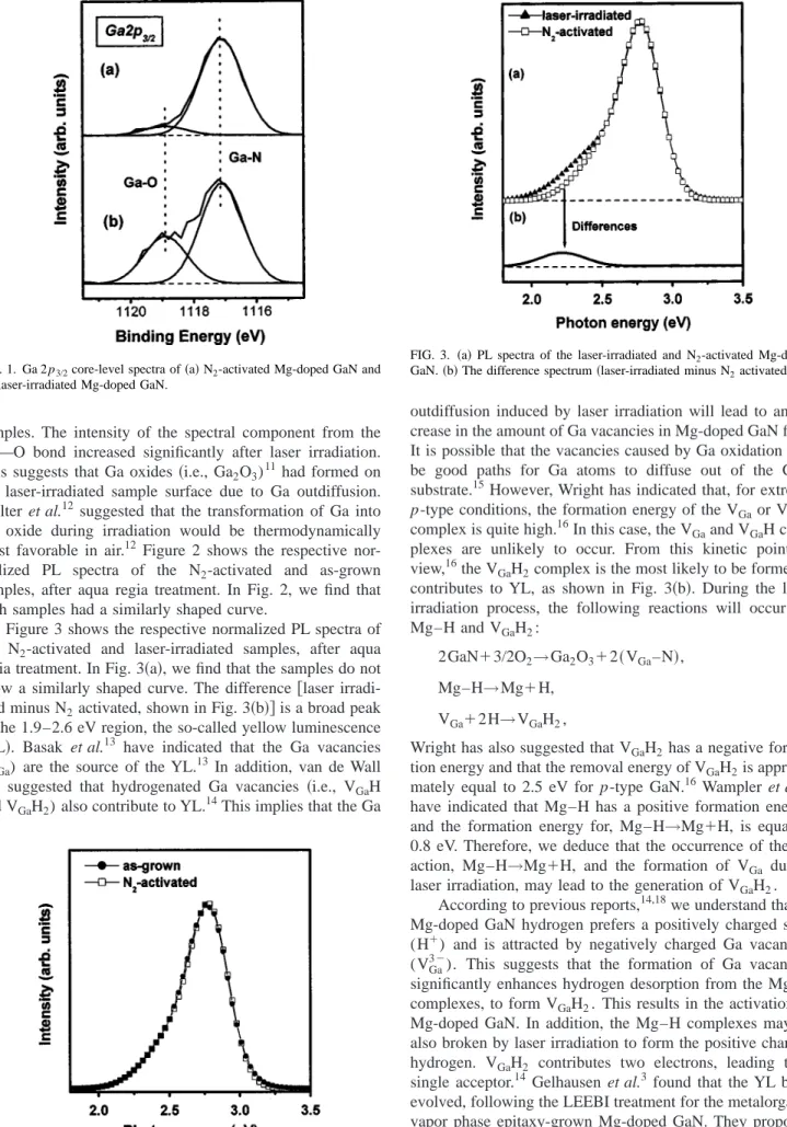
Figure 1 shows the Ga 2 p
3/2core-level spectra of the N
2-activated and laser-irradiated samples, respectively, with- out aqua regia treatment. The Ga—N bonds and Ga—O bonds, were observed in the N
2-activated and laser-irradiated
a兲Author to whom correspondence should be addressed; electronic mail:
APPLIED PHYSICS LETTERS VOLUME 84, NUMBER 14 5 APRIL 2004
2515
0003-6951/2004/84(14)/2515/3/$22.00 © 2004 American Institute of Physics
Downloaded 22 Sep 2008 to 140.116.208.41. Redistribution subject to AIP license or copyright; see http://apl.aip.org/apl/copyright.jsp
samples. The intensity of the spectral component from the Ga—O bond increased significantly after laser irradiation.
This suggests that Ga oxides 共i.e., Ga2O
3)
11had formed on the laser-irradiated sample surface due to Ga outdiffusion.
Wolter et al.
12suggested that the transformation of Ga into Ga oxide during irradiation would be thermodynamically most favorable in air.
12Figure 2 shows the respective nor- malized PL spectra of the N
2-activated and as-grown samples, after aqua regia treatment. In Fig. 2, we find that both samples had a similarly shaped curve.
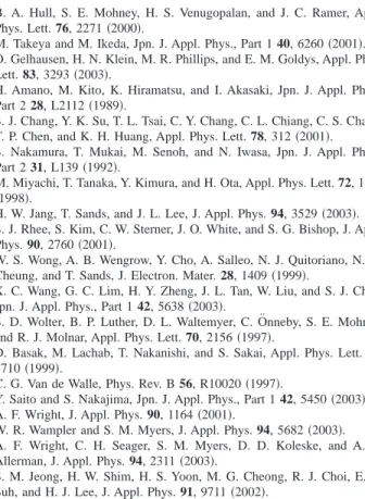
Figure 3 shows the respective normalized PL spectra of the N
2-activated and laser-irradiated samples, after aqua regia treatment. In Fig. 3 共a兲, we find that the samples do not show a similarly shaped curve. The difference 关laser irradi- ated minus N
2activated, shown in Fig. 3 共b兲兴 is a broad peak in the 1.9–2.6 eV region, the so-called yellow luminescence 共YL兲. Basak et al.13 have indicated that the Ga vacancies (V
Ga) are the source of the YL.
13 In addition, van de Wall has suggested that hydrogenated Ga vacancies 共i.e., VGaH and V
GaH
2) also contribute to YL.
14This implies that the Ga
H and V
GaH
2) also contribute to YL.
14This implies that the Ga
outdiffusion induced by laser irradiation will lead to an in- crease in the amount of Ga vacancies in Mg-doped GaN film.
It is possible that the vacancies caused by Ga oxidation will be good paths for Ga atoms to diffuse out of the GaN substrate.
15However, Wright has indicated that, for extreme p-type conditions, the formation energy of the V
Gaor V
GaH complex is quite high.
16In this case, the V
Gaand V
GaH com- plexes are unlikely to occur. From this kinetic point of view,
16the V
GaH
2complex is the most likely to be formed if contributes to YL, as shown in Fig. 3 共b兲. During the laser irradiation process, the following reactions will occur for Mg–H and V
GaH
2:
2GaN ⫹3/2O2→Ga
2O
3⫹2共V
Ga–N 兲, 共1兲
Mg–H →Mg⫹H, 共2兲
V
Ga⫹2H→V
GaH
2, 共3兲
Wright has also suggested that V
GaH
2has a negative forma- tion energy and that the removal energy of V
GaH
2is approxi- mately equal to 2.5 eV for p-type GaN.
16Wampler et al.
17have indicated that Mg–H has a positive formation energy, and the formation energy for, Mg–H →Mg⫹H, is equal to 0.8 eV. Therefore, we deduce that the occurrence of the re- action, Mg–H →Mg⫹H, and the formation of V
Gaduring laser irradiation, may lead to the generation of V
GaH
2.
According to previous reports,
14,18we understand that in Mg-doped GaN hydrogen prefers a positively charged state (H
⫹) and is attracted by negatively charged Ga vacancies (V
Ga3⫺). This suggests that the formation of Ga vacancies significantly enhances hydrogen desorption from the Mg–H complexes, to form V
GaH
2. This results in the activation of Mg-doped GaN. In addition, the Mg–H complexes may be also broken by laser irradiation to form the positive charged hydrogen. V
GaH
2contributes two electrons, leading to a single acceptor.
14Gelhausen et al.
3found that the YL band evolved, following the LEEBI treatment for the metalorganic vapor phase epitaxy-grown Mg-doped GaN. They proposed that electron irradiation will head to the dissociating of hy- drogenated Ga vacancies, resulting in an activation of V
Ga-related complexes, thus inducing the radiative recombi-
FIG. 1. Ga 2 p3/2core-level spectra of
共a兲 N
2-activated Mg-doped GaN and共b兲 laser-irradiated Mg-doped GaN.
FIG. 2. PL spectra of the as-grown and N2-activated Mg-doped GaN samples.
FIG. 3.
共a兲 PL spectra of the laser-irradiated and N
2-activated Mg-doped GaN.共b兲 The difference spectrum 共laser-irradiated minus N
2activated兲.2516 Appl. Phys. Lett., Vol. 84, No. 14, 5 April 2004 Lin, Liu, and Lee
Downloaded 22 Sep 2008 to 140.116.208.41. Redistribution subject to AIP license or copyright; see http://apl.aip.org/apl/copyright.jsp
nation responsible for the observed YL.
3In addition, the in- crease in the number of Ga vacancies caused by Ga outdif- fusion may improve the activation efficiency, by promoting the following reaction Mg
i⫹V
Ga→Mg
Ga(Mg
i: The intersti- tial Mg 兲.19 This is another possible explanation for the in- creased hole concentration. A schematic drawing summariz- ing the laser-irradiation activation mechanisms described above shown in Fig. 4.
In summary, the activation mechanism of laser-irradiated Mg-doped GaN has been investigated for this study. Accord- ing to the experimental results, we deduce that Ga oxidation will lead to an increase in Ga-vacancy-related defects and the acceleration of Ga outdiffusion during laser irradiation.
Therefore, excimer-laser-induced activation is attributed to the dissociation of the Mg–H complexes and the strength- ened formation of the hydrogenated Ga vacancies and/or the Ga vacancies occupied by interstitial Mg.
This project was supported by the National Science Council of Taiwan, the Republic of China, under Contract No. NSC 92-2215-E-035-003. The KrF excimer laser was kindly provided from the Fiber Grating Fabrication Labora- tory, Department of Electrical Engineering, Feng Chia Uni- versity.
1B. A. Hull, S. E. Mohney, H. S. Venugopalan, and J. C. Ramer, Appl.
Phys. Lett. 76, 2271
共2000兲.
2M. Takeya and M. Ikeda, Jpn. J. Appl. Phys., Part 1 40, 6260
共2001兲.
3O. Gelhausen, H. N. Klein, M. R. Phillips, and E. M. Goldys, Appl. Phys.
Lett. 83, 3293
共2003兲.
4H. Amano, M. Kito, K. Hiramatsu, and I. Akasaki, Jpn. J. Appl. Phys., Part 2 28, L2112
共1989兲.
5S. J. Chang, Y. K. Su, T. L. Tsai, C. Y. Chang, C. L. Chiang, C. S. Chang, T. P. Chen, and K. H. Huang, Appl. Phys. Lett. 78, 312
共2001兲.
6S. Nakamura, T. Mukai, M. Senoh, and N. Iwasa, Jpn. J. Appl. Phys., Part 2 31, L139
共1992兲.
7M. Miyachi, T. Tanaka, Y. Kimura, and H. Ota, Appl. Phys. Lett. 72, 1101
共1998兲.
8H. W. Jang, T. Sands, and J. L. Lee, J. Appl. Phys. 94, 3529
共2003兲.
9S. J. Rhee, S. Kim, C. W. Sterner, J. O. White, and S. G. Bishop, J. Appl.
Phys. 90, 2760
共2001兲.
10W. S. Wong, A. B. Wengrow, Y. Cho, A. Salleo, N. J. Quitoriano, N. W.
Cheung, and T. Sands, J. Electron. Mater. 28, 1409
共1999兲.
11X. C. Wang, G. C. Lim, H. Y. Zheng, J. L. Tan, W. Liu, and S. J. Chua, Jpn. J. Appl. Phys., Part 1 42, 5638
共2003兲.
12S. D. Wolter, B. P. Luther, D. L. Waltemyer, C. O¨ nneby, S. E. Mohney, and R. J. Molnar, Appl. Phys. Lett. 70, 2156
共1997兲.
13D. Basak, M. Lachab, T. Nakanishi, and S. Sakai, Appl. Phys. Lett. 75, 3710
共1999兲.
14C. G. Van de Walle, Phys. Rev. B 56, R10020
共1997兲.
15Y. Saito and S. Nakajima, Jpn. J. Appl. Phys., Part 1 42, 5450
共2003兲.
16A. F. Wright, J. Appl. Phys. 90, 1164
共2001兲.
17W. R. Wampler and S. M. Myers, J. Appl. Phys. 94, 5682
共2003兲.
18A. F. Wright, C. H. Seager, S. M. Myers, D. D. Koleske, and A. A.
Allerman, J. Appl. Phys. 94, 2311
共2003兲.
19S. M. Jeong, H. W. Shim, H. S. Yoon, M. G. Cheong, R. J. Choi, E. K.
Suh, and H. J. Lee, J. Appl. Phys. 91, 9711
共2002兲.
FIG. 4. Schematic illustration of the p-type laser-irradiation mechanisms in Mg-doped GaN films.
2517
Appl. Phys. Lett., Vol. 84, No. 14, 5 April 2004 Lin, Liu, and Lee
Downloaded 22 Sep 2008 to 140.116.208.41. Redistribution subject to AIP license or copyright; see http://apl.aip.org/apl/copyright.jsp