行政院國家科學委員會專題研究計畫 期末報告
矽酸鈣為基底之骨填充複合材的前臨床研究
計 畫 類 別 : 個別型
計 畫 編 號 : NSC 101-2221-E-040-004-
執 行 期 間 : 101 年 08 月 01 日至 102 年 07 月 31 日 執 行 單 位 : 中山醫學大學口腔科學研究所
計 畫 主 持 人 : 丁信智
計畫參與人員: 博士班研究生-兼任助理人員:何佳哲 博士班研究生-兼任助理人員:魏忠楷
報 告 附 件 : 出席國際會議研究心得報告及發表論文
公 開 資 訊 : 本計畫涉及專利或其他智慧財產權,2 年後可公開查詢
中 華 民 國 102 年 08 月 21 日
中 文 摘 要 : 矽酸鈣材料因其優異生物相容性及骨形成性,在牙科及骨科 吸引相當多的注意。然在臨床應用之前,某些議題仍須澄 清。本計畫中,首先研究不同溶液 pH 值(7.4 及 4.0)對矽酸 鈣物理化學性質之影響,其次分析不透光劑氧化鉍含量對材 料物理化學性質與細胞效應。結果顯示浸泡 pH 7.4 的試片,
其表面所析出磷灰石顆粒大小大於在 pH 4.0 的環境下;溶液 pH 對徑向拉伸強度無顯著影響;經 30 天浸泡後在 pH 4.0 的 溶液下,重量損失約 0.8%,而於 pH 7.4 的溶液,其試片重 量反而增加約 0.2%。此外,pH 4.0 造成較大的孔隙率。關於 氧化鉍效應,隨著氧化鉍含量增加,硬化時間隨之顯著增 加,試片本身 pH 值及徑向拉伸強度稍微受氧化鉍含量影響;
添加 5、10 及 20 wt%氧化鉍後,試片不透光性顯著提高,其 不透光值相當於 3.3、5.8 及 8.4 mm 厚 Al。不同含量氧化鉍 之試片,其溶解度介在 0.8-1.1%之間,顯著低於白色三氧礦 聚合物(white-colored mineral trioxide aggregate) (1.4%)。20 wt% 氧化鉍的添加導致試片產生較低的細胞增 生、分化與礦化。從硬化時間、不透光性及骨形成性等方 面,可知 10 wt% 氧化鉍的添加可使得矽酸鈣作為根管填充 材白色三氧礦聚合物的另一種替代選擇材。
中文關鍵詞: 矽酸鈣骨水泥、臨床前研究、根管填充材、酸環境、氧化鉍 英 文 摘 要 : Calcium silicates have attracted great attention in
orthopaedics and dentistry because of their excellent biocompatibility and osteogenesis. Prior to the
clinical applications, some issues need to be
clarified. In this project, first we investigated the effect of two solutions differing by pH (7.4 and 4.0) on the physicochemical properties of the radiopaque dicalcium silicate cement. Second, the effects of bismuth oxide (Bi2O3) radiopacifier on the
physicochemical properties and in vitro osteogenic activities of the cements were examined. The results indicated that, after soaking in a pH 7.4 solution for 1 day, the particle size of precipitated apatite spherulites on the cement surfaces was greater than that obtained in a pH 4.0 solution. Solution pH did not result in significant differences (P > 0.05) in diametral tensile strength of cement specimens at the same soaking time-point. On day 30, the sample was associated with a weight loss of 0.8% in a pH 4.0 solution, whereas in a pH 7.4 solution, a weight
increase of 0.2% occurred. A greater porosity of the cement soaked in a pH 4.0 was found compared with that in the solution with pH 7.4. Regarding Bi2O3 effect, the setting time increased significantly (P <
0.05) with increasing Bi2O3 content. The pH value and diametral tensile strength of the cements were
slightly affected by the introduction of Bi2O3. After the addition of 5, 10, and 20 wt% Bi2O3 the
radiopacity of the cements became significantly (P <
0.05) higher, with values equivalent to 3.3, 5.8, and 8.4 mm of Al, respectively. The solubility of the three radiopaque cements ranged between 0.8% and 1.1%, which is significantly (P < 0.05) lower than that of white-colored mineral trioxide aggregate (WMTA) (1.4%). 20 wt% Bi2O3 led to lower cell
proliferation, differentiation, and calcium deposits of MG63 cells on the cement surfaces at all culture times compared those obtained with the other cements.
The addition of 10 wt% Bi2O3 to dicalcium silicate cement improves the setting time, radiopacity, and osteogenic activity, making the cement a potential alternative to WMTA as a root-end filling material.
英文關鍵詞: Calcium silicate cement, preclinical study, root-end filling material, acid environment, bismuth oxide
目錄
1. 中文摘要 --- 3
2. 英文摘要 --- 3
3. Introduction and purpose --- 4
4. Experimental --- 4
4.1. Specimen preparation --- 4
4.2. Soaking in physiological solution --- 5
4.3. Surface morphology --- 5
4.4. Diametral tensile strength --- 5
4.5. Weight change --- 6
4.6. Porosity --- 6
4.7. pH variation of the solution --- 6
4.8. Setting time ---6
4.9. pH variation in the cement---6
4.10. Phase composition---6
4.11. Radiopacity---7
4.12. Solubility---7
4.13. Cell culture---7
4.14. Cell proliferation---7
4.15. Cell differentiation---8
4.16. Calcium quantification---8
4.17. Statistical analysis --- 8
5. Results --- 8
5.1. Surface morphology--- 8
5.2. Diametral tensile strength --- 9
5.3. Weight change --- 9
5.4. Porosity --- 9
5.5. pH variation of the solution ---9
5.6. Setting time --- 9
5.7. pH variation in the cement --- 9
5.8. Phase composition --- 10
5.9. Effect of Bi2O3 on DTS --- 10
5.10. Radiopacity---10
5.11. Solubility--- 10
55.12. Cell proliferation --- 10
5.13. Cell differentiation ---10
5.14. Mineralization ---10
6. Discussion ---11
7. Conclusions --- 13
8. 成果自評--- 14
9. 參考文獻--- 14
Table 1 --- 17
Figure 1--- 18
Figure 2--- 19
Figure 3--- 19
Figure 4--- 20
Figure 5--- 20
Figure 6--- 21
Figure 7--- 21
Figure 8--- 22
Figure 9--- 22
Figure 10 --- 23
1. 中文摘要
矽酸鈣材料因其優異生物相容性及骨形成性,在牙科及骨科吸引相當多的注意。
然在臨床應用之前,某些議題仍須澄清。本計畫中,首先研究不同溶液pH 值(7.4及4.0) 對矽酸鈣物理化學性質之影響,其次分析不透光劑氧化鉍含量對材料物理化學性質與細 胞效應。結果顯示浸泡pH 7.4的試片,其表面所析出磷灰石顆粒大小大於在pH 4.0的環 境下;溶液pH對徑向拉伸強度無顯著影響;經30天浸泡後在pH 4.0的溶液下,重量損失 約0.8%,而於pH 7.4的溶液,其試片重量反而增加約0.2%。此外,pH 4.0造成較大的孔 隙率。關於氧化鉍效應,隨著氧化鉍含量增加,硬化時間隨之顯著增加,試片本身pH 值及徑向拉伸強度稍微受氧化鉍含量影響;添加5、10及 20 wt%氧化鉍後,試片不透光 性顯著提高,其不透光值相當於3.3、5.8及8.4 mm厚Al。不同含量氧化鉍之試片,其溶 解 度 介 在0.8-1.1% 之 間 , 顯 著 低 於 白 色 三 氧 礦 聚 合 物 (white-colored mineral trioxide aggregate) (1.4%)。20 wt% 氧化鉍的添加導致試片產生較低的細胞增生、分化與礦化。
從硬化時間、不透光性及骨形成性等方面,可知10 wt% 氧化鉍的添加可使得矽酸鈣作 為根管填充材白色三氧礦聚合物的另一種替代選擇材。
關鍵詞:矽酸鈣骨水泥、臨床前研究、根管填充材、酸環境、氧化鉍 2. 英文摘要
Calcium silicates have attracted great attention in orthopaedics and dentistry because of their excellent biocompatibility and osteogenesis. Prior to the clinical applications, some issues need to be clarified. In this project, first we investigated the effect of two solutions differing by pH (7.4 and 4.0) on the physicochemical properties of the radiopaque dicalcium silicate cement. Second, the effects of bismuth oxide (Bi2O3) radiopacifier on the physicochemical properties and in vitro osteogenic activities of the cements were examined.
The results indicated that, after soaking in a pH 7.4 solution for 1 day, the particle size of precipitated apatite spherulites on the cement surfaces was greater than that obtained in a pH 4.0 solution. Solution pH did not result in significant differences (P > 0.05) in diametral tensile strength of cement specimens at the same soaking time-point. On day 30, the sample was associated with a weight loss of 0.8% in a pH 4.0 solution, whereas in a pH 7.4 solution, a weight increase of 0.2% occurred. A greater porosity of the cement soaked in a pH 4.0 was found compared with that in the solution with pH 7.4. Regarding Bi2O3 effect, the setting time increased significantly (P < 0.05) with increasing Bi2O3 content. The pH value and diametral tensile strength of the cements were slightly affected by the introduction of Bi2O3. After the addition of 5, 10, and 20 wt% Bi2O3 the radiopacity of the cements became significantly (P <
0.05) higher, with values equivalent to 3.3, 5.8, and 8.4 mm of Al, respectively. The solubility of the three radiopaque cements ranged between 0.8% and 1.1%, which is significantly (P <
0.05) lower than that of white-colored mineral trioxide aggregate (WMTA) (1.4%). 20 wt%
Bi2O3 led to lower cell proliferation, differentiation, and calcium deposits of MG63 cells on the cement surfaces at all culture times compared those obtained with the other cements. The addition of 10 wt% Bi2O3 to dicalcium silicate cement improves the setting time, radiopacity, and osteogenic activity, making the cement a potential alternative to WMTA as a root-end filling material.
Keywords: Calcium silicate cement, preclinical study, root-end filling material, acid environment, bismuth oxide
3. Introduction and purpose
Calcium silicates have attracted great attention in orthopaedics and dentistry because of their excellent biocompatibility and osteogenesis [1-3]. A variety of Portland cement-based materials consisting mainly of calcium silicate have been developed for endodontic use as an alternative to calcium silicate-based mineral trioxide aggregate (MTA), on the basis of reducing setting time [4-8]. In recent studies [9,10], aluminium-free hydraulic and radiopaque dicalcium silicate cement displayed a shorter setting time and better biocompatibility than white-coloured MTA and thus may have the potential to be a root-end filling material.
During clinical practice, the periradicular environment may have varying pH from a neutral pH of 7.4 to an acidic pH as low as 5.0, because of bacterial-induced local metabolic acidosis or tissue inflammation [11]. The root-end filling materials may be exposed to an inflammatory environment with relatively low pH values [12,13]. A low pH could potentially inhibit setting reactions, affect adhesion, or increase the solubility of the materials such as MTA [11-14]. For example, Shie et al. reported that pH 4.0 had a deleterious effect on the morphology of white- colored MTA mixed with water [14]. Hence, it is worthwhile to evaluate changes in the characteristics of the root-end filling material after implantation or after soaking in a physiological solution with different pH values.
On the other hand, MTA powder is basically a mixture of calcium-silicate-based Portland cement and bismuth oxide (Bi2O3). Bi2O3 is introduced to improve radiopacity, which is important for endodontic treatment [15,16]. Endodontic materials should have sufficient radiopacity to be distinguished from the peripheral anatomical structures. ProRoot MTA has 20 wt% Bi2O3 as reported by the manufacturer [17]. However, the addition of radiopacifiers might be detrimental to some of the physical, mechanical, and biological properties of endodontic materials [18]. The addition of Bi2O3 has been questioned as it could influence the properties of the cement. The inclusion of Bi2O3 in Portland cement leads to an increase in the amount of unreacted water in the cement, which in turn increases the porosity of the cement [15]. More importantly, Bi2O3 extends the setting time and reduces the compressive strength of the cement [15,16].
Although a variety of calcium-silicate-based materials have been developed for endodontic treatment [16,7,19,20], calcium silicate cement lacks sufficient radiopacity, limiting its application in endodontic use; therefore, Bi2O3 radiopacifier is added to it to reach an acceptable level of radiopacity.The effects of Bi2O3 on the physical properties, sealing ability, and biocompatibility of Portland cement are well documented [15,16]; however, there have been few systematic studies on its effects on physicochemical properties and in vitro osteogenic activities.
Following previous studies [9,19], the purpose of this study was to examine the physicochemical behaviours of a radiopaque dicalcium silicate cement soaked in physiological solutions with different pH values (4.0 and 7.4). The pH 4.0 condition was selected to simulate clinical conditions that would be considered extreme. The parameters of diametral tensile strength, morphology, porosity and weight change were determined, in addition to the pH of the solution as a function of soaking time. Additionally, the effects of Bi2O3 on the physicochemical properties and osteogenic activities of dicalcium silicate cement were examined systematically.
4. Experimental
4.1. Specimen preparation
Reagent-grade tetraethyl orthosilicate (Si(OC2H5)4; Sigma-Aldrich, St. Louis, MO) and calcium nitrate (Ca(NO3)2·4H2O; Showa, Tokyo, Japan) were used as precursors for SiO2 and CaO, respectively. The catalyst was 2 mol L-1 nitric acid, and absolute ethanol was used as the
solvent. The molar ratio of Ca(NO3)2·4H2O to Si(OC2H5)4 was 3 : 2. General sol-gel procedures, such as hydrolysis and aging, were adopted. A detailed description of the powder’s fabrication has been reported [19]. Briefly, Si(OC2H5)4 was hydrolyzed with the sequential addition of nitric acid and absolute ethanol with 1 h of stirring separately. The required amount of Ca(NO3)2·4H2O was added to the above solution, and the mixed solutions were stirred for an additional hour. The sol solution was sealed and aged at 60 ºC for 1 day.
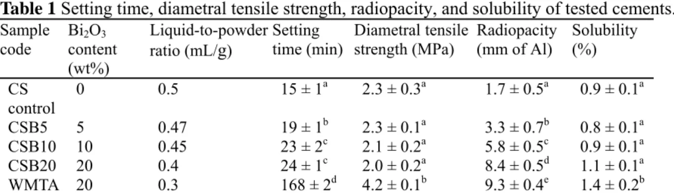
After vaporization of the solvent in an oven at 120 ºC, the dried gel was heated in air to 800 ºC at a heating rate of 10 ºC min-1 for 2 h using a high-temperature furnace and then cooled to room temperature in the furnace to produce a powder. The sintered granules were ball-milled for 12 h in ethyl alcohol using a centrifugal ball mill (Retsch S 100, Hann, Germany) and then dried in an oven at 60 ºC. Bi2O3 (Sigma-Aldrich) was added to the ground powder at 20 wt%, as described by Torabinejad & White [17], using a conditioning mixer (ARE-250, Thinky, Tokyo, Japan). The cement specimens were prepared by hand mixing the powder with distilled water in a liquid-to-powder ratio of 0.4 mL g-1. The cements were placed in a cylindrical Teflon mould to form the cylindrical specimen with dimension of 6 mm (diameter) × 3 mm (height); the specimens were stored in an incubator at 100% relative humidity and 37 ºC for 1 day to set. Regarding Bi2O3 effect, the powder (Sigma-Aldrich) was added to the ground powder at 5%, 10%, or 20% by weight using a conditioning mixer. The cement specimens were prepared by hand mixing the powder with distilled water at liquid-to-powder ratios of 0.5 to 0.4 mL g-1 (Table 1). The specimen codes “CS control”, “CSB5”, “CSB10”, and “CSB20”
represent cements containing 0, 5, 10, and 20 wt% Bi2O3, respectively.
4.2. Soaking in physiological solution
The cement specimens were soaked in 10 mL physiological solution at 37 ºC. Ionic composition of the solution is similar to that of human blood plasma (Chen et al. 2009b), consisted of 7.9949 g NaCl, 0.3528 g NaHCO3, 0.2235 g KCl, 0.147 g K2HPO4, 0.305 g MgCl2·6H2O, 0.2775 g CaCl2, 0.071 g Na2SO4 in 1000 mL distilled H2O and was buffered to either pH 7.4 or 4.0 with hydrochloric acid (HCl; Merck, Darmstadt, Germany) and trishydroxymethyl aminomethane (CH2OH)3CNH2; Sigma-Aldrich). All chemicals used were of reagent grade and used as obtained. The solution in a shaker water bath was not changed daily. After soaking time periods of 1, 3, 7, 15 and 30 days, the specimens were removed from the vials to evaluate the in vitro physicochemical properties.
4.3. Surface morphology
The surface of the cement before and after soaking in the physiological solution was coated with gold using a JFC-1600 (JEOL, Tokyo, Japan) coater and observed under a JEOL JSM- 7401F field emission scanning electron microscope (SEM) operating in the lower secondary electron image mode (LEI) at 3 kV accelerating voltage. The energy dispersive spectroscopy (EDS) was used to perform chemical analyses. Three samples were observed for each test condition.
4.4. Diametral tensile strength
Diametral tensile strength (DTS) testing was performed using an EZ-Test machine (Shimadzu, Kyoto, Japan) at a loading rate of 0.5 mm min1 after weight change and porosity measurements. The strength value of each cement specimen was calculated using the relationship defined in the equation DTS = 2P/πbw, where P is the peak load (N), b is the diameter (mm) and w is the thickness (mm) of the specimen. The peak load at failure was recorded from the load-deflection curves. Twenty specimens were tested for each time point.
Totally 200 specimens were examined.
4.5. Weight change
The degradation behavior of the cement specimens in the physiological solutions with different pH values was also determined through monitoring sample weight change. The samples were dried in an oven at 120 ºC for 3 h before (day 0) and after soaking and then weighed to constant weight using a four-digital balance (AE 240S, Mettler-Toledo AG, Greifensee, Switzerland). Twenty samples were tested for each condition.
4.6. Porosity
The measurement of the porosity was conducted by using a liquid displacement technique, according to the literature [20]. In this method, ethanol was used as the displacement liquid because water was the setting liquid. In order to enhance the precision, twenty samples for each condition were randomly divided into five subgroups. Four specimens were regarded as a subgroup and one measured value was obtained. The average value of five measurements was taken as the porosity of the cement specimens. Before the measurement, the set specimens were dried in an oven at 120 ºC for 3 h. The specimen was immersed in a graduated cylinder that contained a known volume (V1) of ethanol. Afterwards, it was ultrasonically stirred for at least 3 min to force the ethanol into the pores of the specimens until no air bubbles were observed emerging from the specimens. The total volume of ethanol and the ethanol- impregnated specimen was then recorded as V2. The ethanol-impregnated specimen was removed from the cylinder, and the residual ethanol volume was recorded as V3. The volume differences, (V1 – V3) and (V2 − V3), were the pore and total volumes of the cement specimen, respectively. Thus, the porosity of the specimen was obtained by the following equation: Porosity = (V1 − V3)/(V2 − V3).
4.7. pH variation of the solution
The pH values of the solution were measured using a pH meter (SP-701, Suntex, Taipei, Taiwan). The electrode was inserted into the soaking solution at room temperature. Twenty measurements were performed.
4.8. Setting time
The setting time of each cement was tested using a 400-g Gillmore needle with a flat end, 1 mm in diameter, according to ISO 9917-1:2003. Each material was mixed and placed in a cylindrical Teflon mold (diameter = 6 mm and height = 6 mm). The tests were performed in an incubator maintained at 37 ºC with a relative humidity of at least 90%. One minute after mixing, the indenter needle was lowered vertically onto the surface of the test cement for 5 s.
The setting time was recorded as the time that elapsed between the end of mixing and the time when the needle failed to create an indentation of 1 mm in depth in three separate areas of the cement. The setting times of six specimens for each cement group were measured.
4.9. pH variation in the cement
The pH values of the cement specimens during setting were measured with an IQ120 miniLab pH meter (IQ Scientific Instruments, San Diego, CA). Triplicate measurements were used.
4.10. Phase composition
To investigate the phase composition, the cement specimens were characterized using X-ray diffraction (XRD, Shimadzu XD-D1, Kyoto, Japan). Three samples were used for each group.
4.11. Radiopacity
The radiopacity and solubility of various cement specimens were determined according to the method in ISO 6876:2001. The radiopacity of each specimen was measured by irradiating specimens alongside an aluminium step wedge (10 steps, 1 mm per step). An Ashia G610S X- ray unit (Kyoto, Japan) with Kodak dental intraoral E-speed X-ray film (Carestream Health, Rochester, NY) was used, operating at 60 kV, 10 mA, 5 pulses/s, and a focus-surface distance of 200 mm. The developed film was transformed into digital images using a Canon EOS 350D digital camera (Tokyo, Japan). A standard curve of gray-level values versus thickness of aluminum was established to determine the radiopacity value of each specimen using ImageJ software (National Institutes of Health, Bethesda, Maryland). The corresponding gray-level value for each specimen was superimposed on the standard curve and the equivalent thickness of aluminum was recorded. Twelve parallel measurements were performed with the data of every group.
4.12. Solubility
The solubility of the cement was examined according to ISO 6876:2001. After mixing, the cements were placed into a plastic mold (diameter = 20 mm and thickness = 1.5 mm) and covered with a glass plate. The molds were stored in an incubator at 100% relative humidity and 37 ºC for 24 h after which the specimens were removed from the molds. One specimen was placed in a shallow dish and 25 mL of water was added. The dish was then covered and placed in an incubator at 37 ºC for 24 h. The specimens were then removed and rinsed with 3 mL of water; after which, the specimens were discarded. The container and the water were then weighed, followed by evaporation of the water until a constant mass was achieved. The containers were cooled in a desiccator and the weight of the containers before and after the placing of specimens was determined to an accuracy of 0.001 g. The solubility of the materials under test was calculated by recording the difference in mass as a percentage of the original mass of the shallow dish. Six samples were tested for each cement group.
4.13. Cell culture
MG63 human osteoblast-like cells (BCRC 60279; Hsinchu, Taiwan) were used to evaluate cell behavior. They were suspended in Dulbecco’s modified Eagle medium (DMEM; Gibco, Langley, OK) containing 10% fetal bovine serum (FBS) (Gibco) and 1% penicillin (10,000 U/mL)/streptomycin (10,000 g/mL) solution (Gibco) in 5% CO2 at 37 ºC. Prior to cell incubation, the hardened cement specimens were sterilized by soaking in a 75% ethanol solution and exposure to ultraviolet (UV) light for 2 h. MG63 cell suspensions at a density of 5×10³ cells/mL were seeded over each of the cement specimens. Cells cultured on the cement without Bi2O3 were used as a negative control and the CSB20 group was used as a positive control.
4.14. Cell proliferation
The reagent Alamar Blue (Invitrogen, Grand Island, NY) was used for real-time and repeated monitoring of cell proliferation, which is based on the detection of mitochondrial activity. The number of living cells can be estimated via redox reactions between the indicator dye and metabolically active cells. To assess proliferation, cells were cultured for 1, 3, and 7 days. The culture media were changed every 3 days. Briefly, at the end of the culture period, the medium was discarded and the cells were washed with phosphate-buffered saline (PBS) twice. Each well was filled with 350 L at a ratio of 1:99 Alamar Blue:fresh medium and incubated at 37 ºC for 2 h. 100 µL of the solution in each well was transferred to a 96-well
tissue culture plate. Plates were read in a Sunrise Microtiter Reader (Tecan Austria Gesellschaft, Salzburg, Austria) at 570 nm with a reference wavelength of 600 nm. The results were obtained from three separate experiments for each test and represented in terms of optical density (OD).
4.15. Cell differentiation
To evaluate the effect of Bi2O3 content on early cell differentiation, the alkaline phosphatase (ALP) activity assay was carried out using a TRACP & ALP assay kit (Takara, Shiga, Japan) according to the manufacture’s instructions. ALP catalyzes the hydrolysis of the colorless organic phosphate ester substrate, p-nitrophenyl phosphate (pNPP), to p-nitrophenol, a yellow product, and phosphate. To perform the assay, after 7 and 14 days of incubation, the cells were washed with physiological saline (150 mM NaCl) and lysed in 50 μL of lysis buffer (1% NP40 in 150 mM NaCl). For measurement purposes, 50 μL of the substrate solution (20 mM Tris- HCl, 1 mM MgCl2, 12.5 mM pNPP, pH = 9.5) was added to each well and allowed to react at 37 ºC for 30 min in the dark. The reaction was stopped by the addition of 50 μL of 0.9 M NaOH and read at 405 nm using a Sunrise Microplate Reader. Three dependent measurements were made.
4.16. Calcium quantification
The mineralized matrix synthesis was analyzed using an Alizarin Red S staining method, which identifies calcium deposits. After culture for 7 and 14 days, the cells were washed with PBS and fixed in 4% paraformaldehyde (Sigma-Aldrich) for 10 min at 4 ºC. This was followed by staining for 10 min in 0.5% Alizarin Red S (Sigma-Aldrich) in PBS at room temperature. Cells were completely washed with PBS and then observed using an optical microscope (BH2-UMA; Olympus, Tokyo, Japan). To quantify matrix mineralization, the calcium mineral precipitate was destained by 10% cetylpyridinium chloride (Sigma-Aldrich) in PBS for 30 min at room temperature. The absorbance of Alizarin Red S extracts was measured at 562 nm using a Sunrise Microplate Reader. The data were expressed as OD. Mean absorbance values were obtained from three independent experiments. To clarify the material effect, the cement specimens without cell culture (day 0) were also tested as the blank test.
Three measurements were made.
4.17. Statistical analysis
A two-way ANOVA statistical analysis was used to evaluate the significance of the differences between mean values. Scheffé multiple comparison testing was used to determine the significance of the deviations in the data. In all cases, the results were considered statistically significant at a P value <0.05.
5. Results
5.1. Surface morphology
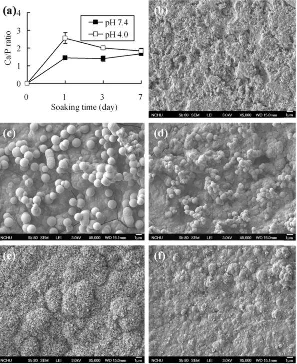
Broad face SEM micrographs of the cements after soaking in a solution with different pH values for 1 and 30 days are shown in Fig. 1, in addition to Ca/P ratio.The Ca/P ratios of the cement specimen after 1 day of soaking in pH 7.4 and 4.0 solutions were 1.44 and 2.56 (Fig.
1a), respectively; on day 7, the Ca/P ratios became 1.68 and 1.84. Before soaking, the cement specimen essentially appeared rather smooth looking with particle entanglement and several micropores (Fig. 1b). After soaking in a physiological solution, i
t was
clear that precipitation took place on the cement surfaces, which were covered with clusters of precipitated apatite spherulites (Fig. 1cf). However, it is worth noting that after soaking in a pH 7.4 solution for 1 day (Fig. 1c), the size of apatite spherulites was greater than that formed in the pH 4.0 solution(Fig. 1d). With increasing soaking time, spherulites coalesced to form a surface apatite layer.
Greater spherule aggregates appeared on the cement surface under a pH 7.4 condition (Fig. 1e) compared to that under a pH 4.0 condition (Fig. 1f).
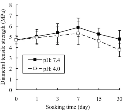
5.2. Diametral tensile strength
The changes in diametral tensile strength value of the cements before and after soaking in a pH 7.4 or 4.0 solution as a function of time are shown in Fig. 2. The cement specimen gradually increased in strength with an increase in soaking time, achieving a maximum on day 7, and thereafter decreased. The values at 0- and 30-day soaking in a pH 7.4 solution were 4.7
± 0.4 and 4.8 ± 0.8 MPa, respectively, indicating no significant difference (P > 0.05). In the case of pH 4.0, day 30 samples could achieve a value of 3.8 ± 0.6 MPa, which was comparable to that obtained in the environment of pH 7.4. Scheffé post hoc tests revealed that the difference between the strength values of cement specimens exposed to pH 7.4 and pH 4.0 was not statistically significant (P > 0.05) at the same soaking time point, although the cement soaked in a pH 7.4 solution had a higher strength than the pH 4.0 solution. Soaking time significantly (P < 0.05) affected the strength of the cement soaked in either pH 7.4 or 4.0 solution.
5.3. Weight change
There were statistically significant differences (P < 0.05) in the weight change among the groups because of solution pH and soaking time. Figure 3 shows the weight changes for the cements after exposure to the physiological solution. In terms of solution pH, the cement gained weight of 2.0% and 1.2% after 3 days of soaking in a pH 7.4 and 4.0 solution, respectively; afterwards, the sample weight reduced to -0.2% and 0.8% on day 30. Not only the initial solution pH values significantly (P < 0.05) affected the weight change of the cement to some extent, but soaking time did also.
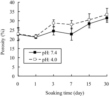
5.4. Porosity
Results shown in Fig. 4 indicate that the porosity increased from the initial 23% to approximately 32% after 30-day soaking; these values were significantly different (P < 0.05).
Concerning solution pH, the cement had a lower porosity in a solution with pH 7.4 at the same soaking time point.
5.5. pH variation of the solution
Figure 5 presents the time-dependent pH changes of the cement-immersed solutions. While soaked in a pH 7.4 solution, the cements caused the pH of the solution to increase at a steady state and attained alkaline values of 8.5 on day 30. In contrast, after soaking in a pH 4.0 solution the cement produced a lower pH value at the same time point. On day 1, the pH (7.3) of the solution originating from the pH 4.0 condition was significantly (P < 0.05) lower than the pH (7.8) of the solution starting from the pH 7.4 condition. The initial solution pH and soaking time significantly (P < 0.05) affected the sequent pH variation.
5.6. Setting time
The setting times (15-24 min) increased significantly (P < 0.05) with increasing Bi2O3
content (Table 1). These values were significantly (P < 0.05) lower than that of WMTA (168 min).
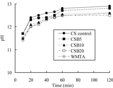
5.7. pH variation in the cement
The pH value of all cements during setting is presented in Fig. 6. After 20 min, all of the
cements reached a pH value of about 12.0. By 1 h, they approached a steady-state pH value.
The variations in pH value slightly decreased with increasing Bi2O3 content.
5.8. Phase composition
Figure 7 shows the XRD patterns of β-Ca2SiO4 cements with and without Bi2O3. The strongest peak at 2θ = 27.4º was ascribed to Bi2O3. It indicates that the products of the hydration process were calcium silicate hydrate (C–S–H) at 2θ = 29.4º overlapping with calcite. In addition, an incompletely reacted inorganic component phase of β-Ca2SiO4 at 2θ between 32−34º was found. The higher the Bi2O3 content, the lower the C–S–H content was in the cement.
5.9. Effect of Bi2O3 on DTS
The relationship between the Bi2O3 content and DTS of the cements is shown in Table 1.
The strength of the cements did not change significantly, although a decreasing trend with increasing Bi2O3 content was found. WMTA had a strength value of 4.2 MPa, which was significantly (P < 0.05) higher than those of all dicalcium silicate cements.
5.10. Radiopacity
The radiopacity (as an equivalent thickness of Al) of the CS control was recorded as 1.7 mm of Al (Table 1). After the addition of 5, 10, and 20 wt% Bi2O3, the radiopacity of the cement became significantly (P < 0.05) higher, with values of approximately 3.3, 5.8, and 8.4 mm of Al, respectively.
5.11. Solubility
The solubility of the four dicalcium silicate cements ranged from 0.8% to 1.1%. The incorporation of Bi2O3 did not significantly (P < 0.05) enhance the solubility of dicalcium silicate cements. WMTA had a solubility of 1.4% and was significantly different with P < 0.05 for all comparisons with dicalcium silicate cements with and without Bi2O3.
5.12. Cell proliferation
Figure 8 shows that the proliferation of MG63 cells cultured on Bi2O3-containing cement surfaces was lower than that on the surface of the cement control at all culture time points. For example, on day 7, the OD value for CSB20 cement was approximately 46% lower than that of the control.
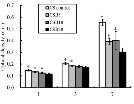
5.13. Cell differentiation
The intracellular ALP level was measured to observe the functional activity of cells. The results are shown in Fig. 9. The ALP level decreased with increasing Bi2O3 content of the cements at all incubation times. On day 7, a significant 21% reduction (P < 0.05) in the ALP level was measured for CSB20 compared to the CS control. The reduction became 24% after 14 days of culture.
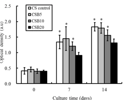
5.14. Mineralization
Quantification of calcium mineral deposits by the Alizarin Red S assay showed that the calcium content in the blank groups (day 0) was much lower than that obtained for the cement seeded with cells due to the lack of positive red staining (Fig. 10). This explains why calcium contents in the material composition did not appreciably affect the quantification of calcium mineral deposits. On day 7, more mineral deposition was found in MG63 cells cultured on the cement specimens than those obtained on day 0. With increasing culture time, mineral
deposition increased for the cells cultured on all cements, but less mineral deposition was found for cements with higher Bi2O3 content. By day 14, a significant 28% reduction (P < 0.05) of calcium content was observed for the 20 wt% Bi2O3-containing cement (CSB20) compared to the CS control.
6. Discussion
An important requirement for any dental material used for permanent treatment is its resistance to degradation (or solubility) when exposed to a host environment for a prolonged time period. Possible factors such as solution pH, soaking time and liquid-to-powder ratio of the cement influence degradation of these materials [21,22]. In this study, the potential variations in the properties of the cement during soaking in a physiological solution with different pH values for different time points were addressed. The two-way ANOVA showed solution pH and soaking time have a significant interactive effect on physicochemical properties (weight change, porosity, strength and pH variation) of the current cement.
Exposure of bioactive material surfaces such as bioactive glass and MTA to a physiological solution elicits the precipitation of a “bone-like” apatite layer, which may support the material’s ability to integrate into living tissue [1,23]. In this study, no matter which pH solution was used, after soaking for 1 day, the formed layer was apatite precipitated from the two physiological solutions, as evidenced previously [19]. Such apatite precipitation indicates high bioactivity of the cement in the presence of phosphate-containing fluids. To further confirm that the observed apatite layer was indeed precipitated from physiological solution, a SEM/EDS analysis was performed on the soaked surface. This precipitate has been identified to be apatite although Ca/P ratios of 1.44 and 2.56 after soaking in pH 7.4 and pH 4.0 solutions for 1 day, respectively. The much higher Ca/P ratio (than the stoichiometric Ca/P ratio, 1.67, of apatite) on 1-day-soaked surface in a pH 4.0 solution was due to the fact that a large quantity of calcium originated from the underlying cement was detected, which also indicated a thinner apatite layer. With an increase in soaking time the Ca/P ratios appeared to approach 1.67 for both pH conditions. However, although having a similar morphology, there were differences in the size of the spherulites size. The apatite precipitation rate on the cement being soaked in a pH 7.4 solution was higher than that in a pH 4.0 solution, which was possibly attributed to the initial higher pH value of the solution. The in vitro bioactivity of the SiO2–CaO-based materials indicates that the presence of PO43 ions in the composition is not an essential requirement for the development of an apatite layer, which consumes the calcium and phosphate ions. This is because PO43 ions originate from the in vitro assay solution [24,25].
Interestingly, the low pH solution did not significantly reduce diametral tensile strength even after 30-day soaking. Watts et al. found that an acidic pH (5.0) in phosphate-buffered saline did not adversely affect compressive strength values of white-colored ProRoot MTA (Dentsply Tulsa Dental, Tulsa, OK) mixed with water after 7 days or 28 days of soaking in comparison to saline with a pH 7.4, which corroborates the present findings [26]. In the present study, the strength data also revealed that the day 7 samples from the two soaking solution systems were connected with an increase in strength compared to the day 0 sample.
During the setting reaction, some of the activated fractions within the set cement did not react, resulting in a weaker entanglement of the cement particles. Thus, the soaking-induced increase in mechanical strength was possibly attributable to the more complete hardening during immersion, which has also been observed in other studies [14,19,27]. Shie et al. reported that white-colored ProRoot MTA mixed with water significantly increased the diametral tensile strength by a factor of 2 after soaking in physiologic solution for 7 days [14]. As for the decreased strength after attaining to the maximum value, the pore formation may be the factor.
The trend in the strength value was similar to the changes in porosity. Porosity has an adverse effect on the properties of ceramic materials, acting as stress raisers and reducing mechanical strength [20]. It was possibly the pH in the solution enhanced the amount of the porosity, which in turn affecting the mechanical behavior of the cements. The increasing acidity results in an extensive porosity [11] and a reduced compressive strength of the set white-colored ProRoot MTA [28]. Shokouhinejad et al. also found that the force needed for the displacement of white-colored ProRoot MTA from root dentin to occur was significantly lower in samples stored at lower pH values [29].
Insolubility or low solubility is a characteristic of root-end filling material that is of utmost importance and necessary for successful results of endodontic therapy. To study the soaking- induced degradation behaviors, a series of measurements of weight change were performed.
The degradation of the current cement was a slow process, and the degree of the degradation was time-dependent and pH-dependent. When soaked in a low pH solution (4.0), the cement was associated with a relative small degree of weight loss of about 0.8% even after a 30-day soaking time; the cement even exhibited a weight increase (0.2%) under a normal pH (7.4) solution. The weight loss may be because of the release of soluble fractions (mainly Ca(OH)2) [14,30]. During the hydraulic reaction, CaO component of the powder would dissolve in the liquid component (water) and become Ca(OH)2, as confirmed by pH increase [23]. On the other hand, the increase in sample weight may be explained by the formation of apatite, which was consistent with the morphology results. These increases in both pH and concentration of Ca2+ ions enhance the supersaturation of a physiological solution with respect to apatite which promotes the precipitation of the apatite layer [24,25]. The results of the higher pH value examined in the cement-soaked solution under a pH 7.4 condition paralleled the apatite precipitation rate. There is a compromise between cement dissolution and apatite precipitation, when soaking in a solution. Last but not least, the current cement was more soluble in the acidic medium but was stable in a pH 7 environment. The soaking time and solution pH exerted appreciably effects on physicochemical properties of the cement.
Radiopacity is required for endodontic treatment. Bi2O3 can be introduced to increase radiopacity due to its high molecular weight [18]. Of note, the presence of radiopacifiers may adversely affect the physicochemical and biological properties. In this study, it was found that with a greater amount of Bi2O3, the diffraction peaks of the hydration product of a C−S−H gel became weaker. This can be explained by the fact that Bi2O3 does not participate in the hydration reaction [31]. It was also found that Bi2O3 significantly prolonged the setting times of the dicalcium silicate cements. This is in agreement with reports from other researchers [16]
who found that Bi2O3 increases setting time. Another concern was whether the incorporation of Bi2O3 into dicalcium silicate cements reduces mechanical strength. Interestingly, the addition of Bi2O3 to dicalcium silicate cement did not significantly affect the diametral tensile strength value, although a decreasing trend was found. The mechanical properties of the hardened cements result from the reaction of the liquid phase, such as water, with the cement components. Cement hydration connects the originally hydrophilic particles together, along with the formation of a C–S–H gel that develops the bonding properties responsible for its hardening [19]. Hence, the formed C−S−H structure is likely to contribute to the overall strength [32].
The setting time and strength of the paste materials depend on factors such as the liquid-to- powder ratio, particle size, powder composition, and liquid phase [1,19,30]. Coomaraswamy et al. used a fixed liquid-to-powder ratio to evaluate the effect of Bi2O3 content on the compressive strength of Portland cement, and found that compressive strength significantly decreased with increasing Bi2O3 content [15]. When the amount of water is higher than that necessary for the stoichiometric setting reaction of the cement to occur, it fills the spaces
among the cement particles; after drying, it evaporates and leads to the formation of pores, which decrease mechanical strength. Since Bi2O3 does not take part in the setting reaction when added, it can be assumed that a smaller amount of water is necessary to mix the powder.
Thus, the liquid-to-powder ratio should be reduced by decreasing the liquid phase in order to prevent the powder mixture from having excess water, when preparing the Bi2O3–containing cements.
Concerning radiopacity, the CS control exhibited a low intrinsic radiopacity (equivalent to 1.7 mm of Al) that is insufficient for distinguishing the cement from tooth and bone. This is consistent with previous studies that reported radiopacity values ranging from 0.9 mm to 1.6 mm of Al for calcium silicate-based cements [9]. Not surprisingly, the radiopacity increased with increasing Bi2O3 content. The cement containing 5 wt% Bi2O3 had a radiopacity slightly higher than the recommended 3 mm of Al. After 10 wt% Bi2O3 addition, the resulting radiopacity increased to 5.8 mm of Al, a value greater than that in the ISO 6876:2001 standards (at least 3 mm of Al). When 20% Bi2O3 was used, the radiopacity value (8.4 mm of Al) was significantly enhanced, making it similar to that for ProRoot WMTA (radiopacity values ranging from 5.9 to 9.3 mm of Al) [9].
The solubility of WMTA obtained in this study was found to be similar to that (1.3%) reported by Islam et al. [27], and significantly greater than that of CSB20. The difference in solubility could be due to the phase composition and structure. However, all values were well within the ISO 6786:2001 requirements, as it did not exceed 3% mass fraction. Insolubility or low solubility is a characteristic of root-end filling material that is of utmost importance and necessary for successful endodontic therapy. The solubility may originate from the release of soluble fractions (mainly Ca(OH)2) [17,21]. During the hydraulic reaction, the CaO component of the powder dissolves in the liquid component (water) and becomes Ca(OH)2 [9], as confirmed by pH changes.
To elucidate the effects of Bi2O3 on osteogenic activities, the biological functions of MG63 cells cultured on cement specimens with various Bi2O3 content levels were evaluated. The numbers of initially attached cells for the CS control and radiopaque cements were different.
Bi2O3 led to decreased cell proliferation. Cell differentiation studies, like cell proliferation assay results, showed a significant impact of Bi2O3, with an emphasis on the importance of material composition. ALP activity decreased with increasing Bi2O3 content. ALP enzyme activity is associated with bone formation; ALP is produced in high levels during the bone formation phase [25]. To more fully assess the role of Bi2O3 in cell function, mineralization ability was examined. Alizarin Red S staining is a common histochemical technique used for detecting calcium deposits in mineralized tissues and cultures. The ability of cells to produce a mineralized matrix and nodules in materials is important for bone regeneration [20]. The calcium in the dicalcium silicate might also stain red. Therefore, a blank test without cell seeding on the cement surfaces was performed. The results confirmed that calcium in the material composition did not affect the OD (day 0). The decreased proliferation, differentiation, and mineralization of MG63 cells showed that the Bi2O3 in the cement was responsible for the reduction in cell growth. Min et al. found that Portland cement with Bi2O3
is significantly more cytotoxic than pure Portland cement in short-term cultures with human dental pulp cells [33].
7. Conclusions
It is concluded that solution pH 4.0 imposed in this study appreciably affect physicochemical properties of the radiopaque dicalcium silicate cement. High in vitro activity and low degradation were the characteristics of the cement. Bi2O3 radiopacifier was added to endodontic materials to obtain an acceptable level of radiopacity. The setting time of the
cement was adversely affected by the introduction of Bi2O3; however, the mechanical strength was not affected. As a general trend, cell proliferation and differentiation and the number of calcium deposits were inversely proportional to the Bi2O3 content. Taking the setting time, radiopacity, and osteogenic activity into account, 10 wt% Bi2O3-containing dicalcium silicate cement is the best choice for a root-end filling material.
8. Evaluation
本計畫目前衍生的成效
項目 成果
SCI 論文 有2 篇 SCI 論文被接受
1. Chiang TY, Ding SJ*. Physicochemical properties of radiopaque dicalcium silicate cement as a root-end filling material in an acidic environment.
International Endodontic Journal 2013;46(3):234-241.
2. Chiang TY, Wei CK, Ding SJ*. Effects of bismuth oxide on physicochemical properties and osteogenic activity of dicalcium silicate cements. Journal of Medical and Biological Engineering 2013;
doi:10.5405/jmbe.1386.
國際會議 1. 2012 年創新生醫材料研討會(Innovations in Biomedical Materials 2012)邀請演講
2. 第 12 屆亞洲生物陶瓷研討會 (ABC2012)秘書長 8. 參考文獻
[1] Ding SJ, Shie MY, Wang CY. Novel fast-setting calcium silicate bone cements with high bioactivity and enhanced osteogenesis in vitro. J Mater Chem 2009;19:1183−90.
[2] Liu WN, Chang J, Zhu YQ, Zhang M. Effect of tricalcium aluminate on the properties of tricalcium silicate-tricalcium aluminate mixtures: setting time, mechanical strength and biocompatibility. Int Endod J 2011;44:41–50.
[3] Shie MY, Chang HC, Ding SJ. Effects of altering the Si/Ca molar ratio of a calcium silicate cement on in vitro cell attachment. Int Endod J 2012;45:337−45.
[4] Chng HK, Islam I, Yap AUJ, Tong YW, Koh ET. Properties of a new root-end filling material. J Endod 2005;31:665−8.
[5] Camilleri J. The physical properties of accelerated Portland cement for endodontic use. Int Endod J 2008;41:151–7.
[6] Gandolfi MG, Perut F, Ciapetti G, Romano Mongiorgi R, Prati C. New Portland cement- based materials for endodontics mixed with articaine solution: A study of cellular response. J Endod 2008;34:39−44.
[7] Chen CC, Ho CC, Chen CH, Ding SJ. Physicochemical properties of calcium silicate cements for endodontic treatment. J Endod 2009;35:1288−91.
[8] Kao CT, Shie MY, Huang TH, Ding SJ (2009) Properties of an accelerated mineral trioxide aggregate-like root-end filling material. Journal of Endodontics 35, 239−42.
[9] Chiang TY, Ding SJ. Comparative physicochemical and biocompatible properties of radiopaque dicalcium silicate cement and mineral trioxide aggregate. J Endod 2010;36:1683−7.
[10] Chen CC, Shie MY, Ding SJ. Human dental pulp cells responses to new calcium silicate- based endodontic materials. Int Endod J 2011;44:83642.
[11] Namazikhah MS, Nekoofar MH, Sheykhrezae MS et al. The effect of pH on surface hardness and microstructure of mineral trioxide aggregate. Int Endod J 2008;41:108–16.
[12] Torabinejad M, Chivian N. Clinical applications of mineral trioxide aggregate. J Endod 1999;25:197–205.
[13] Roy CO, Jeansonne BG, Gerrets TF. Effect of an acid environment on leakage of root- end filling materials. J Endod 2001;27:7–8.
[14] Shie MY, Huang TH, Kao CT, Huang CH, Ding SJ. The effect of a physiological solution pH on properties of white mineral trioxide aggregate. J Endod 2009;35:98−101.
[15] Coomaraswamy KS, Lumley PJ, Hofmann MP. Effect of bismuth oxide radioopacifier content on the material properties of an endodontic Portland cement-based (MTA-like) system. J Endod 2007;33:295-8.
[16] Camilleri J. The physical properties of accelerated Portland cement for endodontic use.
Int Endod J 2008;41:151-7.
[17] Torabinejad M, White DJ. Tooth filling material and method of use. U.S. Patent 5415547, 1995.
[18] Sugawara A, Asaoka K, Ding SJ. Calcium phosphate-based cements: clinical needs and recent progress. J Mater Chem B 2013;1:1081-9.
[19] Chen CC, Ho CC, Chen CH, Wang WC, Ding SJ. In vitro bioactivity and biocompatibility of dicalcium silicate cements for endodontic use. J Endod 2009;35:
1554−7.
[20] Ding SJ, Wei CK, Lai MH. Bio-inspired calcium silicate-gelatin bone grafts for load- bearing applications. J Mater Chem 2011; 21:12793802.
[21] Yanikoğlu N, Yeşil Duymuş Z. Evaluation of the solubility of dental cements in artificial saliva of different pH values. Dent Mater J 2007;26:62–7.
[22] Hu G, Xiao L, Fu H, Bi D, Ma H, Tong P. Study on injectable and degradable cement of calcium sulphate and calcium phosphate for bone repair. J Mater Sci: Mater Med 2010;21:627–34.
[23] Gandolfi MG, Taddei P, Tinti A, Prati C. Apatite-forming ability (bioactivity) of ProRoot MTA. Int Endod J 2010;43:917–29.
[24] Chen CC, Wang WC, Ding SJ. In vitro physiochemical properties of gelatin/chitosan oligosaccharide/calcium silicate hybrid cement. J Biomed Mater Res B: Appl Biomater 2010;95:456–65.
[25] Ding SJ, Shie MY, Wei CK. In vitro physicochemical properties, osteogenic activity, and immunocompatibility of calcium silicate-gelatin bone grafts for load-bearing applications.
ACS Appl Mater Interfaces 2011;3:414253.
[26] Watts JD, Holt DM, Beeson TJ, Kirkpatrick TC, Rutledge RE. Effects of pH and mixing agents on the temporal setting of tooth-colored and gray mineral trioxide aggregate. J Endod 2007;33:970–3.
[27] Islam I, Chng HK, Yap AUJ. Comparison of the physical and mechanical properties of MTA and Portland cement. J Endod 2006;32:193–7.
[28] Kayahan MB, Nekoofar MH, Kazandağ M et al. Effect of acid-etching procedure on selected physical properties of mineral trioxide aggregate. Int Endod J 2009;42:1004–14.
[29] Shokouhinejad N, Nekoofar MH, Iravani A, Kharrazifard MJ, Dummer PMH. Effect of acidic environment on the push-out bond strength of mineral trioxide aggregate. J Endod 2010;36:871–4.
[30] Fridland M, Rosado R, Eng C. Mineral trioxide aggregate (MTA) solubility and porosity with different water-to-powder ratios. J Endod 2003;29:814 –7.
[31] Camilleri J. Hydration mechanisms of mineral trioxide aggregate. Int Endod J 2007;40:
462-70.
[32] Ding SJ, Shie MJ, Hoshiba T, Kawazoe K, Chen G, Chang HC. Osteogenic differentiation and immune response of human bone marrow-derived mesenchymal stem cells on
injectable calcium silicate-based bone grafts. Tissue Eng A 2010;16:2343-54.
[33] Min KS, Chang HS, Bae JM, Park SH, Hong CU, Kim EC. The induction of heme oxygenase-1 modulates bismuth oxide-induced cytotoxicity in human dental pulp cells. J Endod 2007;33:1342-6.
Table 1 Setting time, diametral tensile strength, radiopacity, and solubility of tested cements.
Sample code
Bi
2O
3content (wt%)
Liquid-to-powder ratio (mL/
g)
Setting time (min)
Diametral tensile strength (MPa)
Radiopacity (mm of Al)
Solubility (%) CS
control
0 0.5 15 ± 1
a2.3 ± 0.3
a1.7 ± 0.5
a0.9 ± 0.1
aCSB5 5 0.47 19 ± 1
b2.3 ± 0.1
a3.3 ± 0.7
b0.8 ± 0.1
aCSB10 10 0.45 23 ± 2
c2.1 ± 0.2
a5.8 ± 0.5
c0.9 ± 0.1
aCSB20 20 0.4 24 ± 1
c2.0 ± 0.2
a8.4 ± 0.5
d1.1 ± 0.1
aWMTA 20 0.3 168 ± 2
d4.2 ± 0.1
b9.3 ± 0.4
e1.4 ± 0.2
bValues are mean ± standard deviation. Repeated superscripts in a given column indicate no significant difference (P > 0.05) according to Scheffé’s post hoc multiple comparisons.
Figure 1 after (c
and 30 d
1 Ca/P rati
f) soaking days (e and
io (a) and in a soluti f).
SEM micr on with pH
rographs of H 7.4 (c an
f the cemen d e) or pH
nt specime 4.0 (d and
ens before d f) for 1 (c
(b) and c and d)
0 1 2 3 4 5 6 7 8
0 1 3 7 15 30
Soaking time (day)
D ia m et ra l te ns ile s tr en gt h ( M P a)
pH: 7.4 pH: 4.0
-2 -1 0 1 2 3
0 1 3 7 15 30
Soaking time (day)
W ei ght c ha ng (% )
pH: 7.4 pH: 4.0
Figure 2 Diametral tensile strength of the cement specimens before and after soaking in a solution with pH 7.4 or pH 4.0 for predetermined time durations.
Figure 3 Weight change of the cement specimens before and after soaking in a solution with pH 7.4 or pH 4.0 for predetermined time durations.
0 5 10 15 20 25 30 35 40
0 1 3 7 15 30
Soaking time (day)
Po ro si ty (% )
pH: 7.4 pH: 4.0
0 1 2 3 4 5 6 7 8 9
0 1 3 7 15 30
Soaking time (day)
pH va ul e
pH: 7.4pH: 4.0
Figure 4 Porosity of the cement specimens before and after soaking in a solution with pH 7.4 or pH 4.0 for predetermined time durations.
Figure 5 Variations in the pH of the solution with an initial pH 7.4 or pH 4.0 during soaking.
Figure 6
Figure 7 mixing w
6 Compariso
7 X-ray diff with water. ◘
on of pH ch
fraction patt
◘: CaCO3;
hanges of ce
terns of dica
▼: β-Ca2Si
ements durin
alcium silic iO4; ◊: C−S
ng setting.
cate cements
−H; ●: Bi2O
s with and w O3.
without Bi2O3 after
Figure 8 cement s compare
Figure 9 on dicalc differenc
8 Alamar Bl specimens d to CSB20
9 ALP assay cium silicate ce (P < 0.05
lue assay re at various 0.
y results on e cement sp 5) compared
esults for M time point
n MG63 cell pecimens aft d to CSB20.
MG63 cell pr nts. *Statisti
ls presented fter 7 and 14
.
roliferation ically signi
d as optical 4 days of cu
cultured on ificant diffe
density for ulture. *Stat
n dicalcium ference (P
cell differe tistically sig
m silicate
< 0.05)
entiation gnificant
Figure 1 cultured significan
10 Quantific on dicalciu nt differenc
cation of ca um silicate c ce (P < 0.05
alcium mine cement spec 5) compared
eral deposits cimens afte d to CSB20.
s by Alizari r culture fo .
in Red S ass or 7 and 14
say for MG days. *Stat
G63 cells tistically
出席國際學術會議心得報告
計畫編號 NSC 101-2221-E-040-004
計畫名稱 矽酸鈣為基底之骨填充複合材的前臨床研究
出國人員姓名 服務機關及職稱
丁信智
中山醫學大學 口腔科學研究所 教授
會議時間地點 101/9/10-101/9/13 美國 拉雷
會議名稱 2012 年創新生醫材料研討會
發表論文題目 Development and applications of sol-gel-derived calcium silicate-based bone cements
一、參加會議經過
2012 年創新生醫材料研討會(Innovations in Biomedical Materials 2012)是美國陶瓷學會 在美國北卡羅來納州拉雷(Raleigh) 的 Hilton North Raleigh-/Midtown 的國際會議廳舉行,此屬 於小而美且領域專門的國際會議,而最特別的是生醫材料廠商的參予與報告。發表分口頭(88 篇)及海報貼示(18 篇),當中 plenary speakers 有 Prof. Larry Hench, Prof. Alan J. Russell, Prof.
Delbert Day, Prof. Riad Salem, Dr. Hyun Bae 等知名學者。國際生物陶瓷界大師 Prof. Larry Hench 在其演講中強調下世代生醫材料所面對的問題為何?整個會議研討涵蓋與生醫材料相 關的各種不同議題,如鈦表面處理、生物活性玻璃、電紡材料、磷酸鈣陶瓷、骨取代材、組 織工程、生物相容性研究、藥物制放載體等。主持人大多是醫療器材管理階層的專家,例如 本人的演講的主持人即是 NovaBone (主要產品為 Ca, Si, P 生物活性玻璃)公司的副總裁 Dr.
Gregory Pomrink,接續本人報告之後為 Primus 醫療器材顧問公司創辦人 Dr. Carolyn. Primus
(其本身為牙醫師)的牙科骨泥報告,Dr. Carolyn. Primus 相當欣賞本實驗室在矽酸鈣骨水泥 上的研究成果。此外,另有多場演講是針對材料如何商品化,會場討論氣氛十分熱絡。
二、與會心得
本人有幸被邀請報告,實屬光榮。此次台灣參加的學者另有成功大學材料講座教授 Prof.
Yoshimura 邀請報告新式的金屬表面處理。亞洲生醫材料學者另有日本的 Prof. Kokubo 與印度 Prof. Urooj 等參加。生醫材料研究為一跨領域且理論、應用並重的學門,從與會中所發表的 論文可知仍有相當大的研究空間,但有待臨床醫師與生醫材料研究者雙向交流與合作,才能 更加突破目前所面臨之瓶頸。從國外學者的研究趨勢及發表主題,顯示台灣生醫材料界研究 方向與世界並進、並未偏離。台灣已將醫療器材產業規劃為重點科技,目前有更多的產業與 學者投入生醫研發,因此實有必要強化國際交流與曝光度,有利於醫療器材產業輸出,且如 何商品化。
Development and Applications of Sol-Gel-Derived Calcium Silicate-Based Bone Cements Shinn-Jyh Ding
Institute of Oral Science, Chung Shan Medical University, Taichung City 402, Taiwan E-mail: [email protected]
Cementitious bone repair materials are being used increasingly in minimally invasive clinical applications such as dentistry, vertebroplasty, and orthopedics. Such devices would shorten the surgical operation time, minimize the damaging effects of large muscle retraction, reduce the size of the scars and lessen post-operative pain, allowing patients to achieve rapid recovery in a cost-effective manner. This talk will introduce development and applications of novel calcium silicate-based cements (CSCs) for bone repair and regeneration. The fast-setting CSCs with high bioactivity and osteogenesis have successfully developed by using a sol-gel method, suggesting that CSCs open up new possibilities in the field of self-setting bioactive SiO2−CaO-based bone graft materials. Nevertheless, the ceramic-based cement is difficult to deliver to bone defects with complex structures and is hard to compact because of the brittle nature of the ceramic cement. The presence of natural gelatin polymer appreciably improves the anti-washout and brittle properties of the cements without adversely affecting mechanical strength. The radiopaque CSCs containing bismuth oxide have a shortened setting time in comparison with a commercial ProRoot white-colored mineral trioxide aggregate. The radiopaque CSCs may have the potential to be an endodontic material.
600 N. Cleveland Avenue
26 June 2012
Prof. Shinn-Jyh Ding
Chung Shan Medical University Institute of Oral Science 110, Sec. 1, Jianguo N Road Taichung City, 402
Taiwan
Dear Prof. Ding,
We are pleased to invite you to present your topic entitled Development and Applications of Sol-Gel Calcium Silicate-Based Bone Cements, abstract # 1412333, accepted for presentation at the Innovations in Biomedical Materials 2012 Conference, organized by The American Ceramic Society. The conference is scheduled September 10-13, 2012 at the Hilton North Raleigh-Midtown hotel in Raleigh, North Carolina, USA.
Your presentation will be part of the “Bone Cements” session(s).
Please inform any co-authors of the acceptance of this abstract. The specific details of the day, time and location of your presentation will be communicated by email in July.
The Society will provide a laptop computer, LCD projector, screen, laser pointer and microphone in each technical session room. If your presentation is prepared on a Macintosh computer, please plan to bring your own computer for your presentation. Check in with your session chair 15 minutes before the start of your session. Please bring your presentation on a USB memory stick or CD-ROM and upload your presentation to the laptop at the podium in your session room prior to the start of the session. Presentations may not be loaded while the session is in progress.
All attendees must register for the meeting and pay the appropriate registration fee. Registration and hotel information for the conference is available at http://www.ceramics.org/biomaterials2012. We encourage you to make your hotel reservation early to take advantage of the special conference rate, available only until August 3, 2012. You must mention that you are participating in The American Ceramic Society conference to qualify for the special conference rate.
If it is necessary for you to obtain a travel visa to attend the conference, you may use this letter as an invitation. Please refer to the U.S. government website http://travel.state.gov for official guidelines to obtain a B-1 nonimmigrant travel visa for entry into the U.S. Individuals from Visa Waiver Countries must register with the U.S. Department of Homeland Security’s Electronic System for Travel Authorization (ESTA) program. Immediate application is suggested to allow for the required processing. Information on obtaining a travel visa also appears on the meeting webpage.
Thank you for your interest in participating in the Innovations in Biomedical Materials 2012 Conference. We look forward to seeing you in Raleigh.
Sincerely,
Marilyn Stoltz
Technical Content Administrator Direct Dial: 614-794-5868 Fax : 614-794-5818
國科會補助計畫衍生研發成果推廣資料表
日期:2013/01/08
國科會補助計畫
計畫名稱: 矽酸鈣為基底之骨填充複合材的前臨床研究 計畫主持人: 丁信智
計畫編號: 101-2221-E-040-004- 學門領域: 生醫材料
無研發成果推廣資料
101 年度專題研究計畫研究成果彙整表
計畫主持人:丁信智 計畫編號:101-2221-E-040-004- 計畫名稱:矽酸鈣為基底之骨填充複合材的前臨床研究
量化
成果項目
實際已達成
數(被接受 或已發表)
預期總達成 數(含實際已
達成數)
本計畫實 際貢獻百
分比
單位
備 註
(
質 化 說 明:如 數 個 計 畫 共 同 成 果、成 果 列 為 該 期 刊 之 封 面 故 事 ...等
)
期刊論文 0 0 100%
研究報告/技術報告
1 1 100%研討會論文 0 0 100%
論文著作 篇
專書 0 0 100%
申請中件數 0 0 100%
專利 已獲得件數 0 0 100% 件
件數 0 0 100% 件
技術移轉
權利金 0 0 100% 千元
碩士生 0 0 100%
博士生 2 2 100%
博士後研究員 0 0 100%
國內
參與計畫人力
(本國籍)
專任助理 0 0 100%
人次
期刊論文 2 2 100%
研究報告/技術報告
0 0 100%研討會論文 4 4 100%
論文著作 篇
專書 0 0 100% 章/本
申請中件數 0 0 100%
專利 已獲得件數 0 0 100% 件
件數 0 0 100% 件
技術移轉
權利金 0 0 100% 千元
碩士生 0 0 100%
博士生 0 0 100%
博士後研究員 0 0 100%
國外
參與計畫人力
(外國籍)
專任助理 0 0 100%
人次
其他成果
(無法以量化表達之成
果如辦理學術活動、獲 得獎項、重要國際合 作、研究成果國際影響 力及其他協助產業技 術發展之具體效益事 項等,請以文字敘述填 列。)
與成功大學,台灣大學共同舉辦第 12 屆亞洲生物陶瓷研討會。
成果項目 量化 名稱或內容性質簡述
測驗工具(含質性與量性)
0課程/模組
0電腦及網路系統或工具
0教材
0舉辦之活動/競賽
0研討會/工作坊
0電子報、網站
0科 教 處 計 畫 加 填 項
目 計畫成果推廣之參與(閱聽)人數
0國科會補助專題研究計畫成果報告自評表
請就研究內容與原計畫相符程度、達成預期目標情況、研究成果之學術或應用價 值(簡要敘述成果所代表之意義、價值、影響或進一步發展之可能性)、是否適 合在學術期刊發表或申請專利、主要發現或其他有關價值等,作一綜合評估。
1. 請就研究內容與原計畫相符程度、達成預期目標情況作一綜合評估
■達成目標
□未達成目標(請說明,以 100 字為限)
□實驗失敗
□因故實驗中斷
□其他原因 說明:
2. 研究成果在學術期刊發表或申請專利等情形:
論文:■已發表 □未發表之文稿 □撰寫中 □無 專利:□已獲得 □申請中 ■無
技轉:□已技轉 □洽談中 ■無 其他:(以 100 字為限)
3. 請依學術成就、技術創新、社會影響等方面,評估研究成果之學術或應用價 值(簡要敘述成果所代表之意義、價值、影響或進一步發展之可能性)(以 500 字為限)
本計畫乃將先前發展出的具優異生物相容性及骨形成性矽酸鈣材料,評估其前臨床應用性 質,如在高酸性溶液下之物理化學性質以及不透光劑氧化鉍含量效應。此材料具抵抗酸性 環境能力,且從硬化時間、不透光性及骨形成性等方面,可知 10 wt% 氧化鉍的添加可使 得矽酸鈣作為根管填充材白色三氧礦聚合物的另一種替代選擇材。在臨床實用上,可應用 於牙科、骨科與脊椎外科等骨缺損修補。藉此新材料開發可減少國內醫療器材與裝置的進 口依賴,兼而提升國內醫療產品開發能力,甚至將產品推向國際舞台。