行政院國家科學委員會專題研究計畫 成果報告
吸入性麻醉劑與靜脈麻醉劑對 H9c2 心肌細胞的鉀離子電流
的影響
計畫類別: 個別型計畫 計畫編號: NSC94-2314-B-006-056- 執行期間: 94 年 08 月 01 日至 95 年 07 月 31 日 執行單位: 國立成功大學醫學系麻醉科 計畫主持人: 劉彥青 計畫參與人員: 吳勝男,王雅貞 報告類型: 精簡報告 處理方式: 本計畫可公開查詢中 華 民 國 95 年 6 月 12 日
The Mechanisms of Propofol-Induced Block on Ion Currents in Differentiated H9c2 Cardiac Cells
Yen-Ching Liu1, Ya-Jean Wang2, Sheng-Nan Wu2,3
1
Department of Anesthesiology,
National Cheng Kung University Medical College, Tainan, Taiwan
2
Institute of Basic Medical Sciences,
National Cheng Kung University Medical College, Tainan, Taiwan,
3
Department of Physiology,
National Cheng Kung University Medical College, Tainan, Taiwan.
Running title: Effect of propofol on ion currents in H9c2 cells
Correspondence: Dr. Sheng-Nan Wu
Department of Physiology
National Cheng Kung University Medical College No. 1, University Road
Tainan, 70101 Taiwan Tel: 886-6-2353535-5334 Fax: 886-6-2363660
Summary
Background and purpose: General anesthetic propofol (2,6-bis(isopropyl)-phenol)
possess a chemical structure unrelated to other anesthetic drugs. It block a variety of ion currents. This study is designed to determine the effect of this drug on ion currents in differentiated H9c2 cardiac cells.
Experimental approach: The effects of propofol on ion currents of differentiated
clonal cardiac (H9c2) cells were investigated with patch-clamp and simulation methos.
Key results: Propofol suppressed the amplitude of delayed rectifier K+ current (IK(DR)) in a concentration-dependent manner with an IC50 value of 36 M. This
compound reduced activation time constant and increased current inactivation, although no voltage dependency of propofol-induced block of IK(DR)can demonstrated.
It suppressed L-type Ca2+ current (ICa,L) of cardiac and skeletal types to a similar
extent. Numerical simulations of IK(DR) based on a Markovian model reproduce the
experimental results and show that propofol-induced blockade of IK(DR) is associated
with an decrease in forward rate of the activation process and an increase in transitional rate into the inactivated state. Simulations of human atrial action potential used to mimic the effects of propofol demonstrate that block of ultra-rapid
IK(DR)can elevate the plateau phase of action potential and that concomitant reduction
in ICa,Lcan reverse this effect.
Conclusions and Implications: Propofol can suppress IK(DR) in differentiated
H9c2 cardiac cells in a concentration- and state-dependent manner. These effects can significantly contribute its action on functional activity of heart cells.
Markovian model
Abbreviations: IK(DR), delayed rectifier K+ current; propofol,
Introduction
Propofol (2,6-bis(isopropyl)-phenol) is widely used as an intravenous general anesthetic which is structurally unrelated to other anesthetic drugs. Propofol was reported to block small-conductance Ca2+-actiated K+ channel in reticular thalamic neurons, and to block ATP-sensitive K+channels in rat ventricular myocytes.1,2 This compound can inhibit muscarinic M1 receptor-mediated signal transduction.3 A previous report demonstrated that propofol could inhibit K+ channels in pig articular chondrocytes, in neurons, and in human T lymphocytes.4-6 Propofol-induced inhibition of delayed rectifier K+ current (IK(DR)) in T lymphocytes was previously
proposed to result from open channel block (Mozrzymas et al., 1996).6 This drug was also reported to block Ca2+ and K+ currents in canine cardiomyocytes.7 Although the inhibitory action of propofol on ion currents was thought to contribute to clinical or adverse effects, the mechanism of actions of this compound on ion currents has been incompletely understood.
The H9c2 cell line has been established from embryonic rat cardiac ventricle and it has properties similar to neonatal and adult cardiomyocytes.8 After undergoing differentiation, these cells can functionally express L-type Ca2+ channels of cardiac and skeletal types and ATP-sensitive K+ channels.8,9 H9c2 cells were also reported to express Ca2+-activated K+ current and a Kv2.1-type IK(DR).10,11 The Kv2.1 K+
channel, that exhibits unique gating properties and voltage dependency, has been described to be an important contributor to the delayed rectifier K+current (IK(DR)) in
these cells.10,12
The objective of this study was to investigate the mechanism of inhibitory actions of propofol on ion currents present in differentiated H9c2 cardiac cells. The results showed that this drug could block IK(DR) in a concentration- and
state-dependent fashion. L-type Ca2+ current (ICa,L) of cardiac and skeletal types in
these cells were also blocked by propofol. With the aid of a simulation model, we are also able to duplicate the experimental results and to determine how this drug interacts with the K+ channels to alter the amplitude and kinetics of IK(DR) in these
Materials and Methods Cell culture
The clonal cardiac cell line H9c2, originally derived from embryonic rat ventricles, was obtained from the American Type Culture Collection ([CRL-1446], Manassas, VA). Cells were maintained in monolayer culture in 50-ml culture flasks in Dulbecco’s modified Eagle’s medium (Sigma-Aldrich, St. Louis, MO) supplemented with 10% fetal bovine serum in a humidified atmosphere of 5% CO2
and 95% air at 37 ºC.12 To induce cell differentiation, before confluence, cells were split and then cultured in Dulbecco’s modified Eagle’s medium supplemented with 1% fetal bovine serum. Under this condition, several elongated and multinucleated H9c2 cells were observed. The expression of L-type Ca2+ channel of skeletal type commonly occurred in differentiated H9c2 cells. Therefore, in a separate set of experiments designed to prevent cells from undergoing transdifferentiation into skeletal myoblasts, differentiated H9c2 cells were incubated with retinoic acid (20 nM) for 5-7 days.8 A colorimetric method was often used in examining cell densities in
microtiter plates using a tetrazolium salt
(4-[3-(4-iodophenyl)-2-(4-nitrophenyl)-2H-5-tetrasolio]-1,3-benzene disulfonate; WST) and an ELISA reader (Dynatech, Chantilly, VA).
The clonal strain TC-6 cell line was also obtained from the American Type Culture Collection ([CRL-11506]). This cell line was originally derived from a pancreatic tumor (insulinoma) arising in a transgenic mouse.13 The cells secrete abundant insulin and small amounts of glucagons and somatostatin. Cells were fed twice weakly and maintained in modified Dulbecco’s Modified Eagle’s medium supplemented with 4 mM L-glutamine, 4.5 g/L glucose, 1.5 g/l sodium bicarbonate, and 15% heat-inactivated fetal bovine serum.
Electrophysiological measurements
Immediately before each experiment, H9c2 or TC-6 cells were dissociated and an aliquot of cell suspension was transferred to a recording chamber positioned on the stage of an inverted DM IL microscope (Leica Microsystems, Wetzlar, Germany). Cells were bathed at room temperature (20-25 °C) in normal Tyrode’s solution containing 1.8 mM CaCl2. The recording electrodes were pulled from Kimax-51
glass capillaries (#34500; Kimble Glass, Vineland, NJ) using a PP-830 microelectrode puller (Narishige, Tokyo, Japan) and the tips were fire-polished with an MF-83 microforge (Narishige). For most recordings, the pipette resistance ranged between 3 and 5 M. Patch-clamp recordings were made in the whole-cell configuration using an RK-400 amplifier (Bio-Logic, Claix, France) (Wu et al., 1998).12,14 All potentials were corrected for liquid junction potential, a value that would always develop at the tip of the pipette when the composition of the pipette solution was different from that in the bath.
Data analyses
The signals were displayed on an HM-507 oscilloscope (Hameg, Germany) and on a PJ552-2 liquid crystal display projector (ViewSonic, Walnut, CA). The data were stored in a Slimnote VX3 portal computer (Lemel, Taipei, Taiwan) at 10 kHz
through a Digidata 1322A interface (Axon Instruments, Union City, CA) through a universal serial port. This device was controlled by the pCLAMP 9.0 software (Axon Instruments). Currents were low-pass filtered at 1 or 3 kHz. Macroscopic currents were stored without leakage correction and analyzed with the aid of either the pCLAMP 9.0 software (Axon Instruments), the Origin 7.5 software (Microcal Software, Inc., Northampton, MA) or custom-made macros in Excel 2003 (Microsoft, Redmont, WA).
To calculate percentage inhibition of propofol on IK(DR), cells were bathed in
Ca2+-free Tyrode’s solution, and each cell was depolarized from –50 to +50 mV. The amplitudes in the presence of this compound measured at the end of depolarizing pulses were compared with those after a subsequent application of tetraethylammonium chloride (100 mM). Concentration-dependent relation of propofol on the inhibition of IK(DR) in differentiated H9c2 cells was fitted to the Hill
equation: percentage inhibition = (Emax[C]nh) / (IC50nh+[C]nh), where [C] represents
the concentration of propofol; IC50 and nh are the half-maximal concentration of
propofol required to inhibit IK(DR) (i.e., tetraethylammonium chloride-sensitive K+
current) and the Hill coefficient (i.e., slope factor), respectively; and Emax is
propofol-induced maximal block of IK(DR).
The averaged results are presented as the mean values ± SEM. The paired or unpaired t test and one-way analysis of variance (ANOVA) test with the least significance difference method for multiple comparisons were used for the statistical evaluation of differences among the mean values. Throughout the paper, P < 0.05 was considered significant.
Modeling
Source files for Markovian formulation of IK(DR) adapted to reproduce the
currents and implemented in our laboratory are available at http://senselab.med.yale.edu/senselab/modeldb. The state diagram for this model was shown in figure 1A of the paper by Greenstein et al (2000).15 The detailed
meanings for each model parameter can be found at
http://circres.ahajournals.org/cgi/data/87/11/1026/DC1/1. Basically, this model consists of four closed and four closed-inactivated states, one open state, and one open-inactivated state.15 The solutions to the different set of ordinary differential
equations were approximated using the X-Win32 version of XPPAUT on a Dell Precision 670 workstation (Round Rock, TX).16 This software package is a collection of differential equations solvers used for analyzing and understanding the behavior of physical systems that change over time.16 The information for XPP software is readily available at http://www.math.pitt.edu/~bard/XPP/XPP.html. Using these simulation protocols, the users can study and graphically display the underlying properties of individual ion current to observe how dynamic changes in the different states determine the behavior of measured ion currents. The parameter values that were adapted and modified for numerical simulations of IK(DR) present in
H9c2 cells are listed in Table 1. To simulate the effects of propofol on cardiac action potential, human atrial action potential, source file for which is also available at http://senselab.med.yale.edu/senselab/modeldb, is mathematically reconstructed from ionic processes originally formulated from the paper of Courtemanche et al. (1998).17
Drugs and solutions
Propofol (2,6-bis(isopropyl)-phenol) was obtained commercially as Diprivan® (Zeneca Ltd, Macclesfield, Cheshire, UK), a sterile, nonpyrogenic emulsion containing 10 mg/ml propofol. The solvent for the control experiment is Intralipid (10 %; Lipovenoes), which was purchased from Fresenius AG (Germany). Intralipid contained 10% purified soybean oil, 1.2 % egg lecithin and 2.5 % glycerol. Diazoxide, retinoic acid, tetrodotoxin and tetraethylammonium chloride were purchased from Sigma Chemical (St. Louis, MO) and pinacidil was from Tocris (Bristol, UK). Caffeic acid phenethyl ester was purchased from Cayman Chemical (Ann Arbor, MI). Iberiotoxin and apamin were purchased from Alomone Laboratories (Jerusalem, Israel). Azimilide was a gift from Proctor and Gamble Pharmaceuticals (Cincinnati, OH). All other chemicals were commercially available
and of reagent grade. Tissue culture medium, L-glutamine, penicillin-streptomycin, fungizone, and trypsin were obtained from Life Technologies (Grand Island, NY). The twice-distilled water that had been de-ionized through a Millipore-Q system (Millipore, Bedford, MA) was used in all experiments.
The composition of normal Tyrode’s solution was 136.5 mM NaCl. 5.4 mM KCl, 1.8 mM CaCl2, 0.53 mM MgCl2, 5.5 mM glucose and 5.5 mM HEPES-NaOH buffer,
pH 7.4. To record K+ currents or membrane potential, the recording pipette was backfilled with a solution consisting of 140 mM KCl, 1 mM MgCl2, 3 mM Na2ATP,
0.1 mM Na2GTP, 0.1 mM EGTA and 5 mM HEPES-KOH buffer, pH 7.2. To
measure Ca2+ current, KCl inside the pipette solution was replaced with equimolar
CsCl, and pH was adjusted to 7.2 with CsOH. To record
ether-à-go-go-related-mediated K+ current (IK(erg)), high-K+, Ca2+-free solution
contained a solution consisting of 130 mM KCl, 10 mM NaCl, 3 mM MgCl2, 6 mM
glucose and 10 mM HEPES-KOH buffer, pH 7.2).18 In this condition, inward K+ currents can be enhanced by raising extracellular K+ concentration to increase the available charge carrier. In some experiments, propofol (100 M) was included in the recording pipette. The pipette solution was often filtered on the day of use with a 0.22-m pore size syringe filter (Millipore).
Results
Inhibitory effect of propofol on delayed rectifier K+current (IK(DR)) in differentiated
H9c2 cells
In the first series of experiments, IK(DR) in response to a series of voltage steps
was examined in these cells. Differentiated H9c2 cells were bathed in Ca2+-free Tyrode’s solution containing 0.5 mM CdCl2, in which voltage-dependent Ca2+
currents can be eliminated. As shown in Figure 1, when the cell was held at -50 mV and various potentials ranging from +10 to +50 mV with 10-mV increments were applied, a family of K+ outward currents was elicited. Electrical properties of these currents (i.e., IK(DR)) that were activated at around -10 mV with a half-activation
voltage of around +30 mV were found to resemble those described previously in H9c2 cells.12 Within 1 min of exposing cells to propofol (30 M), these currents evoked by this series of voltage steps was greatly reduced (Fig. 1A). For example, when the cells were depolarized from -50 to +50 mV, current amplitudes measured at the end of depolarized pulses were significantly decreased from 1244 ± 122 to 687 ± 98 pA (n = 8). The time dependence of propofol-induced block of these currents was also observed. The exposure to propofol (30 M) significantly increased activation time constant from 8.1 ± 1.1 to 15.3 ± 3.2 msec (n = 6) and concomitantly reduced the time constant of current inactivation from 202 ± 16 to 156 ± 11 msec (n = 6). This inhibitory effect was readily reversed on washout of propofol. Figure 1B shows the average current-voltage (I-V) relationships for these currents in the absence and presence of 30M propofol. However, application of Intralipid (10 %), a solvent for propofol, did not have any significant effect on IK(DR).
The percentage inhibition of IK(DR) by propofol was also measured at test
difference in percentage inhibition of 30 propofol on IK(DR) across the voltages tested.
For example, at the level of +10 mV, current was significantly blocked by 56 ± 6 % (n = 7), while at +40 mV, inhibition was 55 ± 6 (n = 8). These results suggest that block by propofol of IK(DR) in differentiated H9c2 cells shows little or no voltage
dependence.
The relationship between the concentration of propofol and the percentage inhibition of IK(DR) was further examined. Current amplitudes of IK(DR) during the
exposure to propofol were compared with those after a subsequent application of tetraethylammonium chloride (100 mM). As shown in Fig. 2, this block was found to follow a sigmoidal dose-response kinetics which yielded an IC50 value of 36 M
with a calculated Hill coefficient of 1.2. The results clearly showed that propofol could suppress tetraethylammonium chloride-sensitive IK(DR)in these cells.
Comparisons between effect of propofol and those of apamin, glibenclamide, chromanol 293B and pentobarbital on the amplitude of IK(DR)in differentiated H9c2
cells
The effects of various compounds, including apamin, glibenclamide, chromanol 293B and pentobarbital, on the amplitude of IK(DR) in these cells were examined and
compared. As shown in Fig. 3A, neither apamin (200 nM) nor glibenclamide (30 M) had any effects on IK(DR). Chromanol 293B (30 M), a blocker for the slowly
activating IK(DR)present in cardiomyocytes, suppressed the amplitude of IK(DR)by 72%,
while pentobarbital (30 M), another anesthetic drug structurally distinct from propofol, blocked IK(DR)by 30%. The results suggest that neither small-conductance
Ca2+-activated K+ nor ATP-sensitive K+ currents can contribute to the majority of
IK(DR) under our experimental conditions, although these two types of currents have
pinacidil (100 M) were found to cause no effects on propofol-induced blockade of
IK(DR) (Fig. 3B). Caffeic acid phenethyl ester (30 M), an opener of
large-conductance Ca2+-activated K+ channels,19 also had minimal change on the inhibition of IK(DR)caused by this compound. Thus, it is clear that the ATP-sensitive
K+channels and the small- and large-conductance Ca2+-activated K+ channels are not responsible for propofol-induced block of IK(DR)in H9c2 cells.
Lack of effect of propofol on ether-à-go-go-related-mediated K+current (IK(erg)) in
differentiated H9c2 cells
We have also examined whether propofol affects the amplitude of IK(erg)found in
differentiated H9c2 cells. In these experiments, the cells, which were bathed in a high-K+, Ca2+-free solution, were hyperpolarized from -10 to -100 mV for 1 sec. However, when cells were exposed to propofol (30 M), no significant reduction in the peak amplitude of IK(erg) present in differentiated H9c2 cells could be observed
(595 ± 54 pA [n = 6] versus 592 ± 65 pA [n = 6]) (Fig. 4). In contrast, azimilide (30 M), a blocker of IK(erg),18 significantly decreased the peak amplitude of IK(erg) to 202
± 31 pA (n = 5) (Fig. 4). Thus, it is clear that differentiated H9c2 cells also express other types of K+ currents, such as IK(erg). However, unlike IK(DR), IK(erg) present in
these cells is subject to inhibition by azimilide, yet not by propofol.
Effect of propofol on L-type Ca2+(ICa,L) currents in differentiated H9c2 cells
Previous studies have shown the ability of propofol to block ICa,L.20,21
Functional expression of L-type Ca2+channels of skeletal and cardiac types have been described in differentiated H9c2 cells (Ménard et al., 1999). Therefore, we have further investigated whether this drug can produce any effects on these two types of
ICa,L in H9c2 cells. However, during cell exposure to propofol at a relatively high
evoked from -80 to 0 mV was significantly depressed by about 60%. However, little or no alteration in the configuration of I-V relationship of ICa,L can be demonstrated
between the absence and presence of propofol (Fig. 5). The data show that there appear to be no difference in propofol-induced reduction of ICa,L between these two
types of ICa,L.
Effect of propofol on simulated IK(DR)in differentiated H9c2 cells
To further elucidate the ionic mechanism of inhibitory actions of propofol in these cells, a modified Markovian model used to simulate IK(DR) in these cells was
examined.15 The default parameters for this model are shown in Table 1. Interestingly, as shown in Fig. 6, the inhibitory effect of propofol on simulated IK(DR)
was found to closely resemble the experimental observations. The results show that the inhibitory effect of propofol at a concentration of 30M can be mimicked by both an increase of i0 (i.e., transitional rate into the inactivated states at 0 mV) from
2.825×10⁻⁶ to 2.312×10⁻⁵ msec-1 and a decrease of aa0 (i.e., forward rate of the
activation process at 0 mV) from 0.06212 to 0.03506 msec-1. A progression toward the activated state is slowed in the presence of 30 M propofol by 44%, and transitional rates in the inactivated states from the non-inactivated states were increased by around 10 fold. The simulation results are well fit to the experimental observations showing that during the exposure to propofol (30 M), current amplitudes of IK(DR) in response to a series of depolarizing pulses were decreased,
along with a decrease in activation time constant and an increase in current inactivation. These simulations clearly predict that propofol can have a depressant action on IK(DR) in a state-dependent manner in these cells, although no voltage
dependence of IK(DR) blockade can be demonstrated. It is thus anticipated that this
open state; however, it also increases transitional rate into the inactivated states from the non-inactivated states.
Simulation studies to mimic the effect of propofol on human atrial action potential
It is known that blockade of IK(DR) in atrial cardiomyocytes tends to cause
prolongation of action potential, thereby exacerbating propensity of atrial arrhythmias. Therefore, using the theoretical dynamic model of a human atrial action potential originally developed by Courtemanche et al. (1998),17 we have further investigated how changes in the conductance of ultra-rapid IK(DR)(i.e., IK(UR)) and ICa,Laffected the
configuration of atrial action potential. Previous reports showed that IK(UR)
significantly contributed to the component of IK(DR) in H9c2 cells.22 Therefore,
change in the conductance of IK(UR) was initially chosen to mimic the effect of
propofol. As shown in Fig. 7, after the conductance of IK(UR) was reduced by 80%,
plateau potential of atrial action potential was drastically elevated. However, action potential duration at 90% repolarization was little prolonged and no change in resting potential was observed. Elevation of plateau potential can enhance the activation of
ICa,L. However, a further reduction of ICa,L by 20% which mimics the effect of
propofol (30 M) was found to reverse the elevated plateau potential toward the control. Based on these simulations, it is thus tempting to postulate that the decrease in IK(UR)caused by propofol can be effective in elevating plateau phase of atrial action
potential, and that this drug is able to suppress proarrhythmic effecs of IK(UR)blockade
through its reduction of ICa,L.
Effect of propofol on IK(DR)in pancreaticTC-6 cells
Previous reports have shown the presence of Kv2.1 voltage-dependent K+ channel in pancreatic -cells. The activity of these channels plays a role in controlling glucose-dependent insulin secretion.23,24 For this reason, in a final series
of experiments, we have further investigated whether propofol produces any effect on
IK(DR) in TC-6 cells. In these experiments, TC-6 cells were bathed in Ca2+-free
Tyrode’s solution with 0.5 mM CdCl2. Interestingly, as shown in Fig. 8, when the
cell was depolarized from -50 to +50 mV, the amplitude of outward current was suppressed during cell exposure to 30M propofol. The extent of propofol-induced reduction of IK(DR) in TC-6 cells was similar as compared with that in H9c2 cells.
Figure 8B shows the averaged I-V relationships for IK(DR)in the absence and presence
of 30M propofol. Thus, consistent with the experimental results in H9c2 cells, the data showed that propofol could block IK(DR)present inTC-6 cells.
Discussion
The present results demonstrate that in differentiated H9c2 cells, propofol produces a depressant action on IK(DR)in a concentration- and state-dependent fashion.
It also blocks the amplitude of ICa,Lof skeletal and cardiac types. However, little or
no effect on IK(erg) can be demonstrated in the presence of this drug. Based on a
Markov state model of K+ channel, the simulation results indicate that propofol-induced block is primarily associated with a slowing in the progression to activated state and an increase in transitional rate from the non-inactivated to the inactivated states.
A recent study showed the ability of propofol to block small-conductance Ca2+-activated K+ channels in reticular thalamic neurons.1 However, in our experimental conditions, the IK(DR) in differentiated H9c2 cells was observed in
Ca2+-free Tyrode’s solution and not readily subject to block by apamin, a blocker of small-conductance Ca2+-activated K+ channels. Thus, propofol-induced block of
IK(DR) in H9c2 cells is unlikely to be associated with the inhibition of this type of K+
channel. In addition, the experimental results showed that neither diazoxide, pinacidil, nor caffeic acid phenethyl ester had any effects on propofol-induced block of IK(DR), indicating that the activity of sarcolemmal ATP-sensitive K+ channels does
not contribute to the inhibitory effect.2
Because propofol is a very lipophilic compound, it will cross cell membrane rapidly when it is applied extracellularly. However, intracellular dialysis with 100 M propofol was found to have little or no effect on IK(DR) or ICa,L in H9c2 cells.
Thus, its site of action could be localized mainly on the extracellular side of the channel molecule near the outer channel mouth. The -subunits of voltage-dependent K+ channels are tetrameric membrane proteins, which contain six
transmembrane segments for each subunit (i.e., S1-S6). The S3 segment of Kv2.1 channels is important in determining the gating change and the activation kinetics of these channels (Koopmann et al., 2001; Li-Smerin et al., 2001). This drug is thus likely to interact with the carboxyl terminus of this segment on the extracellular side of the channel molecule near the outer channel mouth. In addition, no voltage dependence of propofol-induced block of IK(DR) could be demonstrated in this study.
The S4 segments present in voltage-dependent ion channels, that are thought to play a pivotal role in voltage-dependent activation,27 are unlikely to the binding site of this drug.
We also implemented a stochastic Markovian model for K+ channel that allow study of complex features of closed- and open-state inactivation. We clearly demonstrated that both the slowing in forward rate of the activation process and the increased transitional rate into the inactivated state is sufficient to account for block by propofol of IK(DR) in differentiated H9c2 cells. It is likely that this drug has a
higher affinity for the closed conformation of K+ channel than for the open conformation, along with evoking open channel block as proposed previously in human T lymphocytes.6 In fact, previous reports showed that the outer vestibule of the Kv2.1 channel might interconvert between these two conformations.28 Nevertheless, there may thus exhibit to be the binding site(s) of this molecule to the non-inactivated states in which both forward rate to the open state and transitional rate into the inactivated states are altered.
In the present experiments, we have also demonstrated the ability of propofol to block ICa,L of skeletal and cardiac types expressed in differentiated H9c2 cells. A
recent study also reported the ability of propofol to block Ca2+ current in hypothalamic paraventricular nucleus neurons (Shirasaka et al., 2004) and in cortical
neurons (Martella et al., 2005) and in cardiomyocytes.7,20,21,29 The binding site for this drug to interact with these two types of L-type Ca2+ channels thus appears to be indistinguishable. In fact, the amino-acid composition of the regions of 1s and 1c
that form Ca2+channels and are essential for the binding of dihydropyridines is quite similar.30 The observed change in the amplitude of skeletal ICa,Lcould partly account
for the effect of propofol on muscle relaxation.31
Based on the present simulations on human atrial action potential, a low concentration of propofol which blocks IK(UR) could be a risk of atrial arrhythmia.
Such a risk may be increased in concomitant use of these drugs with IK(DR) blockers
exists. The results of the simulations also provide the evidence to indicate that the agents known to block ICa,L can circumvent the elevation of plateau phase of action
potential caused by IK(UR) blocker. These observations are consistent with previous
studies showing the ability of this compound at a relatively high concentration to produce a negative inotropic effect on human atrial muscle (Gelissen et al., 1996).32
The half-blocking concentration of propofol required for the inhibition of IK(DR)
with an IC50value of 36M appears to be of the same order of magnitude as propofol
plasma concentration (2-10 g/ml or 10-50 M) employed in anesthesia.33 Both the alterations in the channel gating and the reduction of the currents caused by this drug are likely to be clinically relevant. In addition, dominant-negative functional knockout of Kv family channels was reported to enhance insulin release of rat islets to glucose.24 The magnitude of IK(DR) was proposed to play a role in insulin secretion
from pancreatic-cells.23,24 We also found the ability of propofol to block IK(DR) in
pancreatic TC-6 cells. It would thus be of interest to determine whether propofol used in anesthesia can regulate insulin secretion from the islets through the blockade of K+channels.
With the aid of a simulation model, we have also examined the effect of propofol on atrial action potential. We are able to show clearly that the agents known to block
IK(UR) that has been described in H9c2 cells,22 can elevate the plateau potential of
cardiac action potential. However, a further block of ICa,L can reverse the elevation
by IK(UR) blockade of the plateau phase. Therefore, aggravation of atrial
arrhythmogenicity caused by propofol-induced block of IK(UR) can be ameliorated by
inhibition of ICa,L. In addition, this drug, a strongly hydrophobic substituted phenol,
seems to be an intriguing pharmacological probe for gaining insights into possible mechanisms controlling the gating of the K+channels.
Acknowledgements
The present study was aided by grants from National Science Council (NSC-92-2320-B075B-001, NSC 94-2314-B-006-056-), Taiwan.
References
1. Ying SW, Goldstein PA: Propofol-block of SK channels in reticular thalamic neurons enhances GABAergic inhibition in relay neurons. J NEUROPHYSIOL2005; 93:1935-48.
2. Kawano T, Oshita S, Takahashi A, Tsutsumi Y, Tomiyama Y, Kitahata H, Kuroda Y, Nakaya Y: Molecular mechanisms of the inhibitory effects of propofol and thiamylal on sarcolemmal adenosine trisphosphate-sensitive potassium channels. ANESTHESIOLOGY2004; 100:338-46.
3. Murasaki O, Kaibara M, Nagase Y, Mitarai S, Doi Y, Sumikawa K, Taniyama K: Site of action of the general anesthetic propofol in muscarinic M1 receptor-mediated signal transduction. J PHARMACOL EXP THER 2003; 307:995-1000.
4. Mozrzymas JW, Visintin M, Vittur F, Ruzzier F: Potassium channels of pig articular chondrocytes are blocked by propofol. BIOCHEMBIOPHYSRESCOMMUN 1994; 202:31-7.
5. Friederich P, Urban BW: Interaction of intravenous anesthetics with human neuronal potassium currents in relation to clinical concentrations. ANESTHESIOLOGY1999; 91:1853-60.
6. Mozrzymas JW, Teisseyre A, Vittur F: Propofol blocks voltage-gated potassium channels in human T lymphocytes. BIOCHEMPHARMACOL1996; 52:843-9.
7. Buljubasic N, Marijic J, Berezi V, Supan DF, Kampine JP, Bosnjak ZJ: Differential effects of etomidate, propofol, and midazolam on calcium and potassium channel currents in canine myocardial cells. ANESTHESIOLOGY 1996; 85:1092-9.
L-type calcium channel expression during retinoic acid-induced differentiation of H9c2 cardiac cells. J BIOLCHEM1999; 274:29063-70.
9. Ranki HJ, Budas GR, Crawford RM, Davies AM, JovanovićA: 17-Estradiol regulates expression of KATP channels in heart-derived H9c2 cells. J AMCOLL
CARDIOL2002; 40:367-74.
10. Wang W, Hino N, Yamasaki H, Aoki T, Ochi R: Kv2.1 K+channels underlie major voltage-gated K+ outward current in H9c2 myoblasts. JPN J PHYSIOL 2002; 52:507-14.
11. Wang W, Watanabe M, Nakamura T, Kudo Y, Ochi R: Properties and expression of Ca2+-activated K+ channels in H9c2 cells derived from rat ventricle. AM J PHYSIOL1999; 276:H1559-66.
12. Lo YC, Yang SR, Huang MH, Liu YC, Wu SN: Characterization of chromanol 293B-induced block of the delayed-rectifier K+current in heart-derived H9c2 cells. LIFESCI2005; 76:2275-86.
13. Poitout V, Stout LE, Armstrong MB, Walseth TF, Sorenson RL, Robertson RP: Morphological and functional characterization ofTC-6 cells. in insulin-secreting cell line derived from transgenic mice. DIABETES1995; 44:306-13.
14. Wu SN, Liu SI, Hwang TL: Activation of muscarinic K+channels by extracellular ATP and UTP in rat atrial myocytes. J CARDIOVASCPHARMACOL1998; 31:203-11. 15. Greenstein JL, Wu R, Po S, Tomaselli GF, Winslow RL: Role of the
calcium-independent transient outward current Ito1 in shaping action potential
morphology and duration. CIRCRES2000; 87:1026-1033.
16. Ermentrout GB. Simulating, Analyzing, and Animating Dynamical System: A Guide to XPPAUT for Researchers and Students. Society for Industrial and Applied Mathematics (SIAM), Philadelphia. 2002.
17. Courtemanche M, Ramirez RJ, Nattel S: Ionic mechanisms underlying human atrial action potential properties: insights from a mathematical model. AM J PHYSIOLHEARTCIRCPHYSIOL1998; 275:H301-21.
18. Lo YK, Chiang HT, Wu SN: Effect of arvanil (N-arachidonoyl-vanillyl-amine), a nonpungent anandamide-capsaicin hybrid, on ion currents in NG108-15 neuronal cells. BIOCHEMPHARMACOL2003; 65:581-91.
19. Lin MW, Yang SR, Huang MH, Wu SN: Stimulatory actions of caffeic acid phenethyl ester, a known inhibitor of NF-B activation, on Ca2+
-activated K+ current in pituitary GH3cells. J BIOLCHEM2004; 279:26885-92.
20. Shigemura T, Hatakeyama N, Shibuya N, Yamazaki M, Masuda A, Chen FS, Momose Y, Ito Y: Effects of propofol on contractile response and electrophysiological properties in single guinea-pig ventricular myocyte. PHARMACOLTOXICOL1999; 85:111-4.
21. Martella G, De Persis C, Bonsi P, Natoli S, Cumo D, Bernardi G, Calabresi P, Pisani A: Inhibition of persistent sodium current fraction and voltage-gated L-type calcium current by propofol in cortical neurons: implications for its antiepileptic activity. EPILEPSIA2005; 46:624-35.
22. Shi H, Wang H, Han H, Xu D, Yang B, Nattel S, Wang Z: Ultrarapid delayed rectifier K+ current in H9c2 rat ventricular cell line: biophysical property and molecular identity. CELLPHYSIOLBIOCHEM2002; 12:215-26.
23. MacDonald PE, Ha XF, Wang J, Smukler SR, Sun AM, Gaisano HY, Salapatek AM, Backx PH, Wheeler MB: Members of the Kv1 and Kv2 voltage-dependent K+ channel families regulate insulin secretion. MOL ENDOCRINOL 2001; 15:1423-35.
Wang J, Saleh MC, Chan CB, Tsushima RG, Salapatek AMF, Wheeler MB: Inhibition of Kv2.1 voltage-dependent K+channels in pancreatic-cells enhances glucose-dependent insulin secretion. J BIOLCHEM2002; 277:44938-45.
25. Koopmann R, Scholle A, Ludwig J: Role of the S2 and S3 segment in determining the activation kinetics in Kv2.1 channels. J MEMBRBIOL2001; 182:49-59.
26. Li-Smerin Y, Swartz KJ: Helical structure of the COOH terminus of S3 and its contribution to the gating modifier toxin receptor in voltage-gated ion channels. J GENPHYSIOL2001; 117:205-217.
27. Bezanilla F: The voltage sensor in voltage-dependent ion channels. Physiol Rev 2000; 80: 555-592.
28. Andalib P, Wood MJ, Korn SJ: Control of outer vestibule dynamics and current magnitude in the Kv2.1 potassium channel. J GENPHYSIOL2002; 120:739-755. 29. Shirasaka T, Yoshimura Y, Qiu DL, Takasaki M: The effects of propofol on
hypothalamic paraventricular nucleus neurons in the rat. ANESTHANALG 2004; 98:1017-23.
30. Striessning J, Grabner M, Mitterdorfer J, Hering S, Sinneger MJ, Glossmann H: Structural basis of drug binding to L Ca2+channels. TRENDSPHARMACOLSCI1998; 19:108-15.
31. Abdel-Zaher AO, Askar FG: The myoneural effects of propofol emulsion (Diprivan) on the nerve-muscle preparations of rats. PHARMACOL RES 1997; 36:323-32.
32. Gelissen HP, Epema AH, Henning RH, Krijnen HJ, Hennis PJ, den Hertog A: Inotropic effects of propofol, thiopental, midazolam, etomidate, and ketamine on isolated human atrial muscle. ANESTHESIOLOGY1996; 84:397-403.
Figure Legends
Figure 1. Inhibitory effect of propofol on IK(DR)in differentiated H9c2 cells. Cells
were bathed in Ca2+-free, Tyrode’s solution containing CdCl2 (0.5 mM). A,
Superimposed current traces in the absence (a) and presence (b) of propofol (30M). The cell was depolarized from a holding potential of -50 to +10 mV through +50 mV in 10-mV increments. The uppermost part shows the voltage protocol used. B, Averaged I-V relations for steady-state components of IK(DR) in the absence (closed
symbols) and presence (open symbols) of 30M propofol. Each point represents the mean ± SEM (n = 6-11). C, Bar graph showing little or no change in voltage dependence for propofol-induced inhibition of IK(DR). The percentage inhibition of
IK(DR) by 30 M propofol was shown over the voltage range from +10 to +50 mV
(mean ± SEM, n = 4-9).
Figure 2. Concentration-response curve for propofol-induced inhibition of IK(DR) in
differentiated H9c2 cells. Each cell was depolarized from -50 to +50 mV with a duration of 300 msec. Current amplitude was measured at the end of depolarizing pulses. The gray line represents the best fit to a Hill function as described in Materials and Methods. The values for IC50, maximally inhibited percentage of
IK(DR), and the Hill coefficient were 36 M, 99%, and 1.2, respectively. Each point
represents the mean ± SEM (n = 5-10).
Figure 3. Bar graphs showing comparison of the effect of propofol and those of apamin, glibenclamide, pentobarbital and chromanol 293B on the amplitude of IK(DR)
(A) and lack of effects of diazoxide, pinacidil and caffeic acid phenethyl ester on propofol-induced block of IK(DR) (B) in differentiated H9c2 cells. During each
experiment, the cell was depolarized from -50 to 50 mV and current amplitudes were measured at the end of depolarizing pulses. In A, Apa: apamin (200 nM); Glib: glibenclamide (30 M); Chrom: chromanol 293B (30 M); and Pento: pentobarbital (30 M). Each bar represents the mean ± SEM (n = 5-9). Asterisks indicate that mean values were significantly different from control (P < 0.05). In B, PROP: propofol (30 M); Diaz: diazoxide (30 M); Pina: pinacidil (30 M); and CAPE: caffeic acid phenethyl ester (30 M). Each bar represents the mean ± SEM (n = 5-8).
Figure 4. Lack of effect of propofol on IK(erg)in differentiated H9c2 cells. In these
experiments, cells were bathed in a high K+-Ca2+-free solution. A, Current traces obtained in the absence and presence of propofol (30 M). The cells were hyperpolarized from -10 to -100 mV for 1 sec with an interval of 10 sec. a: control; b: propofol (30 M). B, Bar graph showing effect of propofol and azimilide on
IK(erg). During each experiment, the peak amplitude of K+inward currents evoked by
the hyperpolarizing steps from -10 to -100 mV was measured. PROP: 30 M propofol; Azim: 30M azimilide. Each point represents the mean ± SEM (n = 6-9).
Asterisk indicates that mean values were significantly different from control (P <
0.05).
Figure 5. Inhibitory effect of propofol on L-type Ca2+ currents of cardiac and skeletal types present in differentiated H9c2 cells. To record cardiac ICa,L, cells were
incubated with retinoic acid (20 nM) for 5-7 days. In these experiments, cells were bathed in normal Tyrode’s solution containing 1.8 mM CaCl2 and 1M tetrodotoxin.
shown in the left side of each panel. The depolarizing pulses from -80 to 0 mV were applied to the cell for 300 msec. The right side of each panel shows averaged I-V relations of skeletal (A) and cardiac (B) types, respectively. In each panel, closed and open symbols represent the peak amplitudes of ICa,L obtained during the control
and after application of 100 M propofol, respectively. Each points represents the mean ± SEM (n = 9-11). Of note, propofol can inhibit ICa,L of cardiac and skeletal
types without altering the I-V configuration of this current.
Figure 6. Simulations of propofol block on IK(DR) in simulated models. The
Markovian model for voltage-gated K+currents that were developed by Greenstein et al (2000). The solutions to the ordinary differential equations were implemented in XPP software package. The default values (i.e., in the control) for numerical parameters are shown in Table 1. During identical voltage-clamp protocol shown in Fig. 1, the model cell was depolarized from -50 to various potentials ranging from +10 to +50 mV with 10-mV increments. The left and right sides, respectively, show simulated current traces which closely resemble the observed IK(DR)in the absence and
presence of 30M propofol (PROP). When the values of i0and aa0was changed to
2.312×10⁻⁵and 0.03506 msec-1from a control value of 2.825×10⁻⁶and 0.06212 msec-1, respectively, there was a reduction in current amplitude in combination with the decreased activation time constant and the increased inactivation time constant.
Figure 7. Effects of propofol mimicked by blockade of IK(ur)and ICa,Lon a simulated
action potential of human atria. The model originally developed by Courtemanche et al (1998) was opted to account for the experimental results of propofol. The simulation was performed with a constant cycle length of 1 sec. Potential traces are
illustrated here after 40 driving beats. Reduction in the conductance of IK(ur)and ICa,L
was implemented to examine the effect of propofol at a concentration of 30 M on cardiac action potential. a: control; b: IK(ur) blockade by 80% and c: IK(ur) blockade
by 80% plus ICa,L blockade by 20%. Of note, when IK(ur) is blocked, there is an
elevation in the plateau phase of atrial action potential; however, minimal change in action potential duration at 90% repolarization can be observed.
Figure 8. Inhibitory effect of propofol on IK(DR) in pancreatic TC-6 cells. The
voltage protocols used are similar to those in Fig. 1. A, Currents traces evoked from -50 to 50 mV with an interval of 300 msec were obtained during the control (a) and after application of propofol (30M). B, Averaged I-V relations of IK(DR)obtained in
the absence (closed symbols) and presence (open symbols) of 30 M propofol. Current amplitude was measured at the end of each depolarizing step. Each point represents the mean ± SEM (n = 4-9).
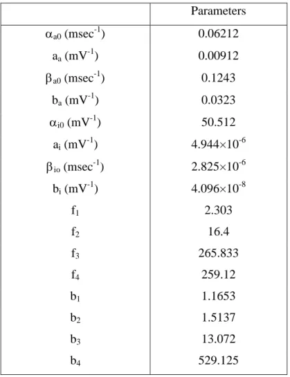
Table 1. Default parameter values modified for the modeling of IK(DR) in differentiated H9c2 cells. Parameters a0(msec-1) 0.06212 aa(mV-1) 0.00912 a0(msec-1) 0.1243 ba(mV-1) 0.0323 i0 (mV-1) 50.512 ai(mV-1) 4.944×10⁻⁶ io(msec-1) 2.825×10⁻⁶ bi(mV-1) 4.096×10⁻⁸ f1 2.303 f2 16.4 f3 265.833 f4 259.12 b1 1.1653 b2 1.5137 b3 13.072 b4 529.125
The forward (a) and reverse (a) rates for the activation are exponential function
of voltage in accordance with Eyring rate theory. Transition rates into (i) and out of
(i) the inactivated states are also expressed as exponential function of voltage. The
forward and reverse rates between non-inactivated and inactivated are assigned the scaling factors (f1-f4and b1-b4).