行政院國家科學委員會專題研究計畫 期中進度報告
眼翼表面之流式細胞儀檢測暨捺壓式細胞學檢查之研究
(1/3)
計畫類別: 個別型計畫 計畫編號: NSC91-2314-B-006-119-執行期間: 91 年 08 月 01 日至 92 年 07 月 31 日 執行單位: 國立成功大學醫學系眼科 計畫主持人: 曾順輝 計畫參與人員: 陳盈廷;曾詩雅 報告類型: 精簡報告 處理方式: 本計畫可公開查詢中
華
民
國 92 年 10 月 2 日
國科會計劃編號: NSC91-2314-B-006-119 計劃名稱: 使用流式細胞儀 檢測暨捺壓式細胞學檢查之研究(1/3) 執行期限: 91 年 8 月 1 日至 92 年 7 月 31 日 主持人: 曾順輝 國立成功大學醫學院 眼科 摘要 關鍵詞: 流式細胞儀,捺壓式細胞學、免疫細胞學檢查, HLA-DR, 眼翼 ABSTRACT
PURPOSE: Human lymphocyte antigen-DR (HLA-DR) antigen is a membrane
receptor that plays a role in the regulation of immune reactions. The expression of HLA-DR antigen was investigated in pterygial epithelial cells as well as conjunctival epithelial cells to explore the inflammatory status in the disease. METHODS: Impression cytology specimens were collected in 18 patients with primary pterygium and in 15 healthy controls: 18 specimens from pterygial surface and 18 specimens from superior bulbar conjunctiva of the same affected eye, as well as 15 specimens from normal conjunctiva. Cells were processed for flow cytometry, by using monoclonal antibodies directed to HLA-DR antigen. Fluorescence intensity levels were quantified and the positive rates of HLA-DR expression were calculated by antibody binding capacity unit of each specimen. RESULTS: HLA-DR antigen expression was found in both normal and pathologic eyes. Quantitation of fluorescence intensity showed a significantly higher expression in pterygia, 32.8% + 19.1%, than in normal controls, 20.1% + 16.5% (P = 0.052). Within the pathologic eyes, HLA-DR expression was still
stronger in pterygia than in superior bulbar conjunctiva, 22.7%+ 15.8% (P = 0.093).
Expression of HLA-DR in the superior bulbar conjunctiva of pathologic eyes was not statistically different from that of the normal conjunctiva. CONCLUSIONS: Flow cytometry with impression cytology showed an upregulation of HLA-DR expression in pterygium, suggesting an inflammatory mechanism in the pathogenesis of pterygium formation.
Key Wor ds: Impression cytology – Immunocytochemistry – Flow cytometry –
Originally believed to be a degenerative disease, another school of thought regarding the pterygium pathogenesis found on the role of inflammation. Our previous study supported by NSC 90-2314-B-006-011 revealed an increased level of Langerhans’ cell expression on pterygial epithelium, which suggested a cell-mediated Type IV hypersentitivity may play a role in the pathophysiology of pterygial formation.
HLA-DR has long been associated with the cell-mediated inflammation immunology. Under normal conditions, Langergans’ cells are known to be the main cell type that has HLA-DR expression and antigen presenting ability on the ocular surface. However, under various inflammatory conditions such as keratoconjunctivitis sicca and Sjögren’s syndrome, conjunctival epithelial cells can also be induced to express HLA-DR, which amplify the inflammation on the ocular surface. Therefore, our previous finding prompted us to investigate the HLA-DR expression on pteryial epithelium to see if inflammation does play an important role in the pathogenesis of pterygium. The method that we employed to quantify HLA-DR on ocular surface was a novel combined technique of impression cytology (IC) and flow cytometry (FCM).
PATIENTS AND METHODS
Patients Selection
A total of 18 eyes (18 patients) with pterygia and 15 eyes (15 normal controls) were recruited in the current study.
Sample Collection and Handling
After the instillation of one drop of topical anesthetic (0.04% oxybuprocaine), two filters 13 x 6.5 mm in size (polyethersulfone filters, 0.20-µm pores, 13-mm in diameter; Supor; Gelman Sciences, MI) were applied to pterygial surface and the superior
conjunctiva of the affected eye with minimal exerting any pressure. Membranes were removed immediately after contact. Approximately 50% to 70% of the total surface of the filter was to be covered by cells. All membranes from each eye were immediately dipped into tubes containing 1.5 ml of cold phosphate-buffered saline (PBS, pH 7.4) with fixative (0.05% paraformaldehyde). Tubes were to be kept at 4°C before impression collection and processed within 1 week. Cells were extracted by gentle agitation for 30 minutes and centrifuged (1600 rpm, 5 mins) for immunocytochemical staining and FCM analysis.
Antibodies and Immunofluor escence Pr ocedur es
Anti-HLA DR antibody and one corresponding negative control were used for assaying. Indirect immunofluorescence procedure for the following label: fluorescein
isothiocyanate (FITC)-conjugated mouse IgG1 anti-HLA DR alpha-chain (clone TAL.1B5, 50 µg/ml; Dako, Copenhagen, Denmark) was used. The FITC-conjugated nonimmune mouse IgG1 was used as a negative isotypic control. Antibodies were used in a 1:50 dilution in 1% bovine serum albumin containing PBS. After 30 minutes of incubation, cell suspensions were washed in PBS by 5-minute centrifugation and reacted with the secondary anti-mouse immunoglobulins in a 1:50 dilution for 30 minutes. At the end of incubations, cells were then centrifuged in PBS (1600 rpm, 5 minutes), resuspended in 100 µml of PBS, and analyzed on a flow cytometer (FACScan; Becton Dickinson, Meylon, France), according to previously validated methods.1
Flow Cytometr y Processing
The linear plot giving granulometry versus cell size consistently revealed a single cell population. Analytic gates were set around this population to exclude cellular debris and aggregates. The number of positive conjunctival cells was then obtained from logarithmic cytograms of mean fluorescence intensities. The superior level of
fluorescence intensity obtained for the isotypic control antibody was considered as the limit of background fluorescence and the threshold of positivity for the tested
antibodies. In each sample, at least 2500 cells were analyzed. The mean fluorescein intensity (MFI) was calculated by standardized arbitrary fluorescence units (AUFs), according to a previously published method2, under the calibration system of DAKO QIFIKIT Set-Up Beads.
RESULTS
Specimen Char acter istics
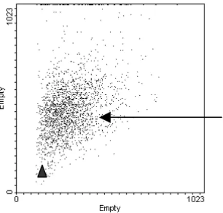
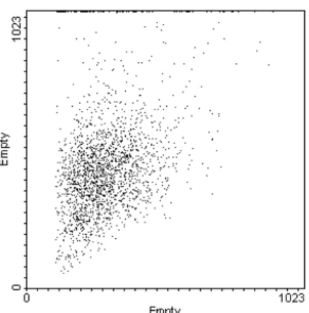
Flow cytometry was performed on 18 IC samples collected from pterygia and 15 IC samples from normal controls. A homogeneous population of conjunctival cells was collected, with few debris or aggregates (Fig.1 and 2). Range of cell numbers collected by IC was 2,500 to 20,000 cells per specimen that reached the level of quality required for reliable analysis.
Flow Cytometr y Results
specimen is summarized in Table 1. A representative cytogram of HLA DR expression in a normal and a pterygium-affected eye is shown given in Fig. 3. HLA-DR expression was found at positive MFI levels above the negative control in all pterygial specimens. HLA-DR–positive cells were 32.8% + 19.1% (mean ± SD) in pterygial specimens. Results of HLA-DR expression from the superior conjunctiva of pterygial patients showed lower percentage of positive cells, 22.7%+ 15.8%, P =
0.093, Mann–Whitney test). As expected, comparison of the pterygium with normal specimens at the mean positive rate of 20.1% + 16.5% showed a marginal significant difference (P = 0.052).
DISCUSSION
The results of the current study provide quantitative information about the degree of HLA-DR expression in pterygium. Together with our previous finding that
pterygium has higher percentage of vimentina-expressing epithelial cells and an increased number of Langerhans’ cells, we presume that type VI hypersensitivity which is a chronic and cell-medicated inflammation may play a vital role in the pathogenesis of pterygium. In order to test our hypothesis, further several important inflammatory marker as well as cell growth regulatory markers including CD95/Fas, the apoptotic marker APO2.7, CD40, and CD40 ligand will be used to investigate pterygium in the part II of NSC project this year with the currently developed flow cytometry model in our laboratory.
Table 1. Positive rates of HLA-DR expression in IC samples Pr imar y pter ygium group
(24 eyes) P (versus control) Pterygium Mean + SD 32.8% + 19.1% 0.052† Superior conjunctiva Mean + SD 22.7%+ 15.8% 0.093 Normal control Mean + SD 20.1% + 16.5% † statistically significant.
Figure 1. Flow cytometry cytogram showing the pterygial cell population obtained by
IC. Cell population appears homogeneous (arrow), with few aggregates (top and right
side), and small size events corresponding to cell debris (arrow head).
Figure 2. Flow cytometry cytogram showing the normal conjuntival cell population
obtained by IC. Similar to pterygium, conjunctival cell population also appears relatively homogeneous, with few aggregates and small size events of cell debris.
Figure 3. Flow cytometric analysis of HLA-DR expressions by pterygial cells.
Cytograms show high expression of HLA. The black histogram is the negative
isotypic control.
REFERENCES
1. Baudouin, C, Brignole, F, Becquet, F, Pisella, PJ, Goguel, A. Flow cytometry in impression cytology specimens: a new method for evaluation of
conjunctival inflammation. Invest Ophthalmol Vis Sci. 1997;8:1458-64
2. Brignole F. Pisella PJ. De Saint Jean M. Goldschild M. Goguel A. Baudouin C. Flow cytometric analysis of inflammatory markers in KCS: 6-month treatment with topical cyclosporin A. Invest Ophthalmol Vis Sci.