Hui-Ming Yu1, 2 Min-Jen Tseng3 Jim-Min Fang4 Suree Phutrakul2 Shui-Tein Chen1
1Institute of Biological Chemistry, Academia Sinica,
Taipei, Taiwan
2Department of Chemistry, Faculty of Science, Chiang Mai University, Chiang Mai, Thailand 3Graduate Institute of Cell
and Molecular Biology, Taipei Medical University, Taipei, Taiwan
4Department of Chemistry, National Taiwan University, Taipei, Taiwan
Capillary electrophoresis using immobilized whole
cells with overexpressed endothelin receptor for
specific ligand screening
A new capillary electrophoresis method using immobilized cells as the stationary phase has been developed. The power of this method is demonstrated by the separa-tion and identificasepara-tion of endothelin antagonists on a capillary column coated by the transfected Chinese hamster ovary (CHO) cells with overexpressing endothelin re-ceptors. The screening results are validated by functional assays suppressing the increase of intracellular calcium concentration induced by endothelin-1. Instead of making efforts in isolating protein receptors, the easily prepared whole-cell capillary column provides a superior tool on the basis of ligand/receptor affinity for a rapid screening of potent drug candidates from compound libraries.
Keywords: Affinity capillary electrophoresis / Endothelin receptor antagonists /
High-through-put screening / Immobilized whole cells / Receptor-ligand interaction
DOI 10.1002/elps.200305804
1 Introduction
Many biological events are triggered by ligand/receptor interactions. For example, endothelin-1 (ET-1), a 21-peptide ligand locally produced in various cell types under different physiological stimuli, has a strong affinity toward endothelin receptor A (ETA) located on the sur-face of an endothelial cell membrane [1–3]. This ligand/ receptor interaction is coupled with G-protein, which triggers a series of biological events to induce an increase of intracellular calcium concentration, [Ca21] i [4]. Antagonism of the endothelin vasoconstrictor is a potential approach to the treatment of a variety of human diseases including hypertension and congestive heart failure [5]. Screening of natural and synthetic com-pounds based on the concept of ligand/receptor re-cognition is an indispensable strategy to search for endothelin receptor antagonists [6–9].
Affinity capillary electrophoresis (ACE) is a powerful sepa-ration method with the advantages of high resolution, small sample requirement, rapid sample throughput, and compatibility to biological conditions [10–14]. The com-ponents in an analyte can be separated by ACE due to their different electrophoresis motilities. By coupling with various sensitive detectors, the ACE technique is widely used to separate bioactive compounds and to determine the biomolecular noncovalent interactions [15–18]. When ACE is applied to screen active ligands in an analyte solution, the target receptor is often immobilized as the stationary phase on the inner wall of a capillary column [10–20]. However, this approach may encounter a prob-lem in isolating of the desired receptors in sufficient quan-tity. Also, many membrane-bound receptors are unstable in isolation, as is the case of ETA. Another problem is that receptors may lose their active conformations upon con-jugation to capillary columns. We therefore investigated the possibility of using whole cells with overexpressing receptors, in lieu of the isolated receptors, as the station-ary phase in ACE for the evaluation of active ligands [21, 22]. In this study, we demonstrate that a whole-cell stationary phase consisting of ETA-overexpressing Chinese hamster ovary (CHO) cells provides a successful ACE protocol for the screening of the ETA-specific ligands. The peptide and non-peptide ETA antagonists (Fig. 1) were satisfactorily resolved on ACE, in accordance to the order of their affin-ity and antagonist potency toward ETA.
Correspondence: Dr. Shui-Tein Chen, Institute of Biological
Chemistry, Academia Sinica, Taipei, Taiwan
E-mail: [email protected] Fax: 1886-2-27883473
Abbreviations: ACE, affinity capillary electrophoresis; CHO cells,
Chinese hamster ovary cells; ETA, endothelin receptor A; ET-1,
endothelin 1; ET-1(16–21), endothelin 1 fragment containing 16–21
residues; FBS, fetal bovine serum
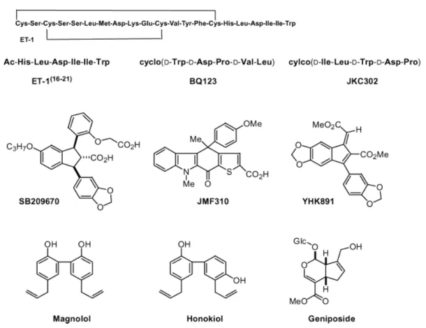
Figure 1. ET-1 and the examined substrates in this study.
2 Materials and methods
2.1 Construction of CHO-K1 cell line
over-expressing ETAand ligand-binding assay
The lipofectin-mediated transfection method described by Tseng et al. [23] was used to construct stable CHO cell lines overexpressing ETA. Cells were grown to 30– 40% confluence in 60 mm dishes and transfected with 1 mg of pcDNA-3 expressing plasmid harboring ETAusing lipofectin reagent for 6–8 h in serum-free medium. Cells were then returned to 5% fetal bovine serum (FBS), cul-tured 36 h, then replated at reduced density in 150 mm plates in the presence of 0.75 mg/mL (active) G418. G-418 resistant colonies were selected and screened for ETA by binding of (3-[125I]iodotyrosyl)endothelin-1 ([125I]ET-1). Binding was conducted to cells plated in 24-well dishes at 2–36105cells/mL the day before the binding assay. For cell-binding assays, [125I]ET-1 (10 pM) was added to HR buffer (5 mM NaCl, 4.7 mM KCl, 1 mM Na2HPO4, 1.28 mMCaCl2, 10 mMHEPES, pH 7.4, with 0.5% bovine serum albumin, and 0.1 mg/mL soybean trypsin inhibitor). Cells were incubated to equilibrium (2 h at 377C), then
washed twice with ice-cold phosphate-buffered saline. The cells were then solubilized with 1 mL of 0.1NNaOH and radioactivity quantified in a g-counter. Nonspecific binding was determined in the presence of 100 nMET-1.
2.2 Preparation of a cell-immobilized capillary column
A fused-silica capillary column (60 cm effective length6 200 mm inner diameter, ,1.88 mL whole volume) was acti-vated by washing successively with MeOH (ca. 20 mL), 1N HCl (ca. 20 mL), deionized water (ca. 20 mL), 1NNaOH (ca. 20 mL), and deionized water (ca. 20 mL). The column was stored in the presence of 1 mM PBS buffer (containing 31.7 mg of NaH2PO4and 206 mg of Na2HPO4at pH 7.3 per liter). The transfected CHO cells harboring ETA (,2.56105 cells/mL) recovered from the culture media were fixed by treatment with formaldehyde (3.7% in water, 5 mL) for 30 min to furnish the desired cross-linkage. The fixed cells were stored at 47C in PBS (1 mM, pH 7.3). For loading of the fixed cells onto the capillary column, the col-umn was washed with EtOH (95%, ca. 40 mL), purged with air in order to dry it, charged with poly-L-lysine (15 000–
CE
and
1036 H.-M. Yu et al. Electrophoresis 2004, 25, 1034–1041
30 000 molecular weight, 0.5 mg/mL in water) for 5 min, and incubated for 30 min [24, 25]. The column was then charged again with poly-L-lysine for 5 min and incubated for 2 h. The column was then dried by airflow for 2 h, the fixed cells in the PBS buffer were purged into the poly-L-lysine-coated column. After 5 min of incubation time, another batch of fixed cells was purged into the column for 30 min of incubation. Then 1% FBS in PBS (ca. 20 mL) was purged to cap the exposed area of poly-L-lysine. The column immobilized with the transfected CHO cells was finally washed with PBS (ca. 40 mL), and stored at room temperature (,25–277C). No apparent degradation was observed after 7 days. Capillary electrophoresis experi-ments were performed on an P/ACE system (Beckman Instruments, Fullerton, CA, USA) at a constant voltage of 10 kV. The sample (,1.5 mL of ,1026to 1027
Msolution in 1 mMPBS, pH 7.0) was introduced into the capillary col-umn by pressure injection at 0.5 psi/3 s. The background electrolyte was PBS (1 mM, pH 7.0). Electrophoresis was monitored by an absorbance detector held at 214 nm. The low concentration of PBS (1 mM) ensured no interference with 214 nm absorbance. The temperature of the capil-lary column was maintained at 257C.
2.3 Samples and reagents
ET-1, the ET-1 fragment ET-1(16–21)containing 16–21 resi-dues [26, 27], and the cyclic peptide antagonists BQ123 [28] and JKC302 [29, 30] were synthesized by using an ABI 433A peptide synthesizer (ABI, Foster City, CA, USA). We also prepared the samples of non-peptide endothelin receptor antagonists SB209670 [31, 32], JMF310 [33], and YHK891 [34]. The active herbal com-ponents, Magnolol, Geniposide and Honokiol, were pur-chased from Wako Pure Chemical Industries (Japan). The 125I-labeled ET-1 ((3-[125I]iodotyrosyl)endothelin-1, 81.4 TBq/mmol) was purchased from NEN Life Science Products (Wilmington, DE, USA). The fluorescent reagent, fura-2 penta(acetoxymethyl) ester, was from Calbiochem-Novabiochem (La Jolla, CA, USA), poly-L-lysine hydro-bromide and G418 antibiotic were from Sigma Chemical (St. Louis, MO, USA). All other chemicals were of analyti-cal grade. Distilled water was used in all experiments. All buffers were filtered through 0.45 mm filters before use.
3 Results and discussion
3.1 Identification of transfected CHO cells
The cultured CHO cells were subjected to transfection with the ETA-expression plasmid DNA using a lipofectin reagent. The efficacy of ETA expression was
demon-strated by the competitive binding assay with a synthetic sample of ET-1 and the radiolabeled [125I]-ET-1 (Fig. 2). The binding affinity of the transfected cell line by the agonist ET-1 was established to have a dissociation con-stant of Kd = 1.52 nM. The receptor density (Bmax) of 6.36105sites/cell was estimated from a Scatchard plot [38, 39]. This result indicated that ETAreceptors were suc-cessfully overexpressed in the CHO cells. The CHO cells harboring ETAwere fixed by treatment with formaldehyde. A fused-silica capillary column (200 mm inner diameter) was first charged with poly-L-lysine, and then purged with the cross-linked CHO cells. After capping the ex-posed portion of poly-L-lysine with FBS, the cell-immobi-lized capillary column was furnished and ready for ACE. It was estimated that about 1–3 cells were attached to the capillary cross-section. Among several possible can-didates of base material, poly-L-lysine turned out to ex-hibit a superb adhesive property for the immobilization of cells on the capillary column. The whole-cell immobilized capillary column could be easily prepared, and no ob-vious decomposition was found on storage with PBS buffer at room temperature for seven days.
Figure 2. [125I]-Endothelin-1 binding assay was per-formed with a synthetic endothelin-1 (ET-1). The binding affinity of transfected CHO cell-line was established with
Kd= 1.52 nM, and Bmaxwas estimated at 6.36105sites/ cell.
3.2 Validation of peptide ligands
In order to validate our ACE method, we first analyzed three known peptide ligands of the ETAreceptor: an ET-1
C-terminal fragment ET-1(16–21)containing six amino acid residues [26, 27] and two cyclic pentapeptides, BQ123 [28] and JKC302 [29, 30]. The behavior of these ligands on three different capillary columns was examined, i.e., an uncoated column, a column coated with poly-L-lysine,
Figure 3. ACE of peptides JKC 302, BQ123, and ET-1(16–21) on a column (a) uncoated; (b) coated with poly-L-lysine; (c) coated with fixed ETA-overexpressing CHO cells. Background electrolyte, 1 mMPBS; absorbance detector at 214 nm. Au, arbitrary unit.
and a column coated with fixed ETA-overexpressing CHO cells. The first set of electropherograms indicated that these three compounds were poorly resolved on an un-coated column, as one would expect. The eluting order of three peptides on the poly-L-lysine-coated column dif-fers from that on the cell-coated column, presumably due to the random electrostatic interactions exerted by poly-L-lysine on the peptide analytes. Complete separation of the mixture of ET-1(16–21), BQ123, and JKC302 was re-alized on a capillary column with the stationary phase of immobilized ETA-overexpressing CHO cells (Fig. 3). The cyclopeptide JKC302 with the longest retention time on the cell-coated column should exhibit the highest affinity toward ETA, whereas the hexapeptide ET-1(16–21)with the shortest retention time should have the least affinity. The speculation of relative affinity JKC302 . BQ123 . ET-1(16–21), as deduced from the ACE experiment, was further supported by the functional assay of their antago-nistic potency against ET-1. It is well known that the bind-ing of ET-1 with ETAwill trigger an increase of intracellular calcium concentration [4]. A control experiment (Fig. 4a) was performed by treatment of ET-1 in 1027Mto the ET
A overexpressing CHO cells, which were first incubated with a calcium-chelating agent fura-2 applied as its pen-ta(acetoxymethyl) ester [39, 40]. The [Ca21]
ichange was monitored by a ratiometric method using dual excitations at 340 and 380 nm wavelengths, and the fluorescence
Figure 4. Fluorescence measurements of intracellular calcium concentration ([Ca21]
i) by addition of tested samples (as shown by the arrow) to the ETA-overexpressing CHO cells in the presence of fura-2. The induced [Ca21]
ichange is taken as a measure of antagonist potency against ET-1: (a) control experiment with addition of ET-1 (1027M), (b) treatment with a mixture of JKC302 (1026M) and ET-1 (1027M), (c) treatment with a mixture of BQ123 (1026M) and ET-1 (1027M), and (d) treatment with a mixture of ET-1(16–21)(1026M) and ET-1 (1027M).
1038 H.-M. Yu et al. Electrophoresis 2004, 25, 1034–1041
emission at 505 nm was measured. The treatment with ET-1 induced a great degree of [Ca21]
i(Fig. 4a). When a mixture of JKC302 (1026M) and ET-1 (1027M) was used in such a functional assay, the ET-1 induced [Ca21]
ichange was entirely suppressed (Fig. 4b). The degree of inhibition against the ET-1 induced [Ca21]
ican serve as a measure of the potency of an antagonist. By comparison of the transient [Ca21]
iassays (Figs. 4b–d), the relative potency JKC302 . BQ123 . ET-1(16–21) in ET
A antagonism is in good agreement with that derived by the eluting order on the capillary column coated with whole cells.
3.3 Validation of nonpeptide ligands
Our present whole-cell coating ACE method is not limited to peptide antagonists; it is also applicable for screening nonpeptide antagonist molecules. For example, a 1,3-di-arylindane-2-carboxylic acid SB209670 is a potent ETA antagonist against ET-1 [31]. The molecular computations together with bioassay of a series of derivatives indicated that SB209670 bears a carboxyl group at the 2-position to mimic the carboxylic terminal of ET-1, and two aryl groups at 4- and 9-positions to mimic the aromatic residues of
Tyr-13 and Phe-14. On the basis of this structural proto-col, a carbazolothiophene-2-carboxylic acid JMF310 [33] was designed as a possible ETAantagonist, and an inde-necarboxylate ester YHK891 [34] was also examined for comparison. Indeed, SB209670 that strongly inhibited the ET-1 induced [Ca21]
i (Fig. 6b) also showed a very long retention time on the whole-cell immobilized capillary col-umn (Fig. 5), as a consequence of its high affinity toward
Figure 5. ACE of a mixture of SB209670, JMF310, and YHK891 on a capillary column coated with fixed ETA -overexpressing CHO cells. Background electrolyte, 1 mM PBS; absorbance detector at 214 nm. A.U., arbitrary unit.
Figure 6. Fluorescence measurements of intracellular calcium concentration ([Ca21]
i) by addition of tested samples (as shown by the arrow) to the ETA-overexpressing CHO cells in the presence of fura-2. The induced [Ca21]
ichange is taken as a measure of antagonist potency against ET-1: (a) control experiment with addition of ET-1 (1027 M), (b) treatment with a mixture of SB209670 (1026M) and ET-1 (1027M), (c) treatment with a mixture of JMF310 (1026M) and ET-1 (1027M), and (d) treatment with a mixture of YHK891 (1026M) and ET-1 (1027M).
the surface endothelin receptors of the transfected CHO cells. The modest antagonist potency of JMF310 (Fig. 6c) was also reflected in the ACE elution profile. On the con-trary, the YHK891 sample having only a marginal inhibi-tory effect against ET-1 (Fig. 6d) was rapidly eluted out from the whole-cell coating column.
3.4 Validation of active herbal components
In addition to the rationally designed molecules, the random screening of active components can also be achieved by the whole-cell ACE method. We have selected several active herbal components that are known to possess bioactivities related to vascular dilation or signal transduction. By ACE we found that an anti-platelet agent, Magnolol [42–44], might function as a new lead compound against ET-1 in ETA receptor binding. Magnolol is the 2,2’-dimer of 4-allylphenol. As shown in the profiles of ACE analysis (Fig. 7) and [Ca21]
i assay (Fig. 8), Magnolol is a stronger ETA antagonist than its structural isomer, Honokiol [45]. Geniposide [46], an iridoid glucoside that exhibits neuritogenic effect on
PC12h cells and enhanced responses of cells to carba-col in terms of cytoplasmic free-calcium concentration, turned out to have a temperate binding affinity and mod-est antagonistic activity against ET-1.
Figure 7. Screening of the Chinese herbal active compo-nents Magnolol, Honokiol, and Geniposide by ACE on a capillary column coated with fixed ETA-overexpressing CHO cells. Background electrolyte, 1 mMPBS; absorb-ance detector at 214 nm. A.U., arbitrary unit.
Figure 8. Fluorescence measurements of intracellular calcium concentration ([Ca21]
i) by addition of tested samples (as shown by the arrow) to the ETA-overexpressing CHO cells in the presence of fura-2. The induced [Ca21]
ichange is taken as a measure of antagonist potency against ET-1: (a) control experiment with addition of ET-1 (1027M), (b) treatment with a mixture of Magnolol (1026M) and ET-1 (1027M), (c) treatment with a mixture of Geniposide (1026M) and ET-1 (1027M), and (d) treat-ment with a mixture of Honokiol (1026M) and ET-1 (1027M).
1040 H.-M. Yu et al. Electrophoresis 2004, 25, 1034–1041
4 Concluding remarks
There are several distinct advantageous features for using whole cells as the stationary phase of ACE (i) isolation of (unstable) receptors is not needed, (ii) the stability of whole cells is improved by immobilization, and (iii) whole cells are easily coated on a fused-silica capillary column
via the guidance of poly-L-lysine template. We have demonstrated this concept of whole-cell immobilized ACE by a protocol using the capillary column coated with the CHO cells harboring overexpressing endothelin receptor A. The capillary column prepared as such exhib-its excellent affinity for separation and identification of endothelin receptor A antagonists (Fig. 9, Table 1). There is a good correlation between the relative inhibition of the
Figure 9. Correlation between the retention time of the examined compound of the capillary column coated with ETA-overexpressing CHO cells and the relative inhibition of the ET-1 induced increase of intracellular calcium ion concentration. In each line, the stronger affinity of a com-pound toward ETA shows a longer retention time and more potent inhibitory effect. (j) From top to bottom are JKC302, BQ123, and ET-1(16–21); (r) from top to bottom are SB209670, JMF310, and YHK891; (m) from top to bot-tom are Magnolol, Geniposide, and Honokiol.
Table 1. Relative inhibition of the ET-1 induced [Ca21] i and retention time of the examined compound on the capillary column coated with ETA -over-expressing CHO cells
Compound Relative inhibition (%) Retention time (min) JKC302 86 69.7 BQ123 61 45.0 ET-1(16–21) 48 24.2 SB209670 91 87.8 JMF310 67 69.7 YHK891 27 10.0 Magnolol 56 61.4 Geniposide 42 34.7 Honokiol 32 24.1
ET-1 induced [Ca21]
i and the retention time of the ex-amined compound on the capillary column coated with ETA-overexpressing CHO cells. This ACE method only requires a small quantity of sample, and offers a reliable assessment of a library of compounds in a relatively short period. The ACE with immobilized whole cells could be developed as a high-throughput screening method based on specific receptor/ligand interactions.
The authors like to thank Prof. Yueh-Hsiung Kuo and Yeun-Min Tsai (Department of Chemistry, National Taiwan University) for kindly providing us the samples of YHK891 and SB209670 and Prof. Sheau-Huei Chueh (Department of Biochemistry, National Defense University) for direct-ing us the [Ca21]
i assay. We also thank the National
Science Council and Taipei Medical University (TMC 87-Y05-A128 to M.-J. T.) for financial support. Support for the research (S.-T. Chen) provided by the Main Subject Projects of Academia Sinica, Taiwan and the National Re-search Program for Genomic Medicine, National Science Council, Taiwan, (NSC 91-3112-13-001-002) are greatly appreciated.
Received September 28, 2003
5 References
[1] Yanagisawa, M., Kurihara, H., Kimura, S., Tomobe, Y., Kobayashi, M., Mitsui, Y., Yazaki, Y., Goto, K., Masaki, T., Nature 1988, 332, 311–315.
[2] Doherty, A. M., J. Med. Chem. 1992, 35, 1493–1508. [3] Schiffrin, E. L., Touyz, R. M., J. Cardiovasc. Pharmacol.
1998, 32, Suppl. 3, 2–13.
[4] Sakurai, T., Yanagisawa, M., Masaki, T., Trends Pharmacol. Sci. 1992, 13, 103–108.
[5] Lerman, A., J. Cardiovasc. Pharmacol. 2001, 38, Suppl. 2, 27–30.
[6] Elliott, J. D., Lago, M. A., Peishoff, C. E., in: Ruffolo, R. R. (Ed.), Endothelin Receptors: From the Gene to the Human, CRC Press, Boca Raton, FL 1995, pp. 79–107.
[7] Doherty, A. M., Drug Discovery Today 1996, 1, 60–70. [8] Webb, M. L., Meek, T. D., Med. Res. Rev. 1997, 17, 17–67. [9] Liu, G., Annu. Rev. Med. Chem. 2000, 35, 73–82.
[10] Fishman, H. A., Orwar, O., Scheller, R. H., Zare, R. N., Proc. Natl. Acad. Sci. USA 1995, 92, 7877–7881.
[11] Chu, Y. H., Dunayevskiy, Y. M., Kirby, D. P., Vouros, P., Kar-ger, B. L., J. Am. Chem. Soc. 1996, 118, 7827–7835. [12] Landers, J. P. (Ed.), Handbook of Capillary Electrophoresis,
2nd ed., CRC Press, Boca Raton, FL 1997, pp. 591–609. [13] Chu, Y. H., Cheng, C. C., Cell Mol. Life Sci. 1998, 54, 663–
683.
[14] Yeung, E. S., J. Chromatogr. A 1999, 830, 243–262. [15] Colton, I. J., Carbeck, J. D., Rao, J., Whitesides, G. M.,
Electrophoresis 1998, 19, 367–382.
[16] Chu, Y.-H., Zang, X., Tu, J., J. Chin. Chem. Soc. 1998, 45, 713–720.
[17] Mito, E., Zhang, Y., Esquivel, S., Gomez, F. A., Anal. Bio-chem. 2000, 280, 209–215.
[18] Kaddis, J., Mito, E., Heintz, J., Plazas, A., Gomez, F. A., Electrophoresis 2003, 24, 1105–1110.
[19] Kaufman, S. E., Brown, S., Stauber, G. B., Anal. Biochem. 1993, 211, 261–266.
[20] Oda, Y., Owa, T., Sato, T., Boucher, B., Daniels, S., Yama-naka, H., Shinohara, Y., Yokoi, A., Kuromitsu, J., Nagasu, T., Anal. Chem. 2003, 75, 2159–2165.
[21] Fishman, H. A., Orwar, O., Allbritton, N. L., Modi, B. P., Shear, J. B., Scheller, R. H., Zare, R. N., Anal. Chem. 1996, 68, 1181–1186.
[22] Miller, K. J., Lytle, F. E., Anal. Chem. 1994, 66, 2420–2423. [23] Tseng, M.-J., Detjen, K., Struk, V., Logsdon, C. D., J. Biol.
Chem. 1995, 270, 18858–18864.
[24] Leif, R. C., Ingram, D., Clay, C., Bobbitt, D., Gaddis, R., Leif, S. B., Nordqvist, S., J. Histochem. Cytochem. 1977, 25, 538–543.
[25] Vieklind, U., Swierenga, S. H., Histochemistry 1989, 91, 81–88.
[26] Kimura, S., Kasuya, Y., Sawamura, T., Shinmi, O., Sugita, Y., Yanagisawa, M., Goto, K., Masaki, T., Biochem. Biophys. Res. Commun. 1988, 156, 1182–1186.
[27] Hashido, K., Gamou, T., Adachi, M., Tabuchi, H., Watanabe, T., Furuichi, Y., Miyamoto, C., Biochem. Biophys. Res. Com-mun. 1992, 187, 1241–1248.
[28] Ishikawa, K., Fukami, T., Nagase, T., Fujita, K., Hayama, T., Niyama, K., Mase, T., Ihara, M., Yano, M., J. Med. Chem. 1992, 35, 2139–2144.
[29] Miyata, S., Hashimoto, M., Fujie, K., Nishikawa, M., Kiyoto, S., Okuhara, M., Kohsaka, M., J. Antibiot. 1992, 45, 83–87. [30] Miyata, S., Fukami, N., Neya, M., Takase, S., Kiyoto, S.,
J. Antibiot. 1992, 45, 788–791.
[31] Elliott, J. D., Lago, M. A., Cousins, R. D., Gao, A., Leber, J. D., Erhard, K. F., Nambi, P., Elshourbagy, N. A., Kumar, C., Lee, J. A., Bean, J. W., DeBrosse, C. W., Eggleston, D. S., Brooks, D. P., Feuerstein, G., Ruffolo, R. R., Wein-stock, J., Gleason, J. G., Peishoff, C. E., Ohlstein, E. H., J. Med. Chem. 1994, 37, 1553–1557.
[32] Clark, W. M., Tickner-Eldridge, A. M., Huang, G. K., Pridgen, L. N., Olsen, M. A., Mills, R. J., Lantos, I., Baine, N. H., J. Am. Chem. Soc. 1998, 120, 4550–4551.
[33] Babu, G., Yu, H.-M., Yang, S.-M., Fang, J.-M., Bioorg. Med. Chem. Lett. 2004, 14, 1129–1132.
[34] Kuo, Y.-H., Wu, C.-H., Wu, R.-E., Lin, S.-T., Chem. Pharm. Bull. 1995, 43, 1267–1271.
[35] Arai, H., Hori, H., Aramori, I., Ohkubo, H., Nakanishi, S., Nature 1990, 348, 730–732.
[36] Sakurai, T., Yanagisawa, M., Takuwa, Y., Miyazaki, H., Kimura, S., Goto, K., Masaki, T., Nature 1990, 348, 732– 735.
[37] Sakamoto, A., Yanagisawa, M., Tsujimoto, G., Nakao, K., Toyooka, T., Masaki, T., Biochem. Biophys. Res. Commun. 1994, 200, 679–686.
[38] Fischli, W., Clozel, M., Guilly, C., Life Sci. 1989, 44, 1429– 1436.
[39] Waeber, C., Hoyer, D., Palacios J. M., Eur. J. Pharmacol. 1991, 176, 233–236.
[40] Grynkiewicz, G., Poenie, M., Tsien, R. Y., J. Biol. Chem. 1985, 260, 3440–3450.
[41] Lin, W.-W., Chuang, D. M., Mol. Pharmacol. 1993, 44, 158– 165.
[42] Teng, C. M., Chen, C. C., Ko, F. N., Lee, L. G., Huang, T. F., Chen, Y. P., Hsu, H. Y., Thrombosis Res. 1988, 50, 757–765. [43] Hamasaki, Y., Kobayashi, I., Zaitu, M., Tsuji, K., Kita, M., Hayasaki, R., Muro, E., Yamamoto, S., Matsuo, M., Ichi-maru, T., Miyazaki, S., Planta Med. 1999, 65, 222–226. [44] Chen, Y.-H., Lin, S.-J., Chen, J.-W., Ku, H.-H., Chen, Y.-L.,
Brit. J. Pharmacol. 2002, 135, 37–47.
[45] Park, E. J., Zhao, Y. Z., Na, M. K., Bae, K. H., Kim, Y. H., Lee, B. H., Sohn, D. H., Planta Med. 2003, 69, 33–37.
[46] Yamazaki, M., Chiba, K., Mohri, T., Biol. Pharm. Bull. 1996, 19, 791–795.
![Figure 2. [ 125 I]-Endothelin-1 binding assay was per- per-formed with a synthetic endothelin-1 (ET-1)](https://thumb-ap.123doks.com/thumbv2/9libinfo/8674772.196476/3.892.465.808.528.797/figure-endothelin-binding-assay-formed-synthetic-endothelin-et.webp)

![Figure 6. Fluorescence measurements of intracellular calcium concentration ([Ca 21 ] i ) by addition of tested samples (as shown by the arrow) to the ET A -overexpressing CHO cells in the presence of fura-2](https://thumb-ap.123doks.com/thumbv2/9libinfo/8674772.196476/5.892.86.688.570.976/figure-fluorescence-measurements-intracellular-concentration-addition-overexpressing-presence.webp)
![Figure 8. Fluorescence measurements of intracellular calcium concentration ([Ca 21 ] i ) by addition of tested samples (as shown by the arrow) to the ET A -overexpressing CHO cells in the presence of fura-2](https://thumb-ap.123doks.com/thumbv2/9libinfo/8674772.196476/6.892.84.683.571.977/figure-fluorescence-measurements-intracellular-concentration-addition-overexpressing-presence.webp)
![Table 1. Relative inhibition of the ET-1 induced [Ca 21 ] i and retention time of the examined compound on the capillary column coated with ET A -over-expressing CHO cells](https://thumb-ap.123doks.com/thumbv2/9libinfo/8674772.196476/7.892.84.424.879.1079/relative-inhibition-induced-retention-examined-compound-capillary-expressing.webp)
