Stimulated emission and lasing of random-growth oriented ZnO nanowires
Hsu-Cheng Hsu, Chun-Yi Wu, and Wen-Feng Hsieh
Citation: Journal of Applied Physics 97, 064315 (2005); doi: 10.1063/1.1862312
View online: http://dx.doi.org/10.1063/1.1862312
View Table of Contents: http://scitation.aip.org/content/aip/journal/jap/97/6?ver=pdfcov
Published by the AIP Publishing
Articles you may be interested in
Polariton lasing in a hybrid bulk ZnO microcavity
Appl. Phys. Lett. 99, 161104 (2011); 10.1063/1.3650268
Exciton related stimulated emission in ZnO polycrystalline thin film deposited by filtered cathodic vacuum arc technique
Appl. Phys. Lett. 88, 191112 (2006); 10.1063/1.2202728
Optical properties of highly faceted ZnO rods
J. Appl. Phys. 99, 033517 (2006); 10.1063/1.2170417
Stimulated emission and optical gain in ZnO epilayers grown by plasma-assisted molecular-beam epitaxy with buffers
Appl. Phys. Lett. 78, 1469 (2001); 10.1063/1.1355665
Room-temperature stimulated emission of excitons in ZnO/(Mg,Zn)O superlattices
Appl. Phys. Lett. 77, 2204 (2000); 10.1063/1.1315340
Stimulated emission and lasing of random-growth oriented ZnO nanowires
Hsu-Cheng Hsu, Chun-Yi Wu, and Wen-Feng Hsieha兲
Department of Photonics and Institute of Electro-Optical Engineering, National Chiao Tung University, 1001 Tahsueh Road, Hsinchu 30050, Taiwan
共Received 5 October 2004; accepted 4 January 2005; published online 14 March 2005兲
We report room-temperature ultraviolet stimulated emission and lasing from optically pumped high-quality ZnO nanowires. Emission due to the exciton-exciton scattering process shows apparent stimulated-emission behavior. Several sharp peaks associated with random laser action are seen under high pumping intensity. The mechanism of laser emission is attributed to coherent multiple scattering among the random-growth oriented nanowires. The characteristic cavity length is determined by the Fourier transform of the lasing spectrum. © 2005 American Institute of Physics.
关DOI: 10.1063/1.1862312兴 I. INTRODUCTION
One-dimensional semiconductors have become the im-portant fundamental building blocks because of their funda-mental physical properties and their potential applications for nanoelectronic and nanophotonic devices.1–3ZnO nanowires are especially interesting because they exhibit large exciton binding energy共60 meV兲, wide band gap 共3.37 eV兲, and low threshold ultraviolet lasing.4 Recent progress in lasing or stimulated emission has been achieved from a variety of low-dimensional ZnO structures such as microcavity,5 nanowires,4,6–9nanorods,10–12and nanoribbons.13For a large diameter such as ZnO microcavity,5 the lasing mechanism results from whispering gallery modes共WGMs兲. The light is trapped proximity to the perimeter of the nanowire by the total internal reflections 共TIR兲. When the diameter of the ZnO nanowire is smaller than the optical wavelength, the WGMs will experience high scattering loss as a result of diffraction. Fabry–Perot resonator has been used to explain the lasing mode in the individual single nanostructure4,13–15 and long whiskers16,17in which two well-aligned end facets of the nanowire serve as the Fabry–Perot cavity.
However, in a disorder system of randomly oriented nanowires, coherent photon may exist during multiple scat-tering among these disordered nanowires. Unlike the con-ventional laser, the random laser emission in disorder media of polycrystalline ZnO films and powders is a result of the coherent photon scattering that did not contain any conven-tional Fabry-Perot cavities.18–20 The key factor to random lasing is the existence of a high-gain medium and efficient light scattering in the samples to provide the necessary co-herent feedback. Due to the high-gain characteristic of the ZnO nanowires,4random lasing also acts in high-density ver-tically aligned ZnO nanorod arrays.21The different resonant cavities formed by multiple scattering could have different lasing directions. In this article, we demonstrate the stimu-lated emission and lasing of randomly oriented disordered ZnO nanowires and discuss their relevant mechanisms. By
Fourier transforming the lasing spectrum, we obtained a characteristic loop length of random lasing cavity.
II. EXPERIMENTAL PROCEDURES
ZnO nanowires were synthesized in a simple vapor transport method mediated by vapor-liquid-solid 共VLS兲 growth. More detail of this growth process has been pub-lished elsewhere.22For continuous-wave共cw兲 photolumines-cence 共PL兲 measurement, we used a He–Cd laser 共325 nm兲 as the excitation source; for pulsed pumping, we used the third harmonic of Nd: YVO4 共yttrium orthovanadate兲 laser
共3.51-eV photon energy兲 with a pulse width of ⬃500 ps and
repetition rate of 1 kHz. The excitation laser beam was di-rected normally and focused onto the sample surface with power being varied with an optical attenuator. The spot size on the sample is about 100m. Spontaneous and stimulated emissions were collected by a fiber bundle and coupled into a 0.32-cm focal-length monochromator with a 1200 lines/ mm grating, then detected by either an electri-cally cooled charge-coupled device 共CCD兲 or a photomulti-plier tube共PMT兲 detector. All of the experiments were per-formed at room temperature.
III. RESULTS AND DISCUSSION
The electron microscopy image provides the general morphology of the as-grown products. As shown in Fig. 1共a兲, the typical scanning electron microscopy共SEM兲 micrograph clearly reveals that there are random-growth oriented ZnO nanowires on the substrate with their lengths normally ex-ceeding 3m and diameters ranging between 60 and 200 nm. Figure 1共b兲 shows the transmission electron micros-copy共TEM兲 image of a single nanowire of 120 nm in diam-eter. The selected-area electron diffraction共SAED兲 pattern of an individual nanowire shown in Fig. 1共c兲 reveals a complete diffraction pattern. It indicates that the ZnO nanowire is single crystalline and can be indexed to the hexagonal ZnO
共110兲 diffraction with growth direction of 关002兴.
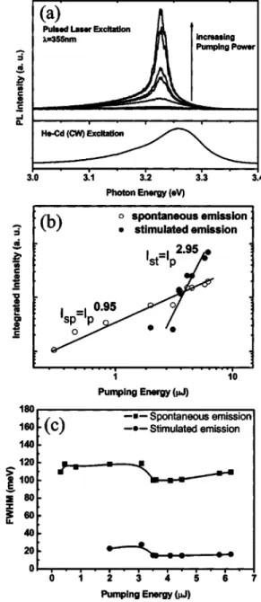
Figure 2共a兲 shows the typical emission spectra of the ZnO nanowires measured at various pumping intensities. As a reference, the cw-PL is also shown at the bottom of Fig. 2共a兲. At low pumping intensity, only a spontaneous emission
a兲Author to whom the correspondence should be addressed, FAX:
⫹886-3-5716631; electronic mail: [email protected]
0021-8979/2005/97共6兲/064315/4/$22.50 97, 064315-1 © 2005 American Institute of Physics
peak at 3.26 eV with a full width at half maximum共FWHM兲 of 110 meV is attributed to the exciton emission. With in-creasing the pumping intensity, a narrow emission appears on the low-energy shoulder of the exciton band. By decom-posing the emission spectrum into a broad spontaneous emis-sion and a sharper peak, we observed that the integrated intensity of the broad spontaneous emission increases almost linearly with pumping intensity 共Isp= IP
0.95兲, illustrated as
close circles in Fig. 2共b兲; however, the integrated intensity of the narrower peak at 3.22 eV experiences a strong superlin-ear dependence 共IST= IP
2.95兲 of the excitation intensity 共open
circles兲. As shown in Fig. 2共c兲, the FWHM of the sharper peak is only 20 meV. These results indicate a clear evidence of stimulated emission. The stimulated emission process was interpreted in terms of inelastic exciton-exciton scattering in which one exciton is scattered into a photonlike polariton state giving rise to luminescence, while the other exciton is scattered into an excited state with a larger quantum number or a totally dissociated state.23
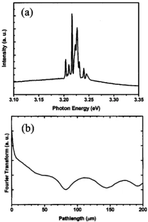
When the pumping intensity exceeds a certain threshold at a specific position of the sample, shown in Fig. 3共a兲, we found that several sharp peaks emerge from 3.20 to 3.25 eV with FWHM⬃2 meV. Note that the redshift of the PL peak with increasing excitation intensity is caused by the band-gap renormalization.
There are two possible optical resonance modes in ZnO nanowires for laserlike action. The first resonance mode is the Fabry–Perot mode of the natural optical cavity formed by the two-end facets of the well-organized nanowires which is a well-accepted reason for explaining the lasing behavior in a single ZnO nanowire4and GaN nanowire.15Another possible lasing mechanism results from light amplification due to photon coherent scattering in the random media.18–20 Com-pared with the previous reports of lasing from ZnO nanowires,4 our samples show an unobvious feature of Fabry–Perot modes. The longitudinal modes spacing can be determined by the equation,
⌬ = 2/共2nL兲. 共1兲
Here L is the laser cavity length, n is the refractive index
共2.45兲, and is the resonant wavelength 共⬃390 nm兲. For a
ZnO nanowire with length of about 3m, the mode spacing between the closest longitudinal modes is expected to be 10 nm. It should exist only a single Fabry–Perot mode in the whole measured PL range 共3.0–3.4 eV兲 and may become a broad lasing spectrum due to averaging out the emission spectra of the single Fabry–Perot modes having different lengths. However, we did observe several sharp lasing modes under high pumping intensity. Therefore, we further consid-ered the other possible lasing action from coherent photon scattering in the random medium.
To confirm the origin of the laserlike emission peaks, we studied the influence of the excitation area on the number of modes in the random lasing. Figure 4 plots the emission
FIG. 1. 共a兲 SEM micrograph of the ZnO nanowires. 共b兲 TEM image of an individual nanowire. 共c兲 SAED pattern showing a single crystalline structure.
FIG. 2. 共a兲 Excitation power dependence of the emission spectra of ZnO nanowires.共b兲 Plot of integrated intensity of the stimulated emission of ZnO nanowires vs the pumping intensity.共c兲 The FWHM of the emission peaks vs pumping intensity.
064315-2 Hsu, Wu, and Hsieh J. Appl. Phys. 97, 064315共2005兲
spectra measured at different s slightly above the lasing threshold. Under the smallest excitation area, 7.9
⫻10−5cm2 no narrow emission peak was observed. With
increasing the excitation area, several sharp laserlike emis-sion peaks appear. In addition, the number of sharp laserlike peaks tends to increase with an increase of the excitation area. According to the previous report on random lasing,18 the laser oscillation would not have occurred if the closed-loop paths were too short to provide enough amplification. This result clearly indicates that the sharp emissions result from the random lasing action. The scattering probability of coherent photon in random-oriented ZnO nanowires is higher than that in well-aligned ZnO nanowires. We found no dis-tinct sharp peaks from the well-aligned ZnO nanowires grown on a quartz glass24with optical pumping. Hence,
ran-dom laser only acted in high-density vertically aligned ZnO nanowires21or disorder-oriented ZnO nanowires.
To further determine the cavity length of the closed-loop random lasing, a simple and powerful technique which is Fourier transform 共FT兲 of the lasing spectrum is used.25,26 The unit of the FT result is in length provided that the unit of the lasing spectrum is in wave number 共1/length兲. For a tra-ditional Febry–Perot laser cavity, the separation of harmonics in FT of the lasing emission is nL /. We assume that the closed-loop paths of the coherent photon are approximately circles, then the optical path 2L is replaced by the circum-ference of the loop, and the resultant FT harmonics appear at the multiples of nD / 2, where D is the loop diameter.
Figure 3共b兲 is the FT of the laser emission spectrum of Fig. 3共a兲. We found a broad peak at 20m and a series of peaks of the harmonics that appear at 56, 112, and 168m, respectively. In fact, the broad peak contains many individual peaks that are belonging to several short laser cavities within the illuminated area. The fundamental resonator of random lasing has a diameter of 46m and the second-harmonic peak is due to the light passing through twice trips of the resonant loop, and so on. The calculated loop diameter of the random laser is smaller than the spot size of the pumping laser beam on the sample.
IV. CONCLUSIONS
In summary, we have shown stimulated emission of ran-domly grown oriented ZnO nanowires under optical pump-ing. Below the lasing threshold, the stimulated emission shows the superlinear dependence of the pump intensity. Above the lasing threshold, several sharp peaks with FWHM
⬃2 meV were observed. The random laser action requires a
long enough path length of closed loop to generate photon amplification via coherent feedback scattering. If the path length of the closed loop was reduced to below a critical length, the laser action stopped. Random lasing is more eas-ily realized in disorder-grown oriented ZnO nanowires than well-aligned ZnO nanowires because of the short mean free path of coherent light scattering in the former case. Finally, the typical cavity length of the random laser can be roughly determined by Fourier transforming the lasing spectrum.
ACKNOWLEDGMENTS
One of the authors 共H.C.H.兲 gratefully acknowledges National Science Council 共NSC兲 of Taiwan for providing a fellowship. This work is partially supported by NSC under Grant No. NSC-93-2112-M-009-035.
1
X. F. Duan, Y. Huang, R. Agarwal, and C. M. Lieber, Nature共London兲 421, 241共2003兲.
2
S. S. Wong, E. Joselevich, A. T. Woolley, C. L. Cheung, and C. M. Lieber, Nature共London兲 394, 52 共1998兲.
3
X. F. Duan, Y. Huang, Y. Cui, J. F. Wang, and C. M. Lieber, Nature
共London兲 409, 66 共2001兲.
4
M. H. Huang, S. Mao, H. Feick, H. Q. Yan, Y. Y. Wu, H. Kind, E. Weber, R. Russo, and P. D. Yang, Science 292, 1897共2001兲.
5
X. Liu, W. Fang, Y. Huang, X. H. Wu, S. T. Ho, H. Cao, and R. P. H. Chang, Appl. Phys. Lett. 84, 2488共2004兲.
6
C. H. Liu, J. A. Zapien, Y. Yao, X. M. Meng, C. S. Lee, S. S. Fan, Y. Lifshitz, and S. T. Lee, Adv. Mater.共Weinheim, Ger.兲 15, 838 共2003兲. 7
J. H. Choy, E. S. Jang, J. H. Won, J. H. Chung, D. J. Jang, and Y. W. Kim, FIG. 3. 共a兲 Typical lasing spectra of the ZnO nanowire. The excitation
intensity is about 2 MW/ cm2;共b兲 Fourier transform of the lasing spectrum shown in Fig. 3共a兲.
FIG. 4. Lasing spectra of the sample of different excitation areas共from top to bottom for decreasing area兲.
Appl. Phys. Lett. 84, 287共2004兲. 8
J. C. Johnson, H. Q. Yan, P. D. Yang, and R. J. Saykally, J. Phys. Chem. B 107, 8816共2003兲.
9
Z. R. Qiu, K. S. Wong, M. M. Wu, W. J. Lin, and H. F. Xu, Appl. Phys. Lett. 84, 2739共2004兲.
10
J. H. Choy, E. S. Jang, J. H. Won, J. H. Chung, D. J. Jang, and Y. W. Kim, Adv. Mater.共Weinheim, Ger.兲 15, 1911 共2003兲.
11
Y. K. Tseng, H. C. Hsu, W. F. Hsieh, K. S. Liu, and I. C. Chen, J. Mater. Res. 18, 2837共2003兲.
12
A. B. Hartanto, X. Ning, Y. Nakata, and T. Okada, Appl. Phys. A: Mater. Sci. Process. 78, 299共2004兲.
13
H. Q. Yan, J. Johnson, M. Law, R. R. He, K. Knutsen, J. R. McKinney, J. Pham , R. Saykally, and P. D. Yang, Adv. Mater.共Weinheim, Ger.兲 15, 1907共2003兲.
14
J. A. Zapien, Y. Jiang, X. M. Meng, W. Chen, F. C. K. Au, Y. Lifshitz, and S. T. Lee, Appl. Phys. Lett. 84, 1189共2004兲.
15
J. C. Johnson, H. J. Choi, K. P. Knutsen, R. D. Schaller, P. D. Yang, and R. J. Saykally, Nat. Mater. 1, 106共2002兲.
16
D. X. Zhao, Y. C. Liu, D. Z. Shen, Y. M. Lu, L. G. Zhang, and X. W. Fan,
J. Appl. Phys. 94, 5605共2003兲. 17
Y. G. Wang, C. Yuen, S. P. Lau, S. F. Yu, and B. K. Tay, Chem. Phys. Lett. 77, 329共2003兲.
18
H. Cao, Y. G. Zhao, S. T. Ho, E. W. Seelig, Q. H. Wang, and R. P. H. Chang, Phys. Rev. Lett. 82, 2278共1999兲.
19
R. K. Thareja and A. Mitra, Appl. Phys. A: Mater. Sci. Process. 71, 181
共2000兲.
20
X. H. H. Wu, A. Yamilov, H. Noh, H. Cao, E. W. Seelig, and R. P. H. Chang, J. Opt. Soc. Am. B 21, 159共2004兲.
21
S. F. Yu, C. Yuen, S. P. Lau, W. I. Park, and G.-C. Yi, Appl. Phys. Lett. 84, 3241共2004兲.
22
H. C. Hsu and W. F. Hsieh, Solid State Commun. 131, 371共2004兲. 23
C. F. Klingshirn, Semiconductor Optics共Springer, Berlin, 1995兲. 24
S. Yang, H. C. Hsu, W. R. Liu, and W. F. Hsieh, Nanotechnology 共submit-ted兲.
25
R. C. Polson, A. Chipouline, and Z. V. Vardeny, Adv. Mater.共Weinheim, Ger.兲 13, 760 共2001兲.
26
R. C. Polson, G. Levina, and Z. V. Vardeny, Appl. Phys. Lett. 76, 3858
共2000兲.
064315-4 Hsu, Wu, and Hsieh J. Appl. Phys. 97, 064315共2005兲