Mycological Society of America
The Genus Akanthomyces on Spiders from Taiwan Author(s): L. S. Hsieh, S. S. Tzean, W. J. Wu
Source: Mycologia, Vol. 89, No. 2 (Mar. - Apr., 1997), pp. 319-324 Published by: Mycological Society of America
Stable URL: http://www.jstor.org/stable/3761089
Accessed: 28/01/2010 22:28
Your use of the JSTOR archive indicates your acceptance of JSTOR's Terms and Conditions of Use, available at
http://www.jstor.org/page/info/about/policies/terms.jsp. JSTOR's Terms and Conditions of Use provides, in part, that unless you have obtained prior permission, you may not download an entire issue of a journal or multiple copies of articles, and you may use content in the JSTOR archive only for your personal, non-commercial use.
Please contact the publisher regarding any further use of this work. Publisher contact information may be obtained at http://www.jstor.org/action/showPublisher?publisherCode=mysa.
Each copy of any part of a JSTOR transmission must contain the same copyright notice that appears on the screen or printed page of such transmission.
JSTOR is a not-for-profit service that helps scholars, researchers, and students discover, use, and build upon a wide range of content in a trusted digital archive. We use information technology and tools to increase productivity and facilitate new forms of scholarship. For more information about JSTOR, please contact [email protected].
Mycological Society of America is collaborating with JSTOR to digitize, preserve and extend access to Mycologia.
S. S. Tzean' W.J. Wu
Department of Plant Pathology and Entomology, National Taiwan University, Taipei, Taiwan 10617, Republic of China
Abstract: Akanthomyces ampullifer, A. aranearum, A. novoguineensis and a new species A. ovalongatus are described from dead spiders in Taiwan. Akanthomyces ovalongatus is characterized by its globose to subglo- bose conidiogenous cells that have an abruptly nar- rowed neck, and obovoid, oblong to ellipsoidal co- nidia. In these features, A. ovalongatus resembles A. aranearum, A. arachnophilus and A. novoguineensis, but it can be readily separated from these species ei- ther by roughness of conidiogenous cells, or by mor- phological characters of conidia. A key to the thir- teen known species of Akanthomyces is provided.
Key Words: Araneae, Clavicipitales, Hyphomyce- tes, systematics, taxonomy
INTRODUCTION
The genus Akanthomyces was established by Lebert in 1858 to accommodate a single species, A. aculeatus Leb., found in France. The same species was later reported from several collections on moths in North and South America (Mains, 1950). Akanthomyces is characterized by producing white, cream or flesh-col- ored cylindrical, attenuated synnemata covered with a hymenium of phialides. These conidiogenous cells are either ellipsoidal, cylindrical, or narrowly cylin- drical and gradually or abruptly tapering to a more or less distinct neck. Conidia are unicellular, hyaline, in short or long chains (Lebert, 1858; Mains, 1950; Vincent et al., 1988). The circumscription of Akan- thomyces was later emended and revised by Mains (1950). He also described in detail and illustrated A. aculeatus Lebert, A. ampullifer (Petch) Mains, and A. aranearum (Petch) Mains. Since then, several species of Akanthomyces associated with insects or spiders have been described by Samson and Evans (1974),
Accepted for publication October 15, 1996. 1 Corresponding author, email: [email protected]
Akanthomyces species described are parasites of in- sects or spiders, except A. johnsonii (Massee) Vincent et al., a saprobe described from decaying leaves and herbaceous litter (Mains, 1950; Samson and Evans, 1974; Samson and Brady, 1982; Vincent et al., 1988). Recently, Hywel-Jones (1996) monographed the ge- nus Akanthomyces on spiders in Thailand, including three new species: A. koratensis Hywel-Jones, A. ciner- eus Hywel-Jones, and A. websteri Hywel-Jones. He also provided a key to the known species of Akanthomyces on spiders.
During a 6 yr survey of insect and spider pathogens in Taiwan that has been in progress since 1989, sev- eral spider cadavers were found to be infested by Ak- anthomyces species. Three species were identified as A. ampullifer, A. aranearum and A. novoguineensis Samson & Brady. A fourth species is reminiscent of A. arachnophilus (Petch) Samson & Evans, but differs from that species in having much larger, oblong to broadly obovate or ellipsoid conidia, and in charac- ters of the conidiogenous cells. It is proposed here as new species.
An account of Akanthomyces species occurring on spiders in Taiwan follows. Cultures were grown on oatmeal agar (Difco) incubated at 25°C in darkness for 7 d to 2 mo. Specimens are deposited in the Plant Pathology and Entomology Department, National Taiwan University, Taiwan, Republic of China. Cul- tures are deposited in the Culture Collection and Re- search Center, Hsinchu, Taiwan. Color nomenclature is from Kornerup and Wanscher (1978).
DESCRIPTIONS OF THE SPECIES
Akanthomyces ampullifer (Petch) Mains, Mycologia 42:
573. 1950. FIG. 1 A-C
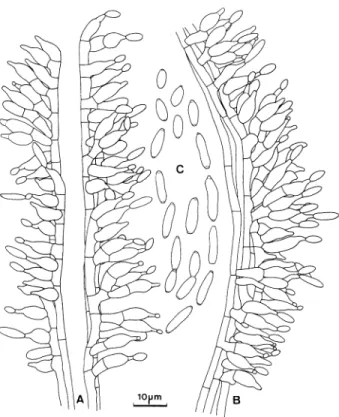
Host covered by floccose, yellowish white, pale yel- low to light yellow (4A2-4) mycelium from which nu- merous synnemata arise. Synnemata of variable length; long synnemata solitary, arising from the cen- tral dorsal part of the host, 5 mm long, ca. 100 txm wide; short synnemata, numerous, arising from the peripheral dorsal part of the host, circular, erect, short, simple, clavate, 480-720 ,tm long, 80-290 jim diam; both kinds of synnemata yellowish gray (4B2), pubescent, pulverulent towards the apex, composed 319
MYCOLOGIA
FIG. 1. A-C. Conidiogenous cells and conidia of Akan-
thomyces ampullifer. On spider Ar. 98.
of compact longitudinal parallel hyphae. Hyphae of
synnema 1.6-2.4 jm wide, septate, hyaline, smooth.
Phialides arising from the outer hyphae or lateral
cells of the synnemata, 6.4-13.9(-15.5) X 3.6-4.4 jLm, forming a hymenial layer, or sparsely arising
from the hyphae or lateral cells of the hyphae, con- sisting of an ellipsoidal to cylindrical venter, 4.8-11.5
X 3.6-4.4 jtm, hyaline, abruptly tapering into a dis-
tinctly long neck, 1.6-3.6(-4.8) X 1.0-1.7 Jim,
smooth. Conidia in a short chains, one-celled, cylin-
drical, 6.0-11.5 X 2.1-3.2 |Jm, occasionally curved,
often with distinctly apiculate ends, hyaline, smooth.
Teleomorph not observed.
Specimen examined. REPUBLIC OF CHINA. TAI- WAN: Taipei County, Sanhsia, Manyuehyuan, on spi- der Ar. 98, 20 Jan. 1995, L. S. Hsieh.
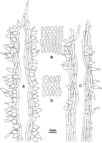
Akanthomyces aranearum (Petch) Mains, Mycologia
42: 574. 1950. FIG. 2 A, B Host covered by white, yellowish white (4A2) to grayish yellow (4B2-3) mycelium from which numer- ous (about 12) synnemata arise. Synnemata arising from all parts of the host, clavate, 0.8-1.8 mm long, slender, 62-74 jIm thick below, simple or occasionally slightly branched, sometimes pubescent below, dark brown (7F5-7) to brown (7E6-7), becoming yellow- ish white (4A2) to orange white (5A2) towards the
oooOO
0
ooQr
bo
B lOum, D
FIG. 2. Conidiogenous cells and conidia of Akanthomyces
aranearum. A, B. on spider Ar 45 and A. novoguineensis.
C, D. on spider Ar 29. Note that conidiogenous cells of A. aranearum are distinctly roughened (A), and A. novogui- neensis are mostly globose to subglobose with a constricted, narrow neck (C).
upper fertile portion, composed of parallel hyphae.
Hyphae of synnema smooth, hyaline to orange
(6A7), dark orange (5A8), sometimes swollen, 3.2-
6.0 pxm wide. Phialides scattered, forming a dense
layer on the hyphae of synnemata, consisting of a
globose to ellipsoid venter, hyaline to greenish white
(30A2), 5.3-12.1 X 3.8-6.3 pJm, asperulate to verru-
cose, abruptly narrowing into a short, smooth neck, 0.8-2.0 pJm long, 0.8-1.6 ptm wide. Conidia narrowly obclavate, often acute at the lower end, narrowing upward, rounded or obtuse at the upper end, one-
celled, hyaline, smooth, catenulate, 4.4-11.4 X 1.9-
2.6 pLm. Teleomorph not observed.
Specimen examined. REPUBLIC OF CHINA. TAI- WAN: Taipei County, Wulai, on spider Ar. 45, 27Jun.
1991, L. S. Hsieh.
Akanthomyces novoguineensis Samson & Brady, Trans.
Brit. Mycol. Soc. 79: 571. 1982. FIG. 2 C, D Host covered by yellowish white to light yellow (4A2-4) mycelium from which numerous (up to 50) 320
aline, smooth or slightly rough, 2.6-5.0 jLm wide. Phialides consisting of a globose to subglobose ven- ter, hyaline, smooth, 5.2-7.1 X 4.8-6.0 iLm, constrict- ed abruptly to a distinct, narrow neck, 1.4-1.8 X 0.8- 1.6 jim. Conidia in short chains, one-celled, hyaline, smooth, narrowly cylindrical to fusiform, sometimes apiculate at one or both ends, occasionally curved, 6.4-11.1 X 1.4-2.9 jim. Teleomorph not observed.
Specimens examined. REPUBLIC OF CHINA. TAI- WAN: Nantou County, Puli, Lienhuachih, on spider Ar. 29, 5 Sep. 1990, Tzy-Mei Lin; same location, on spider Ar. 33, 29 Sep. 1990, WJ. Wu; Nantou County, Luku, Shanlinchi, on spider Ar 43. 10 May 1991, L. S. Hsieh.
Akanthomyces ovalongatus Hsieh, Tzean et Wu, sp.
nov. FIGS. 3, 4
Aranea hospes mycelio pruinoso albo ad aurantio- albo (5A2) cooperta, multis synnematibus aculeatis instructa. Synnemata erecta, undique e hospite en- ascentia, simplicia an raro exigue ramosa, alba ad gri- seo-aurantiaca (5-6B3), 2.2-9.0 mm longa, 112-520
Jim lata. Hyphae hyalinae, septatae, ramosae, (2.3-) 2.5-3.7(-4.2) jm latae. Cellulae conidiogenae phiali- dicae, seu solitariae vel laxe compactae e mycelio en- ascentes, seu e synnematibus disseminatae vel coac- tae; e parte basilari globosa, subglobosa, cylindrica vel ellipsoidea, 6.0-8.7 X 4.0-6.4 ljm, in collum an- gustum, 1.4-3.2 jtm longum, 0.8-1.8 tjm latum, at- tenuatae, glabrae; conidia catenis brevibus adhaer- entia, ellipsoidea, ovata vel oblonga, 6.0-10.3 X 2.4- 4.4 jLm, unicellularia, hyalina, glabra. Status teleo- morphus ignotus.
HOLOTYPUS. REPUBLIC OF CHINA. TAIWAN: Taipei City, Yang-Ming-Shan National Park, on spider Ar. 113, 2 Aug. 1995. L. S. Hsieh (PPH 24, deposited in the herbarium of the Department of Plant Pa- thology and Entomology, National Taiwan University, Taipei, Taiwan, R. 0. C.; Isotype IMI). The ex-type culture PPH 24E was deposited in the Culture Col- lection and Research Center (CCRC 33385), Hsin- chu, Taiwan, R. 0. C.
Spider host covered by powdery white to orange white (5A2) mycelium. Synnemata erect, arising from all parts of the host, numerous, simple or occasion- ally branched, white to grayish orange (5-6B3), 2.2-
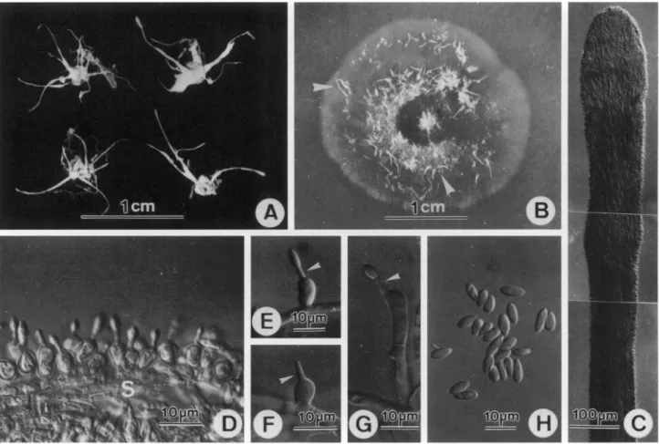
FIG. 3. Akanthomyces ovalongatus. Conidiogenous cells
and conidia. A, B. on naturally infested host. C, D. on oat- meal agar. On spider Ar. 113.
9.0 mm long, 112-520 jLm wide. Hyphae of synnema hyaline, septate, branched, (2.3-) 2.5-3.7 (-4.2) iLm wide. Conidiogenous cells phialidic, either arising from the mycelium and solitary, or loosely compacted into a layer, or arising from synnemata and scattered to crowded over the length of the synnema. Phialides consisting of a globose to subglobose, cylindrical or ellipsoidal basal part, 6.0-8.7jN4.0-6.4 jLm, and an abruptly narrowed neck, 1.4-3.2 Ijm long, 0.8-1.8 Jim wide, smooth. Conidia in short chains, ellipsoid- al, obovate to oblong, 6.0-10.3jN2.4-4.4 jtm, some- times apiculate at one end, one-celled, hyaline, smooth. Teleomorph not observed.
Colonies on oatmeal agar at 25°C in darkness grow- ing slowly, 25-36 mm diam after 2 mo. Mycelium hy- aline to white, plane felty to velutinous. Submerged hyphae septate, smooth, hyaline, infrequently branched, 2.4-6.0 jim wide, pigments grayish orange
(5-6B3), grayish brown to reddish brown (8E3-5). Colony reverse grayish brown, reddish brown to dark brown (9F5). Aerial hyphae septate, smooth, hyaline, branched, swollen near the septa, 2.4-9.1 jtm wide. Exudate in aerial mycelium limited or absent. Syn- nemata erect, semi-erect, or procumbent, straight or
MYCOLOGIA
.3; , . 1 .. S.
B
FIG. 4. Akanthomyces ovalongatus. A. Synnemata arising from infested spiders. B. Synnemata produced on oatmeal agar at 25°C, in darkness after incubation for 2 mo (arrow heads). C. Synnema from spider host, cylindrical and covered with a hymenium of conidiogenous cells. D. Conidiogenous cells subglobose densely disposed on a synnema from spider. E, F. Conidiogenous cells arising from synnemata, with an abruptly attenuated neck (arrow heads). G. Conidiogenous cell from oatmeal agar arising from end of a mycelial hypha with a distinct neck (arrow head). H. Conidia from spider.
slightly curved, white, aculeate, up to 2.7 mm long, 160-400 Ijm wide at base, sometimes forked at base, developing in inconspicuous concentric rings or scat- tered, composed of smooth, parallel, hyaline hyphae 1.8-4.0(-4.8) j,m. Phialides sparse, scattered on the synnemata, or occasionally solitary, arising from the end of an aerial hypha, consisting of a globose, subgl- obose to ellipsoid basal part, 7.1-12.7 X 4.4-6.0 ,pm, and a cylindrical venter that becomes almost abruptly constricted to a short neck, 2.0-5.6 X 1.2-2.2 ,xm, smooth. Conidia in short chains of three or fewer, ellipsoidal, obovate to oblong or broadly obovoid, 6.5-12.7 X 2.8-4.0 ixm, sometimes apiculate at one end, one-celled, hyaline, smooth. Chlamydospores absent. Teleomorph unknown.
DISCUSSION
During a six yr survey conducted from 1989 in Tai- wan, 7 of 117 spider cadavers collected were infested by Akanthomyces, and designated as Ar. 29, 33, 43,
45, 69, 98, 113, respectively. The infested spiders usually inhabited the lower surface of broad leaves herbs or shrubs in humid and shaded areas. Of these, Ar. 69 was immature, and could not be identified to species. Ar. 29, 33, and 43 were identified as A. nov- oguineensis, and Ar. 29was selected as a voucher spec- imen for illustration and diagnosis. The conidial sizes of Ar. 29, Ar. 33 and Ar. 43 were 6.4-11.1 X 1.4-2.9 ,pm, (5.7-) 7.5-14.0 X 1.8-2.9 pim, and 5.6-15.9 (-19.8) X 1.4-2.9 ,xm, respectively. Conidia of Ar. 29 and Ar. 33 are smaller than conidia in the type spec- imen of A. novoguineensis (10.5-17.5 X 1.5-3 ,Im), which was described from Papua New Guinea by Samson and Brady (1982), but comparable to conidia in the specimen (5.7-11.0 X 1.0-1.6 pim) collected in Thailand by Hywel-Jones (1996). Obviously, there is a transition in the conidial size among isolates. The specimen Ar. 45 from Taiwan is a typical A. aranear- um, with verrucose phialides and obclavate conidia. These unusual features were well documented pre- viously (Samson and Evans, 1974; Hywel-Jones, 1996). 322
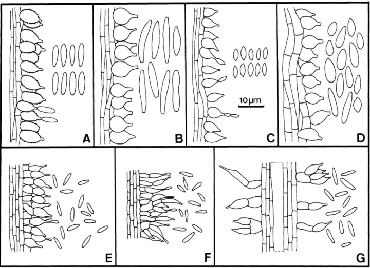
FIG. 5. Illustrations of conidiogenous cells and conidia of 7 species of Akanthomyces parasitic on spiders. A. A. aranearum.
B. A. novoguineensis.C. A. arachnophilus. D. A. ovalongatus. E. A. koratensis. F. A. cinereus. G. A. websteri. A-C and E-G are
redrawn with permission from R. A. Samson and N. L. Hywel-Jones, respectively. All figures are to the same scale. The specimen Ar 98 is A. ampullifer. This is the first
report of A. ampullifer from a spider; it typically oc- curs on dipteran gnats (Mains, 1950).
The collection Ar. 113 is morphologically distinct from all described species of Akanthomyces and is pro- posed as the new species, A. ovalongatus. Akantho- myces ovalongatus can be separated from the six pre- viously described Akanthomyces species that occur on spiders, A. aranearum, A. novoguineensis, A. arach-
nophilus, A. koratensis, A. cinereus, and A. websteri
(Mains, 1950; Samson and Evans, 1974; Hywel-Jones, 1996) by the shape, size and roughness of the phial- ides, and/or by the shape and size of the conidia
(FIG. 5 A-G). Akanthomyces ovalongatus can also be
readily distinguished from A. aculeatus, the type spe- cies of the genus Akanthomyces, which is associated with mosses, by its much larger conidia (6.0-10.3 X 2.4-4.0 ,um vs 3-6 X 2-3 pzm), although both species have similar ellipsoidal or obovoid conidia. Phialides of A. ovalongatus in agar culture are sparse, slender, and cylindrical or ellipsoidal, somewhat reminiscent of A. ampullifer (Mains, 1950). Comparison A. ova-
longatus with the holotype of A. ampullifer (- Hy- menostilbe ampullifera Petch, FH727) demonstrated that phialides and conidia of the latter species were noticeably narrower.
The morphology of synnemata and conidiogenous cells of A. ovalongatus developed on naturally infect- ed spider considerably differed from that found in agar culture. On spiders, the phialides are dense, and form a hymenial layer covering the synnemata, and consist of globose to subglobose basal parts, whereas on agar culture, the phialides are loosely dispersed along the length of the synnemata, and the basal parts of the phialides usually are narrower, ellipsoidal or cylindrical in shape. Moreover, the synnemata on spider hosts are longer and wider than on culture media (2.2-9.0 mm X 112-520 Lm vs 2.7 mm X 160- 400 pLm).
None of the three Akanthomyces species found on spiders in Taiwan has been found correlated with a teleomorph. Hywel-Jones (1996) reported six Akan- thomyces species from Thailand and also found no evidence of a teleomorphic connection. On the other
MYCOLOGIA hand, Samson and Evans (1974), studying Ghanian
specimens, found a constant association of A. pistil- lariiformis (Pat.) Samson & H. C. Evans with an as-
comycetous teleomorph similar to Cordyceps tubercu-
lata (Lib.) Maire but that showed a great variation in perithecial form.
While constructing a key for the known Akantho-
myces species based on the published descriptions, A.
pistillariiformis was found to be extremely similar to
A. angustisporus Mains in morphological characters
of conidiogenous cells and conidia, although the
host, synnema color and branching patterns varied (Mains, 1950). Unfortunately, the holotype of A. an-
gustisporus was not available for loan, because it con- sists of a single larva with a small and fine synnema attached (MICH, R. Fogel, personal communica- tion). Therefore, comparative study of these two spe- cies to clarify the puzzle was not possible. Pending comparison of the respective type specimens, host preference and synnemata are used to distinguish A.
pistillariiformis and A. angustisporus. However, Sam-
son and Evans (1974) indicated that such features may be variable depending on host species and hab- its. Here we key out the thirteen described Akantho- myces species. Some epithets that were orthographi- cally incorrect in gender, have been corrected (In- ternational Code of Botanical Nomenclature Arts. 62.1 and 62.2a).
KEY TO SPECIES OF AKANTHOMYCES
1. Saprobic; conidia broadly fusoid, ends somewhat
truncate, with minute frills ... A. johnsonii
1. Parasitic on spiders or insects; conidia not as above .. 2
2. Parasitic on spiders ... 3
2. Parasitic on insects ... 9
3. Phialides verrucose; conidia obclavate A. aranearum
3. Phialides smooth; conidia not as above ... 4
4. Phialides mostly arising from basal cells perpen-
dicular to the synnemata ... 5
4. Phialides mostly not arising from basal cells per-
pendicular to the synnemata ... 7
5. Phialides often cylindrical, synnemata multiple ....
... A . cinereu s
5. Phialides often ellipsoidal, synnemata simple ... 6
6. Phialides hymenial, covering the synnemata ex-
cept the sterile brown base ... A. koratensis
6. Phialides scattered, covering the length of the
creamish synemmata ... A. websteri
7. Conidia cylindrical, curved, 10.5-17.5 X 1.5-3 p.m
... A . novoguineensis
7. Conidia oblong, fusiform, ellipsoidal, obovoid to cla-
vate, smaller ... 8
8. Conidia fusiform, with acute ends, 4.5-5.5 X
1.5-3.0 pIm ... A. arachnophilus
8. Conidia oblong, obovate or broadly ellipsoidal,
6.0-10.3 X 2.4-4.4 p.m ... A. ovalongatus
9. Conidia often in long chains of more than 3, minute,
2.5-3 X 1-1.6 .m ... A. gracilis
9. Conidia often in short chains of less than 3, longer
than above ... 10
10. Conidia often longer than 6 p.m .. A. ampullifer
10. Conidia often shorter than 6 pLm ... 11
11. Conidia broadly ellipsoidal or obovoid, acute at low-
er end, 3-6 X 2-3 pLm ... A. aculeatus
11. Conidia narrowly fusiform or clavate, 4.5-6 X 1.2-
1.4 (-5) ! m ... 12
12. Synnemata flesh colored, simple or branched, 8-13 X 0.2-0.6 mm; associated with coleopteran
hosts ... A. angustisporus
12. Synnemata white to creamish, often simple, rarely branched, 1-6 X 0.05-0.3 mm; associated
with lepidopteran hosts ... A. pistillariiformis
ACKNOWLEDGMENTS
The research was supported by grants to S. S. T. from the National Science Council (NSC-84-0409-B002-129) and Council of Agriculture, Executive Yuan [84-Ko-chi-1,3-li- ang-31 (8-2)], R. 0. C. The authors thank C. F. Huang and J. Y. Liou for technical assistance. Special thanks are ex- tended to Prof. D. H. Pfister and Dr. E. W. Wood for the loan of the type specimen of Hymenostilbe ampullifer, to Drs. R. A. Samson, H. C. Evans, and B. L. Brady, and Dr. N. L. Hywel-Jones for permission to use their illustrations of A.
arachnophilus, A. aranearum and A. novoguineensis and A. cinereus, A. koratensis and A. websteri, respectively; to Prof.
R. Fogel for indicating the condition of the holotype of A. angustisporus. Special thanks are due to Drs. G. J. Samuels, A. Y. Rossman, R. A. Humber, N. L. Hywel-Jones and W. Gams for critical review of the manuscript and constructive comments and suggestions; and Dr. W. Gams and Mr. P. M. Eckel for preparation of the Latin.
LITERATURE CITED
Hywel-Jones, N. L. 1996. The genus Akanthomyces on spi- ders in Thailand. Mycol. Res. 100: 1065-1070.
Kornerup, A., andJ. H. Wanscher. 1978. Methuen handbook of colour. Eyre Methuen Ltd., London, United King- dom. 252 pp.
Lebert, H. 1858. Ueber einige neue oder unvollkommen gekannte Krankheiten der Insekten, welche durch En- twicklung niederer Pflanzen im lebenden Korper en- stehen. Z. Wiss. Zool. 9: 439-453.
Mains, E. B. 1950. Entomogenous species of Akanthomyces,
Hymenostilbe and Insecticola in North America. Mycolo-
gia 42: 566-589.
Samson, R. A., and B. L. Brady. 1982. Akanthomyces novo- guineensis sp. nov. Trans. Brit. Mycol. Soc. 79: 571-572.
, and H. C. Evans. 1974. Notes on entomogenous fungi from Ghana. II. The genus Akanthomyces. Acta Bot. Neerl. 23: 28-35.
Vincent, M. A., K. A. Seifert, and R. A. Samson. 1988. Ak-
anthomyces johnsonii, a saprophytic synnematous Hy-
phomycete. Mycologia 80: 685-688. 324