國 立 交 通 大 學
生 物 科 技 學 院
生物科技學系
碩 士 論 文
酵素活化自組裝式螢光奈米金粒子檢測平台之建立
並應用於胰臟炎的診斷
Protease Assay by Activated Fluorescent Self-assembled
Gold Nanoparticles Applied in Pancreatitis Diagnosis
研 究 生 :葉芳沅
指導教授 :林志生 博士
酵素活化自組裝式螢光奈米金粒子檢測平台之建立
並應用於胰臟炎的診斷
Protease assay by activated fluorescent self-assembled gold
nanoparticles applied in pancreatitis diagnosis
研 究 生 :葉芳沅 Student:Fang-Yuan Yeh 指 導 教 授 :林志生 博士 Advisor:Chih-Sheng Lin, Ph.D. 國 立 交 通 大 學 生 物 科 技 學 院 生物科技學系 碩 士 論 文 A Thesis
Submitted to Department of Biological Science and Technology College of Biological Science and Technology
National Chiao Tung University in partial Fulfillment of the Requirements
for the Degree of Master in Biological Science and Technology
July 2013
Hsinchu, Taiwan, Republic of China
i
酵素活化自組裝式螢光奈米金粒子檢測平台之建立
並應用於胰臟炎的診斷
研究生:葉芳沅 指導教授:林志生 博士
國 立 交 通 大 學
生 物 科 技 學 院
生物科技學系碩士班
中 文 摘 要
奈米粒子目前廣泛被應用於生物感測,其中尤以奈米金粒子(AuNPs)最常被應用, 此乃 AuNPs 具有易於被生物分子修飾和共振能量轉移的特性。本研究所設計的生物檢 測原理係以 AuNPs 具有遮蔽螢光之特性,其擁有與距離有關的廣效波長遮蔽螢光特性, 據此我們建立了一個快速檢測蛋白酵素(proteinase)活性的平台,並探討胜肽受質的序列 設計,用以獲得較高靈敏度的檢測。我們利用螢光基團 FITC 標記的胜肽受質結合上 AuNPs,此為一活化自組裝式螢光 AuNPs 探針,其被應用於蛋白酵素活性檢測中,原理 是當酵素水解胜肽受質時,其尾端的螢光基團可遠離 AuNPs 表面,使其螢光波長得以 被偵測。本研究首先以具有高活性的蛋白酶 K (proteinase K)為檢測對象,並藉以建立 最佳化檢測條件和受質胜肽序列設計原則。接著我們將上述平台改裝用以胰凝乳蛋白酶 (chymotrypsin)的活性檢測,並運用於胰臟功能的檢測。 為了要增加螢光 AuNPs 探針對酵素的靈敏性,本研究共設計三條胜肽基質用於製 作 AuNPs 探針,其設計著重於降低 AuNPs 表面的立體障礙以利蛋白酵素辨識,並藉由 延伸胜肽的長度或改變特定序列以有效地增加 AuNPs 探針偵測酵素活性的能力。實驗 結果顯示,GPLGLARGGGGGC 之 AuNPs 探針用於偵測蛋白酶 K 與胰凝乳蛋白酶活性, 其較 GPLGLAG(Hyp)C 之 AuNPs 探針用於檢測分別提升了 3 和 10 倍螢光強度的變化; 而利用 GPLGLARDDDDDC 之 AuNPs 探針用於酵素活性的檢測,其偵測極限可以從 ng/mL 下降到 pg/mL 程度,且偵測時間只需 15 分鐘。以上結果證明受質胜肽序列的設 計對螢光 AuNPs 探針應用於檢測蛋白酵素活性上極為重要。ii AuNPs 探針 GPLGLARDDDDDC 進一步被應用於生物樣本的檢測,包括腸液與糞 便檢體中胰凝乳蛋白酶活性的檢測,而在實驗小鼠模式中,單顆小鼠糞便即可簡易地利 用我們所設計的 AuNPs 探針來測定其胰凝乳蛋白酶活性。本研究探討胰凝乳蛋白酶在 小腸中的活性分佈,此實驗為比較在禁食與進食控制下,小鼠小腸液中的胰凝乳蛋白酶 活性變化,結果顯示在不同飲食控制下空腸與迴腸中胰凝乳蛋白酶活性會有顯著的差異, 而其糞便中胰凝乳蛋白酶活性變化也可對應其禁食與進食狀況。本研究也建立利用蛙皮 素(cerulein)腹腔注射誘發急性胰臟炎的小鼠模式,再利用所設計之 AuNPs 探針偵測其 小腸與糞便中胰凝乳蛋白酶活性變化,期可作為評估急性胰臟炎的檢測指標。實驗結果 顯示在急性胰臟炎小鼠的十二指腸腸液與糞便中,其胰凝乳蛋白酶活性分別顯著下降到 正常對照組的 25%與 30%。 本研究所建立的酵素活化自組裝式螢光 AuNPs 檢測平台有潛力成為高靈敏且可快 速偵測的生物檢測平台,也被證實具有臨床應用的可行性。而所建構的 AuNPs 探針可 藉由受質胜肽序列的置換,即可進一步應用於其他酵素活性的檢測,此使本研究之成果 更具可利用性與價值性。 關鍵詞:急性胰臟炎、生物感測、胰凝乳蛋白酶、奈米金粒子、胜肽
iii
Protease assay by activated fluorescent self-assembled gold
nanoparticles applied in pancreatitis diagnosis
Graduate student: Fang-Yuan Yeh Advisor: Chih-Sheng Lin, Ph.D.
Department of Biological Science and Technology
College of Biological Science and Technology
National Chiao Tung University
Abstract
Nanoparticles are usually used in biosensing field and among all of the nanoparticles, gold nanoparticles (AuNPs) are most widely applied. It is because that AuNPs are easy to be conducted with surface biomolecule modification and possess the characteristic of resonance energy transfer. The principle of biosensing platform designed in this study is based on AuNPs provide quenching fluorescence ability and which is wide range wavelength quenching and distance dependence. According to the property, a rapid proteases activity sensing platform was established and the peptide substrates design in order to gain high sensitivity detection platform was also investigated. FITC labeled peptide substrates were used to conjugate onto AuNPs to be an activated fluorescent self-assembled AuNPs (AuNPs probe) that are used for the activity assay of proteases. The detecting mechanism is that proteases could cleavage peptide substrates and then the fluorephore at the end of peptide substrates could diffuse away from AuNPs surface, which the fluorephore emitting
wavelength could be detected. In this study, proteinase K with pretty high specific activity was first used as target proteases to establish optimal detecting conditions and to evaluate the design principle of peptide substrates. After that, the established platforms were used in chymotrypsin activity assay and applied in the estimation of pancreatic function.
For increasing the sensitivity of proteases to AuNPs probe, there were three peptides substrates, i.e. AuNPs probes, designed and evaluated in the present study. The design was emphasized on the decreasing steric barrier on AuNPs surface by extending length of peptide
iv
or changing specific sequences of AuNPs to increase sensitivity of the AuNPs probes used in the assay of proteases activity. The results indicate that the AuNPs probe with
GPLGLARGGGGGC could increase the detecting sensitivity of proteinase K and
chymotrypsin by approximate 3 and 10 folds fluorescent intensity change compared with those by the AuNPs probe of GPLGLAG(Hyp)C, respectively. Moreover, the AuNPs probe with GPLGLARDDDDDC applied in proteases activity assay could lower the detection limit from ng/mL to pg/mL level and the detection time was only 15 min. The results above indicate that the design of peptide sequence plays important role in the AuNPs probe applied in protease assay.
The AuNPs probe with GPLGLARDDDDDC was further applied in the detection of biological samples, including the activity assay of intestinal and fecal chymotrypsin. For the experimental mouse model, simply one feces is needed for chymotrypsin activity assay by the AuNPs probe developed in this study. The chymotrypsin activity distributions in intestinal fluids of mice were investigated by a procedure of fasting/feeding control. The results show a significant difference in jejunum and ileum under diet controls; besides, the activity change of fecal chymotrypsin also corresponds to different situations of fasting and feeding. Acute pancreatitis (AP) mouse model induced by intraperitoneal injection of cerulein was established in this study. The AuNPs probe was applied to evaluate chymotrypsin activity level in intestine and feces with the expectation of being an indicator of AP. The results indicate that chymotrypsin in duodenal fluid and feces significantly decrease and remain only 25% and 30 % activity level compared with those in normal subjects, respectively.
The activated fluorescent self-assembled AuNPs probe established in this study shows the potential to be a biosensing platform with high sensitivity and rapid detection, and has been approved to be available in clinical applications. For wide development by simply replacing the efficient peptide substrates for different protease targets can be rationally expected, it makes the achievements procured this study being more profitably and valuably. Keywords: acute pancreatitis, biosensor, chymotrypsin, gold nanoparticles, peptide
v
誌 謝
光陰匆匆,猶記得甫進交大生科碩班時,老師警惕著其實兩年光陰不算長,幾十個 禮拜將會轉眼間過去,從沒想過兩年會是那麼地五味雜陳,而現在居然就是要畫下句點 的那刻,回首記憶裡的惶恐、挫折、期待、自信、喜悅、悲傷,不禁呀然。 在踏進碩士生涯之前給自己的期許是要盡力地努力一回,選擇了一間可以磨練自己 的實驗室。兩年歲月,多半是沉悶的實驗,幸有許多人們點亮苦悶的生活。首先要感謝 的是棠青學長,如同家裡為晚歸的人所點的那盞夜燈,溫暖、誠懇、值得信賴,大家總 能在各種時候從學長那裡得到支持與安慰。聖壹學長,萬能的小叮噹常常伸出援手來拯 救我這個麻煩的學妹,謝謝你不但給予我許多協助還很肯定我並給我信心。千雅學姊, 實驗室的大姊,說話犀利又不失幽默,大家與爽朗的你談天總能獲得好心情,懷念那些 留到最晚的日子,一起感慨、一起做些小壞事。睦元學長,謝謝你常聽我叨絮實驗上的 困擾,叮嚀我邏輯思維在研究之路上的重要性,協助我破解許多卡關的時刻。戴樂,深 藏不露的助理,相處久了才發現你的細膩、善良都包裝在浮誇的言行之下,其實是個好 人。燕秋,一起奮鬥到最後的好夥伴,我們熬過成長的陣痛,最後終於可以在此時此刻 一同品嘗甜美的果實了! 真的很慶幸有你,這麼一位善良溫暖的女孩,常常一個眼神就 可以讀懂我,這一路相互扶持的情誼會長存在我心中,衷心祝福你在未來一切順利美滿。 琳岡,好姊妹一場,祝福你前途似錦。佩衡,聰穎的女孩,有你和孟融偶而地妝點,實 驗室生活更加精采。一華,意外好用的學弟,其實是璞玉幾經琢磨就可以綻放光采,很 謝謝你忍受我的脾氣,給我很多支持,當我的助手陪伴我度過許多折磨人的時光。意涵, 用心貼心的學妹,常常付出後卻默默受傷的小笨蛋,喜歡我們湊在一塊兒又吵又鬧的日 子。郁彬,同為夜貓子的咖啡友,萬紅叢中一點綠的存在,謝謝你扛起照顧大家的責任。 碧珊,常有脫線演出惹人發笑的糊塗學妹,祝福你未來幸福美滿。酷女孩葛麗和明慧, 兩人意外地都是十足的少女心且時常有意外的笑點,以及精明能幹的采郁,希望你們研 究都可以順順利利。實驗室新血張蓉、日升、子仁,要好好加油喔!不及備載的實驗室 成員們,謝謝你們平日的照顧與對實驗室的付出。另外,謝謝交大校園裡的校狗們,陪vi 伴撫慰相處在這裡的人們,以及跟我回家的貝蒂,默默地守候晚歸的我,療癒我疲憊的 心。論文得以完成,同樣要感謝為科學所犧牲的動物們,謝謝你們的奉獻。 由衷感謝劉典謨 教授與陳思豪 博士,在百忙中抽空對本研究詳加審閱並給予真知 灼見,使本論文能更臻完整。最重要的是要感謝我的指導教授—林志生 教授,老師所 給予的不只是研究上的啟發,訓練我的邏輯思維,培育我獨立思考的能力,並給予自主 發揮的空間,而更多的是做人處事上的態度,夙夜匪懈積極努力的精神,往往讓學生們 自嘆弗如。如今可以蛻變成現在的我,可以自信地說自己在兩年內有所成長,都要感謝 老師悉心地指導並在多方包容學生。另外,感謝袁如馨 老師,老師總是把良善與愛散 播給大家,有幸能被老師所教導,於一年裡奠定了我的學術表達與寫作的基礎,可惜不 能與您分享畢業的喜悅,願您安詳。 人生歷程上受了很多的人的幫助與支持,才可以一路走到現在,謝謝所有在過程中 相知相惜的朋友們。最後,要向我的家人致謝,你們無私的付出才可以讓我無慮地探索 自我,並能夠於此刻順利地完成學業,衷心地感謝你們。我將飛翔。 葉芳沅 謹誌 交通大學生物科技學系碩士班 中華民國一○二年七月
vii
Table of Contents
Chinese Abstract ... i
English Abstract ... iii
Acknowledgment ... v
Table of Contents ... vii
List of Figures ... xi
List of Tables ... xiii
I. Literature Review ... 1
1-1. Introduction ... 1
1-2. Gold nanoparticles ... 2
1-2-1. Citrate reduction method ... 2
1-2-2. Local surface plasmon resonance ... 3
1-2-3. Fluorescence quenching mechanisms by AuNPs ... 7
1-2-4. Derjaguin-Landau-Verwey-Overbeek theory ... 15
1-2-5. Stabilization of AuNPs ... 18
1-2-6. Peptide design for conjugation to AuNPs ... 20
1-3. Enzymes ... 23
1-3-1. Proteinase K ... 23
1-3-2. Chymotrypsin ... 24
1-4. Pancreatitis ... 25
1-4-1. Acute pancreatitis ... 25
1-4-2. Animal model of cerulein-induced acute pancreatitis ... 26
II. Research Strategy ... 35
III. Materials and Methods ... 39
3-1. Instruments ... 39
viii
3-3. Peptide substrates ... 40
3-4. Preparation of the self-assembly AuNPs fluorescence probe ... 40
3-4-1. Synthesis of 15 nm AuNPs ... 40
3-4-2. AuNPs salt stress assays ... 41
3-4-3. AuNPs pH stress assays ... 41
3-4-4. Modification of AuNPs probes ... 41
3-4-5. Effect of peptide substrate charges on AuNPs probes ... 42
3-5. Evaluation of size and morphology change in AuNPs probe ... 43
3-5-1. UV-Vis spectra determination ... 43
3-5-2. Dynamic light scattering determination ... 43
3-5-3. Transmission electron microscopy analysis ... 44
3-5-4. Zeta-potential analysis ... 44
3-5-5. Gel electrophoresis of AuNP ... 44
3-6. Fluorescence assays of proteases activated AuNPs probe ... 45
3-6-1. Conjugation of peptide substrates to AuNPs ... 45
3-6-2. Proteinase activity assay by AuNPs probes ... 45
3-6-3. Effect of stabilizers to proteinase sensitivity to AuNPs probes ... 46
3-7. Effect of peptide design to proteinase sensitivity to AuNPs probes ... 46
3-8. Animal experiments ... 49
3-8-1. Animals ... 49
3-8-2. Sample collection ... 49
3-8-3. Fasting/Feeding experiment ... 50
3-8-4. Cerulein-induced acute pancreatitis mouse model ... 50
3-8-5. Amylase and lipase assays ... 51
3-8-6. Islet isolation from mouse pancreas ... 51
3-9. BTEE assay ... 52
IV. Results ... 53
ix
4-1-1. Characteristic of 15 nm AuNPs ... 53
4-1-2. AuNPs salt stress assays ... 53
4-1-3. AuNPs with stabilizers salt stress assays ... 54
4-1-4. AuNPs pH stress assays ... 54
4-1-5. UV-Vis spectra study on AuNPs probe of various charges peptide substrate ... 54
4-1-6. Effect of peptide substrate charges on AuNPs probes ... 55
4-1-7. Conjugation of peptide substrates to AuNPs ... 56
4-1-8. Zeta potentials of AuNPs and AuNPs probes ... 56
4-2. Optimize the fluorescence assays by AuNPs probes ... 57
4-2-1. Effect of stabilizers to proteinase K sensitivity to AuNPs probe ... 57
4-2-2. pH optimization of proteinase K sensitivity to AuNPs probe ... 58
4-2-3. AuNPs probes concentration optimization ... 58
4-2-4. Proteinase K activity assay by AuNPs probe (7.4A/1466p-FITC) ... 58
4-2-5. Comparison of different AuNPs probes to proteinase K sensitivity ... 59
4-2-6. Proteinase K activity assay by AuNPs probe (5.6A/1482p-FITC) ... 59
4-3. Evaluation of characteristic change in AuNPs probes ... 60
4-3-1. Gel electrophoresis analysis of AuNPs ... 60
4-4. Chymotrypsin assay by AuNPs probes ... 60
4-4-1. pH optimization of chymotrypsin sensitivity to AuNPs probe ... 60
4-4-2. Comparison of different AuNPs probes to chymotrypsin sensitivity ... 60
4-4-3. AuNPs probes (10.0A/1477p-FITC) to chymotrypsin sensitivity ... 61
4-4-4. AuNPs probes (10.0A/1477p-FITC) specificity to chymotrypsin ... 61
4-4-5. BTEE assays of chymotrypsin ... 62
4-4-6. Chymotrypsin activity assay by AuNPs probe (5.6A/1482p-FITC) ... 62
4-5. Animal experiments ... 63
4-5-1. Islet isolation from mouse pancreas ... 63
4-5-2. Effect of fasting/feeding treatments to intestinal chymotrypsin of mouse 63 4-5-3. Effect of fasting/feeding treatments to fecal chymotrypsin of mouse ... 63
x
4-5-4. Cerulein-induced acute pancreatitis mouse model - analysis by plasma
amylase and lipase ... 64
4-5-5. Cerulein-induced acute pancreatitis mouse model - analysis by chymotrypsin in duodenum and pancreas ... 64
4-5-6. Cerulein-induced acute pancreatitis mouse model - analysis by fecal chymotrypsin ... 65
V. Discussion ... 98
5-1. Establishment of activated fluorescent self-assembly AuNPs probes ... 98
5-1-1. Characteristic of AuNPs ... 98
5-1-2. Stability of citrate-capped AuNPs and improvement ... 99
5-1-3. Effect of peptide substrate charges on AuNPs probes ... 101
5-1-4. Conjugation of peptide substrates to AuNPs ... 102
5-1-5. Optimize the sensitivity of AuNPs probes to proteases ... 104
5-1-6. Establishment of proteinase K activity assay by AuNPs probes ... 105
5-2. AuNP and the morphology change analysis ... 107
5-2-1. Gel electrophoresis analysis ... 107
5-3. Establishment of chymotrypsin activated fluorescent self-assembly AuNPs probes ... 108
5-3-1. Establishment of chymotrypsin activity assay by AuNPs probes ... 108
5-3-2. BTEE assays of chymotrypsin ... 109
5-4. Application in animal experiments ... 110
5-4-1. Distributions of chymotrypsin in intestinal fluids ... 110
5-4-2. Cerulein-induced acute pancreatitis mouse model-analysis by plasma amylase and lipase ... 111
5-4-3. Cerulein-induced acute pancreatitis mouse model-analysis by chymotrypsin ... 112
VI. Conclusions ... 115
xi
List of Figures
Figure 1-1. Schematic illustration of citrate-capped AuNPs ... 28
Figure 1-2. Schematic illustration of the deduced process of AuNPs formation ... 29
Figure 1-3. Schematic illustration of two types of surface plasmon resonance ... 30
Figure 1-4. The LSPR of metal NPs exhibits dependence on their size, shape and
material ... 31
Figure 1-5. Parameters affecting the FRET process ... 32 Figure 1-6. Illustration of classical DLVO theory ... 33
Figure 1-7. Proteolytic activation from chymotrypsinogen to α-chymotripsin ... 34
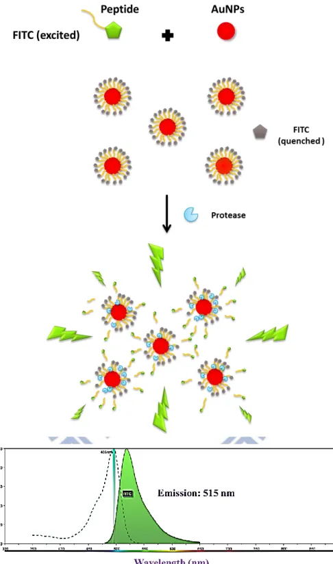
Figure 2-1. A schematic illustration of a protease activated fluorescent self-assembled
AuNPs biosensing platform ... 37
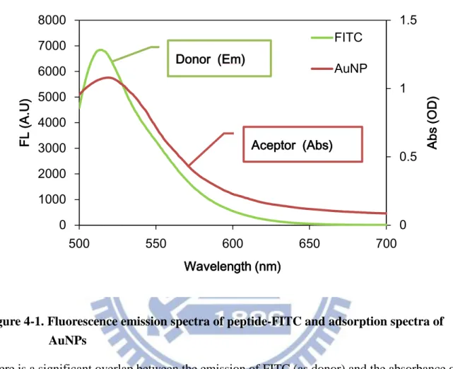
Figure 2-2. The experimental flowchart of research strategy . ... 38 Figure 4-1. Fluorescence emission spectra of peptide-FITC and adsorption spectra of
AuNPs ... 66
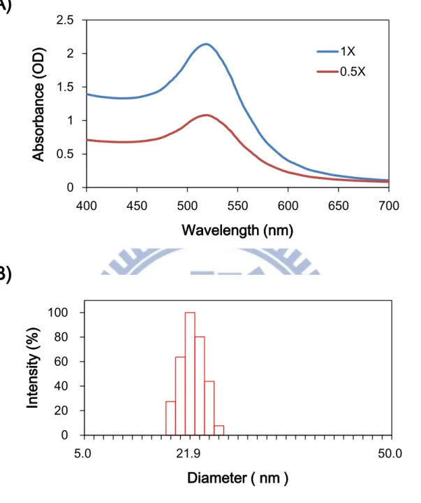
Figure 4-2. Characteristics of citrate-capped AuNPs analyzed by adsorption spectrum
and DLS ... 67
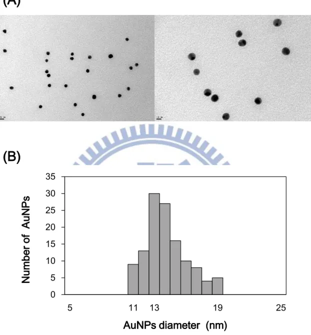
Figure 4-3. Size determination of citrate-capped AuNPs by TEM ... 68
Figure 4-4. Adsorption spectra and aggregation levels of citrate-capped AuNPs under
different salt stress ... 69
Figure 4-5. Aggregation levels of AuNPs with different stabilizers under different salt
stress ... 70
Figure 4-6. Adsorption spectra and aggregation levels of citrate-capped AuNPs under
different pH stress ... 71
Figure 4-7. UV-Vis spectra study on AuNPs probe of various charges peptide
substrate ... 72
Figure 4-8. The aggregation levels of 1466p-FITC in modification and suspension
state of differently functionalized pH ... 73
Figure 4-9. The aggregation levels of 1477p-FITC in modification and suspension
state of differently functionalized pH ... 74
Figure 4-10. Effect of different stabilizers to proteinase K sensitivity by fluorescence
xii
Figure 4-11. pH optimization of proteinase K sensitivity by fluorescence assays of
7.4A/1466p-FITC ... 78
Figure 4-12. Concentration optimization of proteinase K sensitivity by fluorescence assays of AuNPs probes ... 79
Figure 4-13. Fluorescence assays of proteinase K activated by 7.4A/1466p-FITC ... 80
Figure 4-14. Compare two AuNPs probes sensitivity to proteinase K ... 81
Figure 4-15. Proteinase K activity assay by AuNPs probe (5.6A/1482p-FITC) ... 82
Figure 4-16. Comparison between AuNPs and activated AuNPs probe in gel electrophoresis ... 83
Figure 4-17. Gel electrophoresis of AuNP and 5.6A/1482p-FITC probe activated by proteinases ... 84
Figure 4-18. pH optimization of chymotrypsin sensitivity by fluorescence assays of 10.0A/1477p-FITC ... 85
Figure 4-19. Comparisons of two AuNPs probes sensitivity to chymotrypsin assays ... 86
Figure 4-20. Chymotrypsin activity assay by AuNPs probe (10.0A/1477p-FITC) ... 87
Figure 4-21. AuNPs probes (10.0A/1477p-FITC) specificity to serine proteases ... 88
Figure 4-22. BTEE assay of chymotrypsin ... 89
Figure 4-23. Chymotrypsin activity assay by AuNPs probe (5.6A/1482p-FITC) ... 90
Figure 4-24. Isolated mouse islets and beta-cells of islet ... 91
Figure 4-25. The distributions of intestinal chymotrypsin in fasting/feeding mouse analyzed by 5.6A/1482p-FITC ... 92
Figure 4-26. Fasting/Feeding effect to fecal chymotrypsin analyzed by 5.6A/1482p-FITC probe ... 93
Figure 4-27. Plasma amylase and lipase of mouse with induced acute pancreatitis ... 94
Figure 4-28. Chymotrypsin in duodenum and pancreas of mouse with induced acute pancreatitis ... 95
xiii
List of Tables
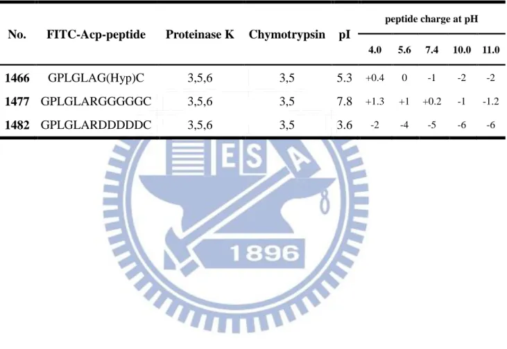
Table 3-1. Peptide sequences and their cleavage sites respective to proteases and
charges at function of pH ... 48
Table 4-1. The function of fluorescence intensity from peptide substrates (pH 8) ... 75
Table 4-2. The ratio of different parts of peptide substrates to total loading peptide
substrates (pH 8) ... 75
Table 4-3. Conjugation ratio of various peptide substrates per 15 nm AuNPs ... 75
1
I. Literature Review
1-1. Introduction
Nowadays, proteases not only catalyze protein degradation by hydrolysis of peptide bonds but also are recognized as exceptionally important molecules that are engaged in
numerous vital life processes. Proteinase activity is considered an important biological marker in various pathologies, because the expression and activity of proteases are significantly different in several pathologies, including inflammatory, cardiovascular diseases, neurological disorders cancer, arthritis, and atherosclerosis [Thobhani et al., 2010]. Thus, the development of proteinase assay has been explored, which usually based on substrate zymography,
radioisotopes, on chromogenic, or fluorogenic substrate. However, these techniques are often time-consuming, expensive, discontinuous, or require specific instruments. More sensitive and convenient proteinase assay is needed; especially methods allow detecting and imaging protease activities in living organisms using different imaging modalities.
Gold nanoparticles (AuNPs) are a potential nanomaterial in biosensor field, account of their unique size- and distance-dependent optical properties and superior performances of being energy acceptors and quenchers [Guarise et al., 2006]. AuNPs interested many biological researchers for their feasible of surface coating and great biocompatibility; hence the applications of AuNPs in proteinase detection have arisen. For the purpose of high sensitivity sensing/detecting, fluorophore conjugated AuNPs protease activatable probes are popular for researchers.
2
1-2. Gold nanoparticles
AuNPs are some of the most widely investigated nanomaterial nowadays, because of their unique characteristic such as: optical, electrical, chemical and catalytic properties. Jans and Huo documented that since 2005, there are more than 5,000 literatures (including journal articles and reviews) were searched with key words “gold nanoparticles” and “detection” using SciFinder Scholar [Jans et al., 2012]. The fact indicated that AuNPs application in detecting field is an arising trend.
1-2-1. Citrate reduction method
The most common chemical route of synthesizing AuNPs starts from Au (III) salts, which are then reduced to Au (0). The gold precursor is usually chloroauric acid (HAuCl4)
dissolved in aqueous and the reducing agents could be sodium citrate, ascorbic acid, sodium boron hydride, or blockcopolymers [Polte et al., 2010]. Since 1981, more than 230 published studies have employed citrate reduction method to generate AuNPs. These researches showed scarce data on non-AuNPs components in the reaction system although some byproducts (such as ketoglutaric acid) in the synthesized AuNP solution have been reported [Kumar et al., 2007; Turkevich et al., 1951].
The sodium citrate reduction technique pioneered by Turkevich et al. in 1951 and refined by Frens in 1973. In brief, an aqueous HAuCl4 reduced by trisodium citrate as the reducing
and stabilizing agent at the boiling point of solution. The stoichiometry of the citrate reduction method is confirmed as follows [Balasubramanian et al., 2010]:
2HAuCl4 + 3Na3C6H5O7 + 1.5 H2O
→ 2Au0
3
The resulting AuNPs acquires a citrate layer on surface, which confers negative charge and stability (Fig. 1-1). The citrate ligand can be easily displaced by several species that form stronger interactions with Au. The size of AuNPs range from 10 to 150 nm in diameter can be easily controlled by the ratio gold precursor (HAuCl4) to reduction agent (Na3C6H5O7) [Fanun,
2010; Frens, 1973]. However, the Turkevich-Frens citrate reduction method produce modestly monodisperse spherical AuNPs suspended in water of only around 10 to 15 nm in diameter; and the larger particles may produce at the loss of monodispersity and shape [Kimling et al., 2006].
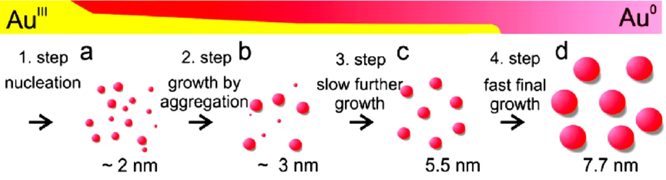
The recently proposed mechanism of gold nanoparticle formation could interpret as a four-step nucleation and growth process (Fig. 1-2). The initial stage is a rapid formation of nuclei and the second stage is that the nuclei coalesce into bigger particles. The third stage is low diffusion growth of AuNPs which comprised by ongoing process of gold reduction and a further coalescence. Finally, AuNPs completely consume their precursor and grow rapidly to their terminal size. This mechanism indicates that the coalescence of small nuclei plays a vital role throughout the AuNPs formation and determines the polydispersity of colloid AuNPs [Polte et al., 2010].
1-2-2. Local surface plasmon resonance
Surface plasmon resonance (SPR) is a physical characteristic that metallic materials such as Ag, Cu, Au, and Al, process a negative real and small positive imaginary dielectric constant over a range of wavelengths [Henry et al., 2011]. When these metallic materials stimulated by electromagnetic radiation, these would form an electron gas that moves away from its
equilibrium position and be displaced by induced surface polarization charges that act as a restoring force. This positive imaginary arises from Coulomb attraction between electrons and nuclei to against the electron gas. The collective oscillation of the conducted electrons is
4
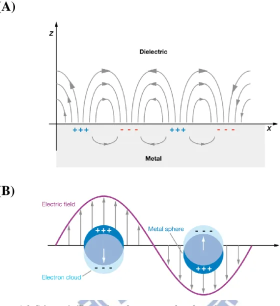
called the dipole plasmon resonance. The oscillation frequency is determined by four factors: the density of electrons, the effective electron mass, and the shape and size of the charge distribution [Jans et al., 2012; Kelly et al., 2003]. The SPR could category to propagating or localized. Propagating surface plasmon is observed on thin metallic films, called surface plasmon polaritons (SPPs); whereas localized surface plasmon is observed on nanoscale structures, which nanoparticles (NPs) are much smaller than the incident wavelength (Fig.
1-3). That plasmon oscillates locally around the nanoparticle with a frequency called local
surface plasmon resonance (LSPR). The LSPR also sensitive to changes in the local dielectric environment like SPR do [Willets et al., 2007].
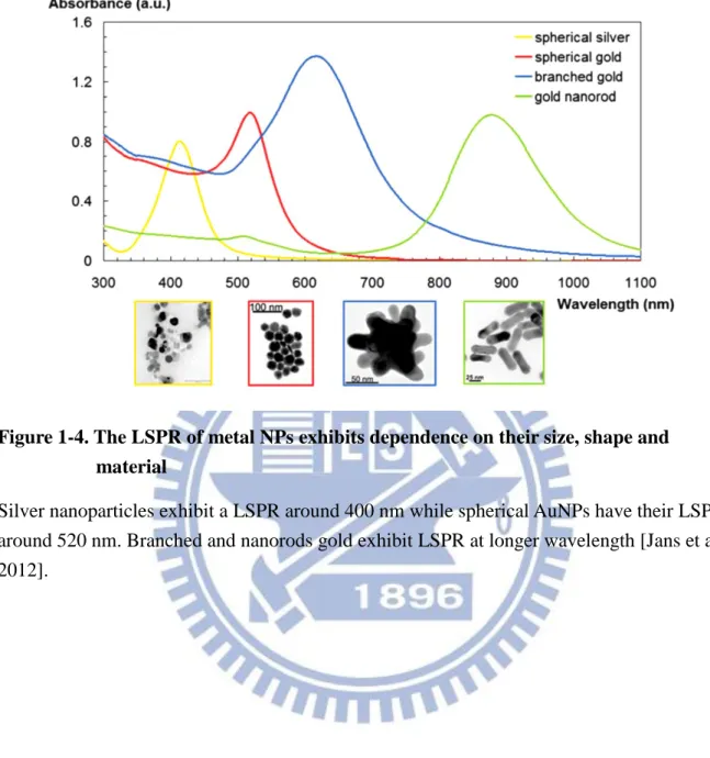
The LSPR spectrum is strongly dependent on the nanoparticle’s size, shape, dielectric constant and the surrounding environment under dielectric constant as mentioned (Fig. 1-4). AuNPs have an optical property which is bulk plasmon resonance in the visible region of the spectrum, while for most other metals this resonance only occurs in the ultraviolet (UV) region. Therefore, the strong absorption or scattering of AuNPs at the visible light region could make them could be observed by naked eyes or be easy to detect color change. Besides, the possibility of tuning the LSPR band of AuNPs (including nanorods, shells, stars, and other shapes) at the near IR region makes them promising materials for in vivo imaging and
analysis [Jans et al, 2012].
LSPR-based sensors are label-free techniques. By LSPR change, there are two types of LSPR-based sensors are conducted. One is based on aggregation of the colloid and results in apparent color change (from red to blue). Aggregation causes a coupling of the colloid plasma modes resulting in a red shift and broadening of the longitudinal plasma resonance in the UV-vis spectrum. Due to dipole—dipole interactions occur, the wavelength of absorption may be varied from 520 nm (effectively isolated particles) through 750 nm (particles that are separated by only 0.5 nm). The resulting spectrum consists of the conventional plasmon
5
resonance due to single spherical particles and the new peak due to particle—particle interactions [Ghosh et al., 2007].
The other type is based on LSPR extinction (or scattering) wavelength maximum, λmax,
which is sensitive to the dielectric constant of the surrounding medium or adsorbents. Thus, extinction wavelength maximum changes in the local environment, for example it should cause a shift in λmax in the presence of an adsorbed species. This relationship is expressed by the following equation:
∆λ𝑚𝑎𝑥 = 𝑚∆𝑛 [1 − 𝑒𝑥𝑝 (−2𝑑𝑙
𝑑 )] (1)
Here 𝑚 is the bulk refractive index response of the nanoparticle, ∆𝑛 is the change in the refractive index induced by the adsorbents, 𝑑 is the effective adsorbent layer thickness, and 𝑙𝑑 is the characteristic EM-field-decay length (approximated as an exponential decay). This relationship is the basis of LSPR wavelength-shift sensing experiments. When molecules bind to a AuNPs the refractive index will change and the LSPR band occurs red-shift. It can be deduced from this equation that the sensitivity towards refractive index changes is distance dependent. Only at close proximity to the nanoparticle surface will give rise to a shift of the LSPR wavelength. This makes the refractive index based biosensor be specific for
interactions close to the NPs surface [Jans et al., 2012; Willets et al., 2007].
Depending on the optical properties of spherical AuNPs as mentioned, the size and concentration of the spherical AuNPs could be determined by UV-Vis spectra. The extinction coefficient is an important parameter that can be used to calculate the NPs concentration or estimating the NPs size. According to Lambert-Beers law, the molar concentration of the solution can be obtained.
6
Liu et al. [2007] used high resolution transmission electron microscopy analysis and UV-vis absorption spectroscopic measurement to determine the extinction coefficients of AuNPs with the size ranging from 4 to 40 nm. An equation was provided that it is
independent to the capping ligands on the NPs surface and the solvent dissolve the NPs, ln 𝜀 = 𝑘𝑙𝑛𝐷 + 𝑎 (3) Where 𝜀 is extinction coefficient in M-1cm-1, 𝐷 is the core diameter of the NPs, and 𝑘 = 3.32, 𝑎 = 10.8. The correlation coefficient is 0.99 and the standard deviation is 0.22.
Haiss et al. [2007] provided equations according to SPR peak that can determine bare spherical AuNPs size and concentration. For particles ranging from 35 to 100 nm can be calculated from the peak position according to eq (4):
𝑑 = ln (
𝜆𝑠𝑝𝑟−𝜆0 𝐿1 )
𝐿2 (4)
Where d is the diameter of the spherical AuNPs, 𝜆𝑠𝑝𝑟 is the wavelength at the peak of the SPR; 𝜆0 =512; L1 = 6.53; and L2 = 0.0216. Haiss et al. found the average of the absolute error is only 3%.
The particle diameter in the size ranging from 5 to 80 nm has a better agreement between theory and experiment as the particle diameter is found if the absorbance ratios are
determined in the wavelength region below 600 nm. Hence the ratio Aspr/A450 should be
particularly suitable to calculate the particle diameter (in nanometers) from 5 to 80 nm, eq (5):
𝑑 = exp (𝐵1𝐴𝐴𝑠𝑝𝑟
450− 𝐵2) (5)
Here, 𝐴𝑠𝑝𝑟 is the absorbance at the SPR peak, A450 is the absorbance at 450nm, B1 = 3.00 and B2 = 2.20. According to eq (5), the particle diameter average deviation is about 11%.
7 N = 𝐴450×1014 𝑑2[−0.295+1.36𝑒𝑥𝑝(−(𝑑−96.8 78.2 ) 2 )] (6)
Where A450 is the absorbance at 450nm, and d is the particle diameter (nm). Haiss et al. found
this equation to be accurate to ~6% [Haiss et al., 2007]. Above equations are also used by NanopartzTM as a tool to determine size and concentration of AuNPs.
1-2-3. Fluorescence quenching mechanisms by AuNPs
Fluorescence activatable probes are comprised of at least two components: the
fluorophore (donor, D) and the quencher (acceptor, A). Any process that causes a decrease in intensity can be considered to be quenching [Lakowicz, 2006]. The fluorescence emission can be altered when the fluorophore is placed near an entity possessing an EM field. Typically metal entities are nano-sized, such as gold, silver, platinum, copper NPs, etc [Kang et al., 2011]. In the development of activatable probes, inorganic AuNPs owns very high sensitivity, which has the highest quenching efficiency (up to 99%) [Swierczewska et al., 2011].
There are two main factors considered to affect the changes on the fluorescence by metal NPs: (1) the plasmon field generated around the particle by the incident light, increasing the excitation decay rate of the fluorophore or enhancing the level of fluorescence emission; and (2) the dipole energy around the nanoparticle reduces the ratio of the radiative to
non-radiative decay rate and the quantum efficiency of the fluorophore, resulting in the fluorescence quenching [Kang et al., 2011]. Plasmon field on AuNPs effect fluorescence by quenching/enhancing was discussed as followings. Mie theory presented a solution to
Maxwell’s equations that describes the extinction spectra (extinction = scattering + absorption) of spherical particles of arbitrary size [Kelly et al., 2003]. According to Mie theory and the size and shape of the particle, the extinction of metal colloids can be due to either absorption or scattering [Yguerabide et al., 1998ab]. Incident energy is dissipated by absorption; and far-field radiation is created by scattering. The radiating plasmon (RP) model provided by
8
Lakowicz [2005] conducted with Mei theory that small colloids are expected to quench fluorescence because absorption is dominant over scattering; while larger colloids are expected to enhance fluorescence because the scattering component is dominant over absorption. The RP model for NPs explains that their induced plasmon can radiate when the scattering cross section rules over the absorption cross-section. The particle cross section for extinction (𝐶𝐸) with a dielectric constant ε1 is dependent on the cross section due to
absorption (𝐶𝐴) and scattering (𝐶𝑠) by:
𝐶𝐸 = 𝐶𝐴 + 𝐶𝑆 = 𝑘1𝐼𝑚(𝛼) +𝑘14
6𝜋|𝛼|
2 (7)
where 𝑘1 is the wavevector of the incident light in medium. Polarizability (𝛼) of a sphere with a radius r is:
α = 4πr3(𝜀𝑚−𝜀1
𝜀𝑚+2𝜀1) (8)
where 𝜀𝑚 is the complex dielectric constant of the metal. The absorption term, 𝐶𝐴 is responsible for quenching, while the scattering term, 𝐶𝑆 can cause fluorescence enhancement. As seen by this model, the NPs size plays a more significant role in 𝐶𝑆 (𝑟3) over 𝐶𝐴 (𝑟3). Therefore, smaller NPs are preferred for quenching. In accordance with the RP model, AuNPs with diameter below 40 nm are more efficient fluorophore quenchers; while larger colloids above 40 nm are expected to enhance fluorescence, because scattering becomes the dominant mechanism [Swierczewska et al., 2011].
Kang et al. [2011] theoretically studied the plasmon field on AuNPs effect to the fluorescence and provided five important factors for designing the quenching and enhancement effect by metal NPs. The normalized enhancement of excitation decay rate (𝛾𝑒𝑥𝑐
𝛾𝑒𝑥𝑐0 ) , which is the main cause for fluorescence enhancement, shows more significant
9 (𝐸𝑝 𝐸0): 𝛾𝑒𝑥𝑐 𝛾𝑒𝑥𝑐0 = ( 𝐸𝑝 𝐸0) 2 (9)
Where the superscript ‘0’ is the value of the system without AuNPs, the plasmon field
strength at a distance (𝛾) to AuNP core is 𝐸𝑝, and an incident light field to AuNP is 𝐸0. The quantum yield (q) indirectly influenced by the plasmon field 𝐸𝑝 can be described as: 𝑞 𝑞0 = 𝛾𝑟 𝛾𝑟0 𝛾𝑟 𝛾𝑟0+ 𝛾𝑎𝑏𝑠 𝛾𝑟0 + (1−𝑞0) 𝑞0 (10)
where 𝛾𝑟 is the radiative decay rate, 𝛾𝑎𝑏𝑠 is the additional non-radiative decay rate resulted from the radiated energy absorbed by the particle, and 𝑞0 is the intrinsic quantum yield of the fluorophore. For a spherical particle with a quasi-static polarizability, 𝛾𝑟
𝛾𝑟0=
𝛾𝑒𝑥𝑐
𝛾𝑒𝑥𝑐0 .
The fluorescence enhancement rate (φ) is, therefore, the combined effect of the enhancement of the excitation decay rate and the change in the quantum yield, both influenced by the plasmon field.
φ =𝛾𝑒𝑥𝑐
𝛾𝑒𝑥𝑐0
𝑞
𝑞0 (11)
Kang et al. [2011] provided five important factors for designing the quenching and enhancement effect by metal NPs: (1) The metal type of the particle for example: AuNP, which the dielectric permittivity of the metal determines the plasmon field distribution. (2) The NPs size, i.e., field strength and the enhancement of the excitation decay rate depends on the particle size, (eq (9)). (3) The fluorophore to be used, which determines the wavelength. The field strength depends upon the excitation wavelength, and the level of absorption of the emission light by the NPs varies depending upon the emission wavelength, (eq (10)). (4) The
10
intrinsic quantum yield of the flurophore: It is one of the major factors that determine the quantum yield of the fluorophore placed near the nanoparticle, (eq (10)). (5) The placement of a shell on the surface. The plasmon field distribution may change significantly depending on the material properties of the shell on the particle [Kang et al., 2011].
(2) The radiative and non-radiative energy transfer of fluorophore to AuNPs
Quenching efficiency depends on the measure of the fluorescence decay rate (Rfluo),
radiative decay rate (Rrad), nonradiative decay rate (Rnonrad), and the fluorescence quantum
efficiency (η). The fluorescence decay rate is the inverse of the fluorescence lifetime (t), Rfluo = 1/τ and can be expressed as the sum of the radiative and nonradiative decay rates
[Dulkeith et al., 2002]:
Rfluo = Rrad + Rnonrad (12)
η = Rrad/ Rfluo (13) The process of the dye molecule releases a photon returning to the ground state is called radiative decay; while the excited photon cannot return to its ground state due to various processes (such as intersystem crossings or heat dissipation) is called non-radiative decay. Radiative and nonradiative rates depend on the size and shape of the NPs, the distance between the fluorophores and NPs, the orientation of the dye molecule binding onto the AuNPs, and also on the overlap of the fluorophore’s emission and NP absorption [Swierczewska et al., 2011].
At those small distances, the large fluorescence quenching efficiency of 99.8% is due to two effects of equal importance: first, the AuNPs increase the non-radiative rate (Rnonrad ) of
the molecules due to energy transfer, and second, the radiative rate (Rrad) of the molecules is
decreased because the molecular dipole and the dipole induced on the AuNP radiate out of phase if the molecules are oriented tangentially to the AuNPs surface [Dulkeith et al., 2005].
11
Three mechanisms used to explain energy transfer between a donor and AuNP of fluorescence quenching are discussed here, such as Gersten–Nitzan (GN), Fluorescence Resonance Energy Transfer (FRET) and Nanometal Surface Energy Transfer (NSET) models. In general, the quantum efficiency (𝜑𝐸𝑛) of energy transfer efficiency can be written as:
𝜑𝐸𝑛 = 1
1+(𝑅0𝑅)𝑛 (14)
Where 𝜑𝐸𝑛 is dependent on the distance between the donor and acceptor (𝑅), and the 50% quenching distance (𝑅0) [Ray et al., 2007].
The Gersten-Nitzan model (GN Model) describes a coupling of the fluorophore induced plasmon and strong electric field of AuNPs. Both radiative (fluorescence enhancement) and non-radiative (fluorescence quenching) rates are taken into account under this model. In eq (14), n = 6, and 𝑅0 is: 𝑅0𝐺𝑁 = [2.25 𝑐3 𝜔𝐷3𝜑𝐷𝑎 3 (𝜀1+2)2+𝜀22 𝜀2 ] 1 6 ⁄ (15)
where 𝜔𝐷 is the frequency of the donor dye, 𝜑𝐷 is the quantum yield of the donor, a is the radius of the metal nanoparticle, 𝜀1 and 𝜀2 are the real and imaginary components of the dielectric constant of the metal, respectively, and 𝑐 is the speed of light. The GN model is able to show that a small dipole from the fluorophore can induce a large dipole in the NPs. Such an enhancement in the dipole increases energy transfer efficiencies by 104 ~ 105.
However, such strong interactions may underestimate the quenching abilities of AuNPs due to the rapid damping of the electric field on their surface [Swierczewska et al., 2011].
Dulkeith et al. [2005] applied GN model to find out how the influence of AuNPs on the quantum yield of fluorophores ceases when the separation between the two species is
gradually increased. They proved that the energy transfer rate is 2 orders of magnitude smaller than that calculated using the GN model and that the distance independent (2.2 ~ 16.2 nm) is
12
opposite expected from the Förster theory. Implying energy transfer does not play an important role in quenching with longer distance. In other words, the reduced quantum efficiency may be due to the reduced radiative rate and not energy transfer. This discrepancy is attributed to the fact that the GN model does not take into account nonlocal effects but the Förster theory does [Dulkeith et al., 2005].
FRET is an electro-dynamic phenomenon, occurring between a donor (D) molecule in the excited state and an acceptor (A) molecule in the ground state. The donor molecules typically emit at shorter wavelengths that overlap with the absorption spectrum of the acceptor (Fig. 1-5A). The term resonance energy transfer (RET) is preferred because the process of long range dipole— dipole interactions between the donor and acceptor does not involve emission and reabsorption photons. The theory of energy transfer is based on a
fluorophore acts as an oscillating dipole, which can exchange energy with another dipole with a similar resonance frequency. Hence RET is similar to the behavior of coupled oscillators and is a non-radiative energy transfer.
The rate of transfer for a donor and acceptor separated by a distance r is given by
𝑘𝜏(𝑟) =𝜑𝐷𝜅 2 𝜏𝐷𝑟6( 9000(𝑙𝑛10) 128𝜋5𝑁𝑛4) ∫ 𝐹𝐷(𝜆)𝜀𝐴(𝜆)𝜆4𝑑𝜆 ∞ 0 (16)
where 𝜑𝐷 is the quantum yield of the donor in the absence of acceptor, n is the
refractive index of the medium, N is Avogadro's number, 𝑟 is the distance between the donor and acceptor, and 𝜏𝐷 is the lifetime of the donor in the absence of acceptor. The refractive index (n) is typically assumed to be 1.4 for biomolecules in aqueous solution. 𝐹𝐷(𝜆) is the corrected fluorescence intensity of the donor in the wavelength range λ to λ + Δλ with the total intensity (area under the curve) normalized to unity. 𝜀𝐴(𝜆) is the extinction coefficient of the acceptor at λ, which is typically in units of M–1 cm–1. The term 𝜅2 is a factor
13
𝜅2 is usually assumed to be equal to 2/3, which is appropriate for dynamic random averaging of the donor and acceptor.
The overlap integral (𝐽(𝜆)) expresses the degree of spectral overlap between the donor emission and the acceptor absorption:
𝐽(𝜆) = ∫ 𝐹𝐷(𝜆)𝜀𝐴(𝜆)𝜆4𝑑𝜆 =∫ 𝐹𝐷(𝜆)𝜀𝐴(𝜆)𝜆 4𝑑𝜆 ∞ 0 ∫ 𝐹0∞ 𝐷(𝜆)𝑑𝜆 ∞ 0 (17)
The rate of energy transfer from a donor to an acceptor 𝑘𝜏(𝑟) is given by
𝑘𝜏(𝑟) = 𝜏1 𝐷( 𝑅0 𝑟) 6 (18)
where 𝜏𝐷 is the decay time of the donor in the absence of acceptor, 𝑅0 is the Förster distance, and 𝑟 is the donor-to-acceptor distance. Hence, the rate of transfer is equal to the decay rate of the donor (1/𝜏𝐷) when the D-to-A distance (𝑟) is equal to the Förster distance (𝑅0), and the transfer efficiency is 50%. At this distance (𝑟 = 𝑅0) the donor emission would be decreased to half its intensity in the absence of acceptors. The rate of RET depends strongly on distance, and is proportional to 𝑟 = 6 [Lakowicz, 2006].
The distance at which RET is 50% efficient is called the Förster distance (𝑅0) , which is typically in the range of 20 to 60 Å . At this distance, half the donor molecules decay by energy transfer and half decay by the usual radiative and non-radiative rates. With 𝑘𝜏(𝑟) = 1/𝜏𝐷 obtains: 𝑅0𝐹𝑅𝐸𝑇 = [9000(𝑙𝑛10)𝜅2𝜑𝐷𝐽(𝜆) 128𝜋5𝑁𝑛4 ] 1 6 ⁄ (19)
In FRET, the acceptor, AuNPs, are estimated to be molecular with little disruption placed on it by the donor. Therefore, this energy transfer model does not describe the strong effect of dipole interactions towards the AuNPs SPR.
14
physical origin for NSET is attributed to the interaction of the electromagnetic field of the donor dipole interacting with the nearly free conduction electrons of the accepting metal. These conduction electrons behave like a Fermi gas and will interact most strongly with the oscillating dipole if they travel near and perpendicular to the metal surface. Because the AuNPs electrons are homogenously oriented, the constraint on dipole—dipole coupling has been greatly relaxed and thus gives rise to energy transfer efficiency at much larger distances. NSET does not require a resonant electronic transition, which is fundamental to a Förster theory. The dipole does not interact with a discrete resonant electronic transition, but rather an interaction with the electronic continuum levels of a metallic system [Yun et al., 2005].
According to the Persson and Lang model, the quenching mechanism can be explained as interactions of oscillating electronic dipoles of a dye with plasmon bands of a metal surface [Persson and Lang, 1982]. Plasmon bands consist of free conduction electrons and result in quenching when interacting with oscillating dipoles of the dye in close proximity to the metal surface. NSET models energy transfer from a molecular dipole to a nanometal surface at twice the FRET range[Rahul et al., 2009].
In eq (14), 𝑛 = 4 and 𝑅0 is replaced with 𝑑0, because the distance is between the donor and surface, not the acceptor. The value is calculated as:
𝑑0𝑁𝑆𝐸𝑇 = 0.525 ( 𝑐3𝜑𝐷 𝜔𝐷2𝜔𝐹𝑘𝐹) 1 4 ⁄ (20)
where 𝜑𝐷 is the quantum yield of the donor, 𝜔𝐷 is the angular frequency of the donor emission, and the other values are constants: c = 3 × 10⁸ m s−1, the speed of light,
𝑘𝐹 = 1.2 × 108 cm-1
, and 𝜔𝐹 = 8.4 × 10¹⁵ rad s-1. This model takes into account the limitation that FRET has in explaining fluorescence quenching by metal NPs, namely the small length of energy transfer [Swierczewska et al., 2011]. Distinguishing NSET from FRET is in two significant aspects: (1) the distance dependence changes from 1/R6 to 1/R4, which
15
extends the usable distances for the measurement; and (2) the same NP is able to quench dyes of different emission wavelength, spanning the visible range into the near infrared. Therefore, NSET can be used for the studies in which distances are expected to extend beyond 10 nm, or the studies with multiple dyes needed to be quenched [Ray et al., 2007].
Rahul et al. [2009] applied NSET mechanism to modeling the functional AuNPs (5 and 10 nm) with two dyes (Cy3 and Cy5) spacing from the particle surface by a rigid DNA spacer allows precise for determining of the distance-dependent effect of the metal NPs on
fluorescence intensity. Fluorescence is quenched significantly for distances somewhat larger than the particle diameter, in good agreement with the predictions of an NSET model based on interacting dipoles. The distance dependence of surface energy transfer behavior, i.e. quenching efficiency, is proportional to 1/d4, which involves no consideration of the size of
the particle and the spectral overlap of the dye and AuNPs. This surface energy transfer model is found qualitatively and agrees with the NSET model, though the exponent is greater than 4 for the smaller NPs (5 nm), and smaller than 4 for the larger NPs (10 nm).
1-2-4. Derjaguin-Landau-Verwey-Overbeek theory
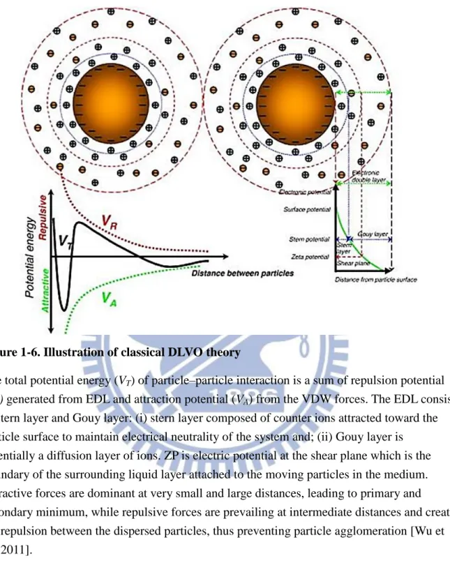
Derjaguin-Landau-Verwey-Overbeek (DLVO) theory is usually applied to explain the mechanism of stabilization of colloidal materials in water. The DLVO theory assumes that the forces acting on the colloidal particles in a medium include repulsive electrostatic forces and attractive van der Waals (VDW) forces. VA is determined by the Hamaker constant, particle size and inter-particulate distance while VR depends on particle size, distance between the
particles, zeta potential, ion concentration and dielectric constant of the medium. The repulsive forces are originated from the overlapping of electrical double layer (EDL) surrounding the NPs in the medium, and thus preventing colloidal agglomeration. The EDL consist of two layers: stern layer and Gouy layer. Zeta potential (ZP) is electric potential at the
16
shear plane which is the boundary of the surrounding liquid layer attached to the moving particles in the medium. ZP is a key parameter widely used to predict suspension stability; which the higher the ZP, the more stable the suspension is [Kim, 2004]. Attractive forces are dominant at very small and large distances, leading to primary and secondary minimum, while repulsive forces are prevailing at intermediate distances and create net repulsion between the dispersed particles, thus preventing particle agglomeration (Fig. 1-6). VR is extremely
sensitive to ion concentration in the medium. As the ion strength is increased in the medium, the thickness of EDL decreases due to screening of the surface charge. This causes decrease in
VR, increasing the susceptibility of the dispersed particles to form aggregates [Wu et al., 2011].
The DLVO theory explains that the total interaction potential (𝑉𝑇) between two AuNPs can be expressed as the sum of electrostatic repulsion due to the so-called double layer of counter ions (𝑉𝑒𝑙𝑒𝑐) and the van der Waals attraction (𝑉𝑣𝑑𝑤) [Derjaguin et al., 1941; Verwey, 1947].
𝑉𝑇 = 𝑉𝑒𝑙𝑒𝑐+ 𝑉𝑣𝑑𝑤 (21) Depending on the particle size and the double layer thickness, the electrostatic repulsion potential (𝑉𝑒𝑙𝑒𝑐), between two colloidal particles of radii 𝑅1 and 𝑅2 can be expressed in the following two different forms: [Hunter et al., 1992]
𝑉𝑒𝑙𝑒𝑐 = 4πϵ𝜑02 𝑅1𝑅2 𝑅1+𝑅2𝑙𝑛(1 + 𝑒 −𝜅𝑥) (which κR > 5) (22) 𝑉𝑒𝑙𝑒𝑐= 4πϵ𝑅1𝑅2𝑌1𝑌2(𝑘𝐵𝑇 𝑒 ) 2 𝑒−𝜅𝑥 𝑥+𝑅1+𝑅2 (which κR < 5) (23) Which, κ = [1000𝑒2𝑁𝐴(2𝐼) 𝜖𝑘𝐵𝑇 ] 1 2 ⁄ (24)
17 𝑌𝑖 = 8𝑡𝑎𝑛ℎ(𝑒𝜑0⁄(4𝑘𝐵𝑇)) 1+[1−2𝜅𝑅𝑖+1 (𝜅𝑅𝑖+1)2𝑡𝑎𝑛ℎ 2(𝑒𝜑0⁄(4𝑘𝐵𝑇))] 1 2 ⁄ (25)
Where ϵ is the permittivity of the medium, κ is the potential at the particle surface, 𝜑0 is the inverse Debye length, 𝑥 is the distance of closest approach between two colloidal particle surfaces, 𝑒 is the electronic charge, 𝑘𝐵 is the Boltzmann’s constant, 𝑇 is the temperature, 𝑁𝐴 is the Avogadro number, and 𝐼 is the ionic strength of the solution. The potential at the particle surface can be estimated from the ξ potential measurements.
Assuming the particles to be spherical and the surface potential and the background ionic strength to be constant, the van der Waals attraction potential, 𝑉𝑣𝑑𝑤 [Verwey et al., 1955]:
𝑉𝑣𝑑𝑤= −𝐴6𝐻[𝑑2−(𝑅2𝑅11𝑅+𝑅22)2+ 2𝑅1𝑅2 𝑑2−(𝑅1−𝑅2)2− 𝑙𝑛 𝑑2−(𝑅 1+𝑅2)2 𝑑2−(𝑅1−𝑅2)2] (26)
The van der Waals interaction (𝑉𝑣𝑑𝑤) is the dominantattraction and is dependent on the particle radii: 𝑅1 and 𝑅2, the center-to-center separation distance, 𝑑, and the Hamaker constant, 𝐴𝐻, which plays an important role in the description of attraction energy between the particles. The Hamaker constant (𝐴𝐻) of gold particles from literature varies in the range (1 − 4 × 10⁻¹⁹) J, so the average value, 2.5 × 10⁻¹⁹ J is common used [Evans et al., 1999; Ghosh et al., 2007].
The generation of electrostatic repulsion between charged conjugates is the most common strategy to keep NPs separated in aqueous medium [Bastús et al., 2008].
Citrate-capped AuNPs are considered less stable which of better capping agents should better be neutrally charged and high molecular weight [Stankus et al., 2011]. AuNPs are surrounded by an electrical double layer due to adsorbed citrate and chloride anions. An increase in ionic strength of the medium compresses the double layer and shortens repulsion range leading to an irreversible particle aggregation.
18
Changes in AuNPs solution environment usually lead to change of the dispersions; nevertheless, excess citrates and non-AuNPs components (e.g. chloride ions) are expected to remain in the product solution but should be removed before use [Balasubramanian et al., 2010]. Therefore, modified AuNPs are often synthesized with the existence of byproducts, which the environment is electrostatic stabilized. On biological issue, it is crucial to remove undesired components such as non-AuNPs components and excess peptide substrates, both of which can cause misleading results. For example, during an in vitro assessment of cytotoxicity, the presence of citrate, rather than AuNPs, reduced viability and impaired proliferation of human alveolar cells [Uboldi et al., 2009]. However, purification process like centrifugation will destine to change the environment of AuNPs suspension leading to aggregation. The surface characteristic and the aqueous medium are very crucial to stability of AuNPs suspension.
1-2-5.
Stabilization of AuNPs
Stabilization of AuNPs by ligands or capping agents is not only important for their long-term stability but also important applications in biomedicines and biotechnology. The citrate-capped AuNPs can be functionalized by thiol ligands to form monolayer-protected gold clusters (MPCs). Thiol ligands including straight chain alkanethiolates of different length [Mrksich et al., 1996; Sellers et al., 1993], glutathione [Schaaff et al., 1998], mercaptophenol [Brust et al., 1995], tiopronin [Cui et al., 2012; Kohlmann et al., 2001], thiolated poly
(ethylene glycol) [Manson et al., 2011; Wuelfing et al., 1998] or peptides [Harkness et al., 2012]. AuNPs also have been stabilized by proteins, DNA and carbohydrate moieties [Housni et al., 2008]. Some proteins, particularly antibodies, can adsorb strongly to AuNPs to form stable conjugates with retaining its’ biological property. A major drawback of the affinity of proteins to AuNPs is undesired or non-specific labeling to other components in the biological
19
system. Hence, AuNPs must be stabilized with an inert macromolecule such as bovine serum albumin, gelatin, or polyethylene glycol. Stabilization can usually be done through washing with buffer containing the macromolecules after the absorption of the desired conjugates onto the AuNPs [Thobhani et al., 2010].
There are two capping agents are conducted in this thesis. One is polyethylene glycol (PEG) which is an ideal neutral polymer, also known as polyethylene oxide (PEO) or
polyoxyethylene (POE), depending on its molecular weight. PEG is soluble in many different solvents, ranging from water to many organic solvents such as toluene, methylene chloride, ethanol and acetone. PEG can improve thermal and mechanical stability, which also could reduce sensitivity to pH and salts [Chapman, 2002]. PEG can resist opsonization and effectively make hydrophobic AuNPs be “stealth particles” with extended circulation times [Niidome et al., 2006]. The cytotoxicity results of PEG stabilized AuNPs prove the
biocompatibility of this molecule [Pan et al., 2007]. It is known that PEG-8000 could greatly stabilize NPs [Guarise et al., 2006; Juewen et al., 2006; Wang et al., 2010].
The other is bovine serum albumin (BSA) which is one of the most widely used proteins. Serum albumin is the most abundant protein in blood plasma, serving as a vehicle for
intracellular transportation; besides, it is important in pharmacology that albumin conjugated to drugs could decrease toxicity. There are many researches showed that BSA conjugated NPs show improved stability against flocculation, increased quantum yield and low toxicity [Brewer et al., 2005; Housni et al., 2008]. Brewer et al. [2005] used the ξ-potential
measurements to show that the stabilization of AuNPs by BSA has a significant contribution from a steric mechanism because AuNPs are stable even at their isoelectric point (pI 4.6). The interaction between BSA and a citrate-coated gold surface or a nanoparticle is not a displacement but electrostatic interactions which must consist of salt-bridges, for example, of the carboxylate-ammonium type, between the citrate and the lysine on the protein surface.
20
The binding mechanism can be verified when: (1) at low adsorbed concentrations, BSA exhibited a side-on conformation blocking colloid deposition; (2) at high adsorbed
concentrations, BSA adapted to an end-on conformation promoted colloid deposition; and (3) colloid deposition on the BSA layer may progressively generate end-on molecules (sites) by conformation change of side-on BSA, resulting in sustained increasing deposition rates [Yang et al., 2012].
1-2-6.
Peptide design for conjugation to AuNPs
The basic design principle was to create a ligand that would readily attach to the surface of the gold particle and form a well-packed layer with a hydrophilic terminus. Ligands like peptides could be used to form MPCs; however, only a relatively small subset of all possible polypeptides does not flocculate under physiological conditions. The main concept of peptide design is to avoid electrostatic aggregation, which may cause by attractive interactions
between ammonium and carboxylate moieties. This is best achieved by ensuring that the particles have a positive or negative net charge, leading to overall repulsive interactions [Lévy et al., 2004]. An electrolyte-induced aggregation in peptide-stabilized AuNP could depend on variety, such as the concentration, sequence, length, hydrophobicity, and peptide charge; the AuNP size and solution pH [Fanun, 2010].
The conjugation of a peptide with biological activity to one AuNP is by means of spontaneous reaction via a thiol (Cys) or an N-terminal primary amine with AuNP surface. Thiols are the most important type of stabilizing molecules for AuNPs of any size, leading to the formation of strong Au–S bonds [Kogan et al., 2007].
Lévy et al. [2004] reported a rational design of peptide-capping ligands for AuNPs: pentapeptide CALNN (cysteine-alanine-leucine-aspargine-aspargine). They discussed peptide length and the sequence as: anchor residue in position 1, core residue of hydrophobic and
21
hydrophilic in position 2 and 3, and terminal residue in position 4 and 5. Increasing the length of the peptide (from CA to CAL, CALN, and to CALNN) could increase stability of the peptide-capped AuNPs under higher concentrations of NaCl. The thiol group in the side chain of the N-terminal cysteine (C) is able to form a covalent bond to the AuNP surface. Even using amino group supposed to bind on AuNP as anchor residue like lysine (K) and the substitution sequence is KALNN; however, the aggregation is observed. It clearly indicated that the thiol group plays a major role in stabilization [Lévy et al., 2004]. Bellino et al. [2004] reported that when a positively charged ammonium group presences in the vicinity of the thiol will significantly accelerates the adsorption kinetics onto citrate-stabilized AuNPs. Alanine (A) and leucine (L) in positions 2 and 3 possess hydrophobic side chains and were chosen to promote peptide self-assembly. The leucine side chain is larger than that of alanine taking into account nanoparticle curvature. Asparagine (N) in positions 4 and 5 is an uncharged, but hydrophilic amino acid due to the amide group on the side chain, and the C-terminal asparagine in position 5 can bear a negative charge due to the terminal carboxylic group [Lévy et al., 2004].
The peptide designs conducted by Olmedo et al. [2008] that they verified the position of Asp (D) in CLPFFD-NH2 conjugate. Olmedo et al. [2008] demonstrated that the peptide
sequence, steric effect, and charge and disposition of hydrophilic and hydrophobic residues are crucial parameters when considering the design of AuNP peptide conjugates for
biomedical applications. They conclude that the presence of a carboxylate group belonging to the D residue situated near the surface seems to induce the exclusion of D residues accounted to citrate carboxylates repulsive force. But the exposition of D residues could increase
repulsion between the AuNPs and ensure colloidal stability [Olmedo et al., 2008].
Yang et al. [2011] modified AuNP with hexapeptide consisting of a cysteine (C) residue as anchor residue and has four alanine residues (AAAA) as the core region which promotes
22
peptide assembly into a densely packed monolayer on the gold surface. The functional end group is a single amino acid, which can be positively charged arginine (R) or lysine (K), negatively charged glutamic acid (E), neutral hydrophilic serine (S), or hydrophobic
tryptophan (W). Only the peptide with negatively charged end residue, which is CAAAAE, does not aggregate at the physiological ionic condition (150 mM NaCl).
Above all researches emphasize on the residue selection and the position of
amine/carboxylate group to comprise a good stabilizer peptide for AuNPs. But Willett et al. [2005] suggested that the electrostatic charge of the peptides may play a role in the binding affinity between various inorganic surface and the peptides selected. Tullman et al. [2007] proposed that aggregation can be controlled by the electrostatic charge of peptide-modified AuNPs. They investigate four peptides (Biotin-KHKHFHF, Biotin-KHKHWHW,
Biotin-AHAHAHA, and Biotin-FHFHFHF) under a variety of peptide concentrations, modification and re-suspension buffer pH. Finally, they excluded the possibility of over loaded peptides make bridging flocculation but concluded that the electrical double layer (EDL) is disrupted when positively charged peptide modified on AuNPs and can lead to irreversible particle aggregation. However, at pH above the peptide’s pI, which the peptide is negatively charged will make AuNPs remain stable in solution, and peptides remain bound to the particles possibly through amine coordination of gold [Tullman et al., 2007].
23
1-3. Enzymes
1-3-1. Proteinase K
Proteinase K (also known as endopeptidase K or Pro-K; EC 3.4.21.64) is an extracellular endopoptidase isolated from a culture filtrate of the fungus Tritirachium album limber. This fungus is uniquely able to grow on and degrade Keratin as the sole source of carbon and nitrogen and therefore given the 'K designation'. Therefore, this enzyme was named “proteinase K” with respect to its keratin hydrolyzing activity [Ebeling et al., 1974].
Proteinase K is a member of the class of serine proteases (S8). Proteinase K is a subtilisin-like serine protease with the classic catalytic triad of serine (224), histidine (69) and aspartic acid (39) at its active site. Thus, proteinase K is a member of a new subfamily of the subtilisins [Jany et al., 1986]. A specificity of the enzyme for peptide bonds adjacent to the carboxylic group of aliphatic and aromatic amino acids was observed. The enzyme is stable over a broad range of pH (pH 4 ~ 12.5) and temperature (37 ~ 65°C) . Also, proteinase K is stable under wide range of salt concentration and is active in the presence of strong detergents, such as SDS. It has a molecular weight of 28.9 kDa [Berman et al., 2000; Betzel et al., 1988; Larson et al., 2009].
Proteinase K is commonly used in molecular biology to digest protein and remove contamination from preparations of nucleic acid. Proteinase K retains active under the presence of chemicals that denature proteins such as SDS and urea, chelating agents such as EDTA, sulfhydryl reagents, as well as trypsin or chymotrypsin inhibitors; therefore, makes it highly-suited to use in nucleic acid preparations. Proteinase K is used for the destruction of proteins in cell lysates (tissue and cell cultured) and for the release of nucleic acids, since it very effectively inactivates DNases and RNases [Goldenberger et al., 1995]. Proteinase K has also been used for selective protein digestion in order to identify prion proteins [Petrotchenko et al., 2012].
24
1-3-2. Chymotrypsin
Chymotrypsin (EC 3.4.21.1) is produced in the acinar cells of the pancreas as the inactive precursor, chymotrypsinogen. α-Chymotrypsin is the predominant form of active enzyme produced from it's zymogen, Chymotrypsinogen A. Two predominant forms of
chymotrypsin, A and B, are found in equal amounts in cattle pancreas; although they are about 80% similar in sequence but have significantly difference in proteolytic characteristics
[Hartley, 1964; Meloun et al., 1966]. In vivo, the rate of hydrolysis of the zymogen by trypsin and by autolysis produces varying amounts of α, π, δ and γ variants. α-Chymotrypsin is a serine endopeptidase of the peptidase S1 family consisting of 241 amino acid residues , which the molecular weight is 25.6 kDa and has optimal pH about 7 to 9 [Wilcox, 1970].
Chymotrypsin is activated through cleavage of the bond between arginine and isoleucine (R15 and I16) by trypsin, causing structural modifications and formation of the substrate binding site. The molecule has three peptide chains: A chain (13 residues), B chain (131 residues), and C chain (97 residues) as shown in Fig. 1-7.
Specificity of chymotrypsin for large hydrophobic residues can be explained by a hydrophobic S1 binding pocket formed by residues of amino acid 189 ~ 195, 214 ~ 220, and 225 ~ 228 [Vajda et al., 1976]. α-Chymotrypsin from bovine pancreas selectively catalyzes the hydrolysis of peptide bonds on the C-terminal side of tyrosine, phenylalanine, tryptophan, and leucine. A secondary hydrolysis will also occur on the C-terminal side of methionine, isoleucine, serine, threonine, valine, histidine, glycine, and alanine [Burrell, 1993].
Chymotrypsin is usually used as an indicator for evaluating pancreatic function [Bermudes et al., 2011; Kadhim et al., 2010] and related to pancreatic diseases [Goldberg, 2000; Piotrowski et al., 2003; Shimada et al., 2000].