On-Line Microdialysis-Graphite Furnace Atomic
Absorption Spectrometry in the Determination of Brain
Magnesium Levels in Gerbils Subjected to Cerebral
Ischemia/Reperfusion
Ming-Cheng Lin, MS, Yeou-Lih Huang, PhD, Hong-Wen Liu, MD, Dar-Yu Yang, MD, Chien-Pin Lee, MS, Lin-Lan Yang, Fu-Chou Cheng, PhD
Department of Medical Technology, Chung-Tai Institute of Health Sciences and Technology (M.C.L.), Department of Emergency (D.Y.Y.), Medical Research (C.P.L., L.L.Y., F.C.C.), Taichung Veterans General Hospital, Department of Applied Chemistry, Providence University (C.P.L.), Taichung, Taiwan Graduate Institute of Medicine, (M.C.L.), School of Technology for Medical Sciences (Y.L.H.), Kaoshiung Medical University, Department of Internal Medicine, Kaoshiung Medical University Hospital (H.W.L.), Kaoshiung TAIWAN
Key words: cerebral ischemia, gerbil, magnesium, microdialysis, graphite furnace atomic absorption spectrometry Objectives: Description of use of equipment for on-line microdialysis (MD) coupled with graphite furnace atomic absorption spectrometry (GFAAS) system, for dynamic monitoring of extracellular Mg in gerbils subjected to transient focal cerebral ischemia.
Methods: Gerbils’ right middle cerebral artery (MCA) and common carotid artery (CCA) were occluded for 60 minutes, and then reperfused for 60 minutes with Ringer’s solution, after which extracellular fluid samples were collected via a microdialysis probe inserted into the right cortex before, during and after inducing ischemia. Reperfusion was at a rate of 2L/min through the microdialysis probe, on-line diluted with measured water injected onto the GFAAS via an autosampler for Mg analysis.
Results: The detection limit of the Mg concentrations has ranged from 0.50 to 3.00g/L; our detection limit was 0.03g/L. We applied this on-line system to monitor extracellular Mg levels in the cortex during focal cerebral ischemia. Mg concentrations significantly decreased to 41% of baseline during cerebral ischemia and gradually returned to 67% of baseline after 60 minutes of reperfusion.
Conclusions: We presume that derangement of Mg homeostasis could be important in brain cell injury and is closely associated with cerebral ischemia event. The described analytic system permits autosampling in the brain and allows for continuous determination of Mg and trace minerals in minute sample volumes in a living system.
INTRODUCTION
Increasing evidence indicates that cerebral ischemia not only results in delayed neurological morbidity and mortality but also is a major cause of death in older adults [1]. Further-more, cerebral ischemia remains the third leading cause of death in the United States and the number of cerebral ischemic patients continues to increase every year [2].
The study of any disease process is advanced by means of
the availability of an optimum experimental model for labora-tory investigators. Focal cerebral infarction has been difficult to reproduce experimentally in many animal models because of the efficiency of the collateral cerebral circulation. Specifically, due to genetic variation, Mongolian gerbils possess unique physiology that allows a unilateral hemispheric infarction to be easily induced [3,4]. This unique characteristic is due to the absence of connecting arteries between the basilar and carotid circulatory systems. An incomplete circle of Willis is formed
Address reprint requests to: Dr. Fu-Chou Cheng, Department of Medical Research, Taichung Veterans General Hospital, Taichung 40705, TAIWAN. E-mail: [email protected]
Journal of the American College of Nutrition, Vol. 23, No. 5, 561S–565S (2004) Published by the American College of Nutrition
and each hemisphere therefore has an independent blood sup-ply [5,6]. Thus, Mongolian gerbils have been widely employed as an animal model of cerebral ischemia that is analogous to some forms of human stroke [3,4].
The essential mineral magnesium (Mg) is widely present in plants and animals, and its biological importance for living organisms has been described elsewhere [7]. In living organ-isms, the major biological function of Mg is as a cofactor in more than 300 enzymatic reactions. Mg modulates the activity of adenosine-triphosphatases (ATPases), which have central importance in energy metabolism [8].
Mg deficiency is correlated with a number of diseases and an inverse association between diseases and low blood Mg levels has been documented [9 –11]. In addition, Mg may have an influence in enhancing cerebral blood flow to ischemic areas [12]. A previous study indicated that Mg levels in the cortex remained unchanged in severe head injury patients [13], there-fore, further investigation is necessary to elucidate the role of Mg during cerebral ischemia.
In clinical laboratories today, conventional methods such as colorimetric assay, flame photometry, and ion-selective elec-trodes are commonly used for the measurement of Mg in a variety of biological samples [14 –16]. However, the above analytical technics cannot facilitate analysis and quantitative determination of the extremely small volumes and/or lower concentrations of Mg in the brain, and are therefore inappro-priate in investigating brain Mg involvement during cerebral ischemia. On the other hand, atomic absorption spectrometry (AAS) specifically flame AAS (FASS), is the most widely used technic for detection of Mg in trace levels and is considered to be a reference method. However, there are many problems that need to be overcome to apply this form of spectroscopy to determination of Mg in the brain. One problem is the extremely small sample volume available for spectrometry analysis. The other problem is the difficulty of assembling an autosampling system that can sense dynamic changes in situ. Thus, develop-ing an appropriate autosampldevelop-ing system in analysdevelop-ing Mg throughout an animal experiment is necessary and is beneficial to eliminate the above problems.
Within the past decade, microdialysis (MD) has become a standard in vivo sampling technic for extracellular fluids in discrete compartments of living systems [17–20]. Through a hollow fiber via a semi-permeable membrane with a selectable molecular weight cut-off, macromolecule-free samples can be obtained from extracellular fluids of tissues, organs or directly from body fluids. The major advantage of the MD technic is its autosampling ability. Physiological functions and the anatom-ical structure being studied remain intact, permitting a single animal to be used, without the necessity to sacrifice it to perform the experiment.
Previous studies of Mg concentrations in the brain were performed with conventional measurement technics and micro-dialysis, which lack real-time assay [21]. Therefore, the role of extracellular Mg in the brain during cerebral ischemia has
remained controversial. The purpose of the present study was to develop an on-line system to investigate dynamic Mg levels in the gerbil during focal cerebral ischemia/reperfusion.
MATERIALS AND METHODS
An on-line MD coupled with graphite furnace atomic ab-sorption spectrometry (GFAAS) to determine of extracellular Mg levels in the brain of gerbils subjected to cerebral ischemia/ reperfusion is shown in Fig. 1. The MD system consists of a microinjection pump (CMA/100, Carnegie Medicin, Stock-holm, Sweden) and a metal-free microdialysis probe with a 0.5mm diameter polycarbonate membrane (4 mm in length, a cut-off at 20 kDa, CMA/20, Carnegie Medicin, Stockholm, Sweden).
A Perkin-Elmer Model Analyst 300 atomic absorption spec-trometer (Perkin-Elmer, Uberlingen, Germany) was used for analyzing extracellular Mg concentrations. For atomization of Mg, the temperature was controlled at 1700°C and Mg was detected at a wavelength of 285.2 nm.
Six male gerbils (65– 85 g) were obtained from the Labo-ratory Animal Center at the Taichung Veterans General Hos-pital of the Republic of China (Taichung, Taiwan). These animals were allowed to acclimatize to their environmentally controlled quarters (25°C and 12:12 h light-dark cycle) before the experiments. The gerbils were anesthetized intraperitone-ally (i.p.) with chloral hydrate (400 mg/kg body weight), and with additional chloral hydrate (200 mg/kg) when needed
Fig. 1. A schematic diagram of the on-line microdialysis coupled graphite furnace atomic absorption spectrometry system for the deter-mination of extracellular Mg levels in a gerbil subjected to cerebral ischemia. A⫽ microinjection pump, B ⫽ microdialysis probe, C ⫽ stereotaxic apparatus, D⫽ diluent, E ⫽ atomic absorption spectropho-tometer, F⫽ graphite tube, G ⫽ integrated computer system.
throughout the experimental process. The body temperature was maintained at 37°C with a heating pad (CMA/150).
The right common carotid artery (CCA), exposed through a ventral midline incision in the neck, was carefully separated from the vago-sympathetic trunks and loosely encircled with sutures for later occlusion. The gerbil’s head was mounted on a stereotaxic apparatus (Stoelting, IL, USA) with the nose bar positioned 4.0 mm below the horizontal line. Following a midline incision, the skull was craniectomized to expose the right middle cerebral artery (MCA). An 8-0 suture (blue mono-filament polypropylene, DG, Davis-GECK, Wayne, N.J.) was positioned so that it encircled the middle cerebral artery for later ligation. The microdialysis probe (4 mm in length, CMA/ 20, Carnegie Medicin, Stockholm, Sweden) was stereotaxically implanted into the cortex (AP 0 mm, ML 5 mm, DV - 5.0 mm from Bregma).
Transient focal ischemic lesion was induced by simulta-neous occlusion of the right CCA and the right MCA for 60 minutes followed by 60 minutes of reperfusion [20]. The dial-ysis probe was perfused with Ringer’s solution (147 mM Na⫹; 2.2 mM Ca⫹⫹; 4 mM K⫹; pH adjusted to 7.0) at 2L/min using a CMA/100 microinfusion pump. Dialysate samples col-lected over the first 1 hour were discarded to prevent any interference from acute effects of the surgical procedures and the probe implantation. All reagents used were of analytical grade and were purchased from E. Merck. All containers were soaked with 20% of nitric acid, rinsed with water and then were dried in a clean room for later use.
RESULTS
A schematic diagram of on-line MD-GFAAS system is shown in Fig. 1. The optimum operating conditions for the GFAAS system were achieved in our previous study [21]. The calibration curve ranged from 0.50 to 3.00g/L (correlation coefficient value ⬎ 0.995) and the detection limit was 0.03g/L in the present assay. A recovery of 102% relative to an aqueous standard for Mg was observed.
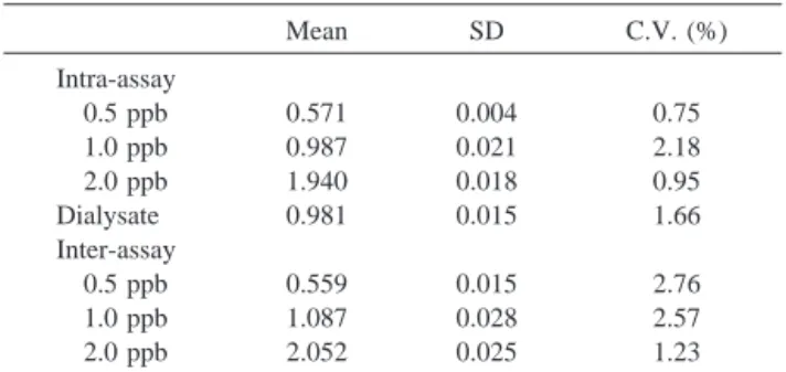
The precision and accuracy were tested using standard mixtures and pooled dialysate samples as shown in Table 1. The intra- (n⫽ 10) and inter-assay (n ⫽ 9) correlations were assessed and expressed as means and coefficients of variation (C.V. %). The C.V. values for detection of Mg were less than 3% in standard mixtures and pooled brain dialysates. The inter-assay variability of assessments of Mg over six consecu-tive days was less than 3%. The mean concentration of Mg after an on-line dilution in the basal dialysate was 1.5g/L. During cerebral ischemia, the mean Mg level significantly decreased to approximately 41% of the baseline levels and gradually returned to about 67% of baseline 1 hour after reperfusion (Fig. 2).
DISCUSSION
Cerebral ischemia can result from a wide range of distur-bances and is associated with several intra- and extracellular events leading to neuronal cell death. Many hypotheses have been proposed to explain the pathological and biochemical mechanisms underlying ischemic brain damage, including al-teration of energy metabolism, imbalance of metal ions, in-creased excitotoxicity, calcium overload, and free radical for-mation [22].
Mg is an allosteric activator of many enzyme systems and plays an important role in oxidative phosphorylation and gly-colysis [23]. Within the cell, Mg is bound primarily to proteins and negatively charged molecules; 80% of cytosolic Mg is bound to ATP. Mg has been proposed to possess neuroprotec-tive properties in several experimental models of ischemia. The possible mechanisms of neuroprotection include non-competi-tive blockade of NMDA receptors, inhibition of calcium entry into cells through leakage, enhancing cerebral blood flow to ischemic areas, and recovery of cellular energy metabolism after reperfusion [22]. Furthermore, extracellular Mg accounts for about 1% of the total body Mg content and provides for the maintenance of intracellular Mg levels.
Most ATP is used for maintenance of intracellular ho-meostasis and ATP-driven pumps for stabilization of trans-membrane concentration gradients of Mg, sodium, potassium and calcium [24].
Fig. 2. Time profiles of the changes in Mg concentrations (expressed as % of basal values) in the dialysates from gerbil cortices during 60 min CCA⫹ MCA occlusion and 1 h reperfusion. Data are presented as mean⫾ SEM (n ⫽ 6).
Table 1. Analytical Precision (Coefficient of Variation, C.V.
%) on Intra-Assay (n⫽ 10) and Inter-Assay (n ⫽ 9, in Nine Working Days) Stabilities of Mg Standard Solutions and Brain Dialysates in the GFAAS System
Mean SD C.V. (%) Intra-assay 0.5 ppb 0.571 0.004 0.75 1.0 ppb 0.987 0.021 2.18 2.0 ppb 1.940 0.018 0.95 Dialysate 0.981 0.015 1.66 Inter-assay 0.5 ppb 0.559 0.015 2.76 1.0 ppb 1.087 0.028 2.57 2.0 ppb 2.052 0.025 1.23
In general, stable basal levels of Mg in the cortex were obtained 1 hour after the start of measurement by the MD-GFAAS system. The mean concentration of Mg after an on-line dilution in the basal dialysate was 1.50g/L. During cerebral ischemia, the mean Mg level significantly decreased to approx-imately 41% of the baseline levels and gradually returned to about 67% of baseline 1 hour after reperfusion. An increase of Mg influx and/or a decreased energy-coupled efflux might have caused the decrease of Mg levels observed in the present study. Previous studies of biochemical processes have mainly been based on total Mg concentrations in brains obtained from experimental animals [25]. Total Mg rises in cells in a poor energy state with less ATP. Mg influx into the cell mainly occurs by means of diffusion from the higher free concentration in the extracellular space. The gradient over the cell membrane is a respiration-driven Mg influx or efflux depending on the intra- and extracellular Mg concentrations. We speculate that during cerebral ischemia, Mg influx may increase and an en-ergy-coupled efflux is depressed. Derangement of Mg ho-meostasis, that is closely associated with cerebral ischemia, can be an important factor in brain cell injury.
CONCLUSION
Conventional clinical methods for measuring Mg in living systems have many problems including precision, accuracy, specificity, and interference and therefore need to be improved to detect ultra trace levels and/or even the free form of extra-cellular Mg in brain. As demonstrated here, the on-line MD-GFAAS method was applied in the present study to measure extracellular Mg in extremely low concentrations and small sample volume in the brain of living Mongolian gerbils.
Accordingly, on-line MD-GFAAS system can provide great precision and accuracy for determination of Mg. This system is useful not only in autosampling in situ throughout the experi-mental process but also in monitoring the dynamics changes of minerals in extremely low concentrations and small sample volumes.
ACKNOWLEDGMENTS
This study was supported by grants from Taichung Veterans General Hospital (TCVGH-917306C & 917311D) and the Na-tional Science Council (NSC-91-2113M-075A-002), Taiwan, Republic of China.
REFERENCES
1. Hemphill JC: Ischemic stroke: Clinical strategies based on mech-anisms and risk factors. Neurology 55:42–52, 2000.
2. Yanigihara T: Experimental stroke in gerbils: Correlation of clin-ical, pathological and electroencephalographic findings and protein synthesis. Stroke 9:155–159, 1978.
3. Crockard AF, Iannotti AT, Hunstock RD, Smith RD, Harris RJ, Symon L: Cerebral blood flow and oedema following carotid occlusion in the gerbil. Stroke 11:494–498, 1980.
4. Kahn K: The natural course of experimental cerebral infarction in the gerbil. Neurol 22:510–515, 1972.
5. Levine S, Payan H: Effect of ischemia and other procedures on the brain and retina of the gerbil (Meriones unguiculatus). Exp Neurol 16:255–262, 1966.
6. Levy DE, Brierley JB: Communications between vertebral basilar and carotid artery circulations in gerbils. Exp Arch Neurol 28:503–508, 1973. 7. Wacker W, Parisi A: Magnesium metabolism. N Engl J Med
278:658–663, 1968.
8. Romani A, Scarpa A: Regulation of cell magnesium. Arch Bio-chem Biophy 298:1–12, 1992.
9. Haupt H, Scheibe F: Preventive magnesium supplement protects the inner ear against noise-induced impairment of blood flow and oxygenation in the guinea pig. Magnes Res 15:17–25, 2002. 10. Bussiere FI, Mazur A, Fauquert JL, Labbe A, Rayssiguier Y,
Tridon A: High magnesium concentration in vitro decreases human leukocyte activation. Magnes Res 15:43–48, 2002.
11. Dilsiz N, Olcucu A, Cay M, Naziroglu M, Cobanoglu D: Protective effects of selenium, vitamin C and vitamin E against oxidative stress of cigarette smoke in rats. Cell Biochem Funct 17:1–7, 1999. 12. Beyenbach KW: Transport of magnesium across biological
mem-branes. Magnes Trace Elem. 9:233–254, 1990.
13. Goodman JC, Valadka AB, Gopinath SP, Uzura M, Grossman RG, Robertson CS: Simultaneous measurement of cortical potassium, cal-cium, and magnesium levels measured in head injured patients using microdialysis with ion chromatography. Acta Neurochir. 75:35–37, 1999. 14. Abarca A, Canfrance E, Sierra I, Marina ML: A validated flame AAS method for determining magnesium in a multivitamin phar-maceutical preparation. J Pharmaceut Biomed 25:941–945, 2001. 15. Pasternak K: Tissue concentrations of magnesium in rats receiving
various dosages of Ethanol. Magnes Res 12:167–170, 1999. 16. Rob PM, Dick K, Bley N, Seyfert T, Brinckmann C, Hollriegel V,
Friedrich HJ, Dibbelt L, Seelig MS: Can one really measure magnesium deficiency using the short-term magnesium loading test. J Int Med 264:373–378, 1999.
17. Tseng WC, Yang MH, Chen TP, Huang YL: Automated, contin-uous, and dynamic speciation of urinary arsenic in the bladder of living organisms using microdialysis sampling coupled on-line with high performance liquid chromatography and hydride gener-ation atomic absorption spectrometry. Analyst 127:560–564, 2002. 18. Delgado JM, DeFeudis FV, Roth RH, Ryugo DK, Mitruka BM: Dialytrode for long-term intracerebral perfusion in awake mon-keys. Arch Int Pharmacodyn 198:9–21, 1972.
19. Cheng FC, Yang DY, Wu TF, Chen SH: Rapid on-line microdi-alysis hyphenated technique for the dynamic monitoring of extra-cellular pyruvate, lactic acid, and ascorbic acid during cerebral ischemia. J Chromatogr B 723:31–38, 1999.
20. Yang DY, Tsai TH, Cheng CH, Lee CW, Chen SH, Cheng FC: Simultaneous monitoring of extracellular glucose, pyruvate, lac-tate, and glutamate in gerbil cortex during focal cerebral ischemia by dual-probe microdialysis. J Chromatogr A 913:349–354, 2001. 21. Chen CJ, Cheng FC, Liao SL, Chen WY, Lin NN, Kuo JS: Effects
of naloxone on lactate, pyruvate metabolism and antioxidant en-zyme activity in rat cerebral ischemia/reperfusion. Neurosci Lett 287:113–116, 2000.
22. Lee M, Zipfel GJ, Choi DW: The changing landscape of ischaemic brain injury mechanisms. Nature 399:A7–14, 1999.
23. Kristian T, Siesjo BK: Calcium in ischemic cell death. Stroke 29:705–718, 1998.
24. Ito U, Spatz MJ, Walker TJ, Klatzo I: Experimental cerebral
ischemia in Mongolian gerbils. Acta Neuropathol 32:209–223, 1975.
25. Saris NEL, Mervaala E, Karppanen H, Khawaja JA, Lewenstam A: Magnesium: An update on physiological, clinical and analytical aspects. Clin Chim Acta 294:1–26, 2000.