On-line microdialysis sampling coupled with flow injection
electrothermal atomic absorption spectrometry for in vivo monitoring
of extracellular manganese in brains of living rats
Wei-Chang Tseng,
aYuh-Chang Sun,
bMo-Hsiung Yang,
cTyen-Po Chen,
dTe-Hsien Lin
eand Yeou-Lih Huang*
ea
Graduate Institute of Medicine, Kaohsiung Medical University, Kaohsiung, Taiwan
b
Nuclear Science and Technology Development Center, National Tsing-Hua University, Hsinchu, Taiwan
c
Department of Nuclear Science, National Tsing-Hua University, Hsinchu, Taiwan
d
Department of Internal Medicine, Kaohsiung Medical University Hospital, Kaohsiung, Taiwan
e
School of Technology for Medical Sciences, Kaohsiung Medical University, Kaohsiung, Taiwan. E-mail: [email protected]
Received 20th May 2002, Accepted 22nd November 2002
First published as an Advance Article on the web 6th December 2002
An on-line microdialysis sampling coupled with flow injection (FI) electrothermal atomic absorption spectrometry (ETAAS) method has been developed for in vivo monitoring of extracellular diffusible Mn in brains of living rats. Microdialysates perfused through implanted microdialysis probes were collected with a sample loop of an on-line injection valve and directly introduced into the atomizer of the ETAAS by a FI system. Ultrapure saline solution (0.9% NaCl) was used as the perfusion solution at a flow rate of 1 ml min21
through the microdialysis probe. The 25 ml of chemical modifier [0.4 g l21of Mg(NO3)2] and 25 ml of
microdialysate were on-line mixed at a micro-Tee and loaded into a sample loop. A six-port on-line injection valve equipped with a 50 ml of sample loop, a peristaltic pump and an autosampler arm of ETAAS for direct introduction of the sample into the graphite tube were employed to act as a homemade FI system. The optimized conditions of ETAAS were performed for measurement of Mn in microdialysate. The performance characteristics of the on-line microdialysis-FI-ETAAS system for Mn were as follows: linearity range, 0–20 ng ml21; detection limit (3s, n ~ 7), 0.43 ng ml21; recovery (n ~ 3), 98%–114%; precision (RSD, n ~ 20), 7.2%. The accuracy of the proposed on-line method was checked by four spiked artificial cerebrospinal fluid samples and compared with conventional ETAAS. The use of an on-line microdialysis-FI-ETAAS system permitted the in situ, dynamic and continuous in vivo monitoring of extracellular diffusible Mn in brains of living anaesthetized rats after administrated with MnCl2with a temporal resolution of 25 min.
Introduction
In recent years, microdialysis has become an important tech-nique for the in vivo sampling of the extracellular fluid in discrete compartments of living systems. Interest in microdialysis has increased significantly over the past decade as discussed in several reviews.1–4In the field of inorganic chemistry, micro-dialysis has been applied to monitor metals in different tissues by electrothermal atomic absorption spectrometry (ETAAS).5,6 These studies used off-line collection of the microdialysis sample and subsequent analysis by ETAAS. However, the sample volumes collected are only a few microliters and evaporation and contamination may be problems with manipulation off-line. In addition, conventional off-line procedures are usually time-consuming and tedious. The on-line analysis, on the con-trary, avoids some of the sample handling problems resulting from off-line collection. On-line microdialysis sampling coupled with ETAAS provides many advantages such as in situ sampl-ing and measurement, simplified sample preparation, rapid analyses and dynamic monitoring of trace elements in living systems.
To date, on-line microdialysis coupled with various analytical methods has been addressed in more than 100 original publica-tions. On-line microdialysis sampling coupled to liquid chromato-graphy,7capillary electrophoresis,8electrospray ionization mass
spectrometry,9electrochemical detection,10chemiluminescence systems11and tandem mass spectrometry12has been described. However, to our knowledge, no studies have been made to investigate the technique of on-line microdialysis coupled with ETAAS for continuous determination of trace elements in living systems. The key point of developing this on-line system is the interface between the microdialysis system and ETAAS. The interface between on-line sampling and measurement usually consists of a sample loop, an on-line injection valve and a peristaltic pump. Moreover, an important concept of flow injection (FI) is employed to develop the interface system.
Difficulties in the direct coupling of FI on-line to ETAAS are due to the discrete non-flow-through nature of ETAAS and the limited sample volume capacity of the graphite tube.13,14 Furthermore, complex samples cannot be directly processed by ETAAS owing to severe matrix interference. Nevertheless, extensive work has been successfully carried out in the last few decades on this subject, showing that there is great interest in developing automated FI-ETAAS on-line system. Many studies13,14have been aimed at automation of sample prepara-tion in recent years. Some innovaprepara-tions suitable for processing different kind of samples on-line in a continuous way in FI systems have been developed in order to automate (or at least to simplify) analytical procedures and to solve various specific analytical problems. The implementation of automated on-line 38 J. Anal. At. Spectrom., 2003, 18, 38–43 DOI: 10.1039/b204860d
Downloaded on 24 May 2012
sample pretreatment systems play a major role in the delicate stage of analyte transfer to the graphite tube.15 The on-line
coupling of FI-ETAAS has been proved to be a powerful technique for trace determination of a variety of elements.13–19 Microdialysis, shown to be a powerful technique for in vivo systems, is a continuous sampling method. Microdialysates are collected over a fixed time interval to provide the required sample volume, and each sample represents an average con-centration value obtained over this time interval. The use of a microdialysis probe in living systems allows efficient sampling of small target molecules while precluding large interfering species (e.g., proteins). Therefore, it is possible to couple microdialysis directly to FI-ETAAS because microdialysis can provide discrete samples and clean microdialysis samples are protein-free. Thus, the microdialysates can be allowed direct injection into the graphite tube through FI-ETAAS.
Manganese (Mn) is one of the essential trace elements required for living organisms for certain physiological func-tions such as brain development and metabolism.20 Mn is also a metal of toxicological concern mainly because chronic overexposure may lead to toxic effects in the central nervous system (CNS).21The deficiency or excess of Mn is known to cause neurotoxicity in experimental animals and man. Previous reports demonstrated that abnormally high amounts of Mn in the brain might lead to clinical signs resembling Parkinson’s disease.22,23 However, the exact mechanism of Mn toxicity in the CNS is not yet clearly understood, probably owing to the difficulty in measuring extracellular fluids in the brain. In the present work, taking Mn as a model element and magnesium nitrate [Mg(NO3)2] as the chemical modifier,24a novel
analy-tical system of on-line microdialysis sampling coupled with FI-ETAAS was developed for in vivo monitoring of extracellular Mn in brains of living rats.
Experimental
Apparatus
The microdialysis system was purchased from the Carnegie Medicine Associates (CMA, Stockholm, Sweden). The micro-dialysis sampling system consists of a microinjection syringe pump (CMA/100) and a 24 mm long microdialysis probe (CMA/20) with a 4 mm long and a 0.5 mm diameter poly-carbonate membrane, which is metal-free and has a molecular mass cut-off of 20 kDa. Connections of the microinjection syringe pump to the inlet of the microdialysis probe and the outlet of the microdialysis probe to the micro-Tee (Valco Instruments Co., Houston, TX, USA) were both accomplished with fluorinated ethylene polypropylene (FEP) tubing (internal volume of 1.2 ml per 100 mm length) (50 cm long 6 0.12 mm id) (CMA, Stockholm, Sweden). Another connection of the micro-injection syringe pump to the micro-Tee for introducing a chemical modifier was 100 cm long and 0.12 mm id FEP tubing. The micro-Tee was connected to the sample loop using 50 cm long and 0.12 mm id FEP tubing. The total dead volume of FEP tubing from probe to sample loop is approximately 12 ml. All connections of FEP tubing to the microinjection syringes, probe and the other FEP tubings were accomplished by tubing adaptors (CMA, Stockholm, Sweden), which ensure tight and zero internal volume connections.
The line FI interface was performed with a six-port on-line injection valve (Omnifit smart actuator, Cambridge, UK) equipped with a 50 ml of sample loop (PerkinElmer B050-6714), a peristaltic pump (Bio-Rad, USA) and an autosampler arm of ETAAS for introducing sample into the graphite tube. The on-line injection valve has an inert metal-free Teflon body designed for avoiding contamination of metals. The connections and conduits were made of polytetrafluoroethylene (PTFE) con-necting tubes (11 cm long 6 1.0 mm id, PerkinElmer B019-1058). The pump tubing (20 cm long 6 0.8 mm id, Bio-Rad,
USA) was employed for propelling the samples and air. A 20 cm delivery tube was used to connect the FI system with the sampling capillary of autosampler arm.
An atomic absorption spectrometer (PerkinElmer Model Zeeman 5100 PC), equipped with a graphite furnace (HGA-600), Zeeman-effect background correction, and an auto-sampler (AS-60) were used. The furnace was flushed with argon. The radiation source was a Mn hollow cathode lamp operated at 279.5 nm and a low 0.7 nm spectral slit width. Pyrolytic graphite coated graphite tubes (PerkinElmer) with an inte-grated platform were used. The system was operated through a DECpc 325sxLP personal computer and the associated AA Lab Benchtop software, version 7.0. Peak height (absorbance), peak area (integrated absorbance) and statistical data were printed out using an Epson LQ-570C printer.
Reagents and vessels
High-purity water (18.3 MV cm) was prepared with a deionized water system (Milli-Q, Millipore) and used throughout the work. All reagents used were of the highest available purity and at least of analytical grade. The perfusion solution (ultrapure saline) was prepared by dissolving 0.9 g of sodium chloride (Merck, ultrapure grade) in 100 ml of high-purity water. Chemical modifier, magnesium nitrate (Ultra Scientific, IMM-003, North Kingstown), was tested for optimal measurement of Mn in microdialysate. Stock solutions (1000 mg l21) of Mn were purchased from Merck. The working aqueous standards were prepared fresh daily with ultrapure saline. The test samples were all dissolved in artificial cerebrospinal fluid (aCSF) consisting of 147 mM NaCl, 2.7 mM KCl, 1.2 mM CaCl2, 0.85 mM MgCl2and 5.9 mM glucose.
Before each use, all containers and pipette tips were scrubbed in 20% nitric acid (Riedel-de Hae¨n, Germany) overnight, then were cleaned with high-purity water five times. Tubings used for connections of all apparatus were perfused with high-purity water, until the contamination was eliminated. In order to avoid contamination of Mn, metal-free syringes (Model 81320, Hamilton, USA) were used throughout this work as the perfusion syringes. Because no commercially metal-free needles were found, the polyether ether ketone (PEEK) tubing (0.12 mm id) was adhered to a syringe to make a homemade metal-free needle.
In vivo experimentation
Adult male Sprague–Dawley rats (350–450 g) were obtained from the Laboratory Animal Center at National Science Council of Republic of China (Taipei, Taiwan). These animals were specifically pathogen-free and were allowed to acclimate to their environmentally controlled quarters (25uC and 12 : 12 h light–dark cycle) for at least 5 days before experimentation and fasted overnight prior to sacrifice.
In order to be able to estimate the in vivo concentrations of Mn, it was necessary to determine the in vivo recovery of the microdialysis probe. A retrodialysis technique was used for the assessment of in vivo recovery. The microdialysis probe was inserted into the rat brain under anesthesia with urethane (ethyl carbamate). Perfusion solution containing Mn (10 ng ml21) was perfused through the probe at a constant flow-rate (1 ml min21) using the microinjection syringe pump. After a
2 h stabilization period after the surgical procedure, the perfusate (Cperf) and microdialysate (Cdial) concentrations of
the Mn were determined by ETAAS. The relative loss of Mn during retrodialysis (Lretro) or in vivo recovery (Rin vivo) by
dialysis, was then calculated as follows:25Lretro~ Rin vivo ~
[(Cperf2 Cdial)/Cperf].
The rats were initially anesthetized with urethane (1200 mg kg21 body weight, i.p.), and remained anesthetized throughout the experimental period. Following a midline incision of skull, the
J. Anal. At. Spectrom., 2003, 18, 38–43
Downloaded on 24 May 2012
brain cortex was exposed and a microdialysis probe (CMA/20, 4 mm dialysis membrane) was inserted. The microdialysis probe was implanted into the brain cortex (0 mm anterior, 6 mm lateral to the bregma and 4 mm from the brain surface). The probes were perfused with a saline solution. The flow rate of the perfusate was 1 ml min21. Microdialysates collected over the first 2 h were discarded to prevent acute effects from the surgical procedures. The probe was connected with on-line analytical system approximately 2 h after surgery. Basal Mn levels were monitored for at least 100 min prior to Mn administration. After intraperitoneal injection of manganese chloride (100 mg kg21 body weight), Mn was continuously
monitored every 25 min.
Results and discussion
Optimization of the on-line microdialysis-FI-ETAAS system Microdialysis system. Most microdialyser designs used for in vivo monitoring involve the use of short dialysis fiber membrane lengths of a few millimeters to facilitate direct inser-tion of dialysis probes into the living systems. The membrane is perfused with a solution of similar ionic strength as the surrounding fluid or tissue. Extremely low perfusion flow rates are therefore required to achieve acceptable recovery. In this work, a commercial metal-free probe (CMA/20) incorporating a relatively short microdialysis membrane of 4 mm was used because of the consideration of the depth of rat brain. In order to reduce the complexity of the matrix and avoid the con-tamination of Mn, the implanted probe was perfused with a ultrapure saline solution (0.9% NaCl) at a flow rate of 1 ml min21.
Moreover, all microdialysis equipment involving perfusion syringes, needles, tubings, microdialysis probe and micro-Tee were designed to be metal-free. The addition of a modifier in the microdialysate using another syringe on the same micro-injection syringe pump and a micro-Tee effectively pools two equal volumes of liquid into a sample loop of the on-line injection valve.
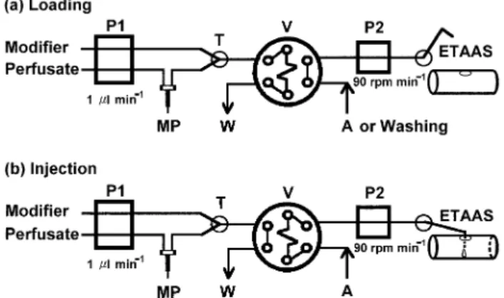
On-line FI interface operation. Instead of transporting the microdialysate off-line to the ETAAS, a homemade FI system was employed to act as an on-line sampling interface. FI is currently broadly accepted as the ideal sample introduction mode for ETAAS;15the coupling to microdialysis for on-line monitoring should further demonstrate its versatility. The FI manifold for the on-line microdialysis coupled with ETAAS is shown schematically in Fig. 1. Parameters used in this FI interface system for introducing the sample was mainly adapted from those reported by Ba¨ckstro¨m and Danielsson26 with minor modifications. In this system design, the FI
interface between the microdialysis sampling system and ETAAS analysis system must be considered for a variety of functions. Firstly, it must convert the continuous sampling stream of the microdialysis system into discrete samples for ETAAS analysis. Secondly, the delivery tube and sampling capillary of FI interface must be washed to expel the residual sample after introducing sample into atomizer. Finally, the interface must also match up the sampling time of the autosampler arm to introduce sample into graphite tube without loss of sample. The on-line interface described here performs all three functions.
An on-line injection valve can convert the continuous sampling stream of the microdialysis system into discrete samples. The sample, consisting of chemical modifier and microdialysate, flows directly into the sample loop on an on-line injection valve. A microdialysis syringe pump shown as pump 1 in Fig. 1 controls the flow. In the loading step [Fig. 1(a)], the microdialysis flow rate of 1 ml min21is converted
into a 50 ml sample by the on-line injection valve. This sample trapped in the sample loop is injected into the transfer tubing at timed intervals. The loading sequence is performed in parallel with the ETAAS measurement of the previous sample. Meanwhile, the 0.2% HNO3washing solution is run to expel
the residual sample in the delivery tube and sampling capillary. After a 25 min loading period, the on-line injection valve was switched to the injection position by a remote controlled handset.
In this work, automatic injection of small volumes retained in the collecting loop was performed by air displacement. When the microinjection syringe pump has delivered the preset volume, the on-line injection valve is switched to the injection position by a remote controlled handset. In the injection step [Fig. 1(b)], the air is introduced into the sample loop to propel the sample into the capillary tubing of the autosampler by a peristaltic pump. Meanwhile, the spectrometer computer is actuated to run the sampling step of ETAAS program. The autosampler was simultaneously actuated to put the tip of the sampling capillary into the injection hole of graphite tube. By adjusting flow rate of peristaltic pump, the 50 ml of sample (25 ml of microdialysate and 25 ml of modifier) is successfully introduced into the graphite tube. During the injection period, the microdialysate is led to waste without any increase of back pressure in the probe. Consequently, a part of this second fraction, corresponding to the injection time, is wasted. It is therefore important to have an injection time as short as possible. In this study, the valve is switched back to the load position after the preset 5 s of injection time. The total time for a single determination was 25 min containing about 2 min for detection.
ETAAS parameters for determination of Mn in microdialy-sate. Recently, a conventional ETAAS procedure has been developed to determine Mn in brain tissue.27 In that work, many chemical reagents were studied as effective chemical modifiers and calcium hydroxide was selected throughout the experiment. In the present study, magnesium nitrate was used as a chemical modifier in place of calcium hydroxide. Additionally, the conventional ETAAS procedure was com-pared with the proposed on-line microdialysis-FI-ETAAS system for an accuracy test.
Determination of trace elements by ETAAS must consider some important parameters, such as temperature program (Table 1), chemical modifier and matrix interference. Pyrolytic graphite coated graphite tubes with an integrated platform were used for Mn determination. The integrated platform is curved allowing the pipetting of larger sample volumes of up to 50 ml without difficulty. No problems were encountered in pipetting 25 ml of microdialysate and 25 ml of modifier into the graphite tube. A 20 ng ml21Mn standard dissolved in micro-dialysate matrix was employed in all instances.
Fig. 1 FI manifold for on-line microdialysis coupled with ETAAS. For details see text. P1, microinjection syringe pump; MP, microdialysis probe; T, micro-Tee; W, waste; V, on-line injection valve; A, air; P2, peristaltic pump; ETAAS, electrothermal atomic absorption spectro-meter. Valve position: (a) loading; (b) injection.
J. Anal. At. Spectrom., 2003, 18, 38–43
Downloaded on 24 May 2012
The 160 uC drying temperature is enough to remove solvent from the microdialysate. However, solvent was observed to spray to the wall of graphite tube when only one step was used to suddenly reach 160uC. Therefore, two drying steps (at 90 and 160uC) and long drying ramp time (10 s) were necessary to avoid sputtering of sample.
The effects of the pyrolysis temperature and chemical modifier were studied to remove the matrix and keep Mn in the microdialysate sample (see Fig. 2). Without a modifier, the background signal was lowest at 1200uC wherein the highest Mn signal appeared. This indicates that the bulk of the NaCl, the main inorganic salt in the microdialysate, was completely driven off near 1200uC. On the other hand, the use of mag-nesium nitrate as modifier (25 ml volume of sample and 25 ml of 0.4 g l21chemical modifier) kept the integrated absorbance of Mn remaining constant on varying the pyrolysis temperature from 1100 to 1500 uC. The high pyrolysis temperature used (1500 uC) favoured separation of Mn from more volatile concomitants present in the microdialysate sample, thus reducing the background signal.
Matrix modification serves a dual purpose. It allows pyrolysis temperatures to be increased without analyte loss, and it delays the atomization signal to ensure the analyte does not vaporize until the interior of the furnace is temporally nearly isothermal. Thus it is important to select a correct modifier for the analyte of interest. The purpose of a matrix modifier in conjunction with the platform was to delay the atomization until the graphite tube approached a stable temperature. The choice of pyrolysis temperature is critical since at too high a temperature Mn was lost and at too low a temperature potential interferents remained. Manning and Slavin28 found that there was less interference on Mn in seawater than in an equivalent concentration of NaCl. They could char a seawater sample at 1400 uC with little loss of
Mn, while Mn in aqueous sample solutions was lost above 1200uC. The effect of seawater on the Mn determination was due to Mg salts present in seawater. Magnesium, as Mg(NO3)2,
has been added to solution to be analyzed for Mn to permit charring at higher temperature. Slavin and colleagues24believe that Mn and Mg are reduced to the oxide in the solid phase, prior to vaporization of the oxide. The function of the Mg addition is to imbed the analyte in a matrix of Mg oxide, delaying vaporization of the analyte until the Mg oxide is vaporized. The bulk of the NaCl, on the other hand, was driven off at char temperatures near 1200uC. The outcome of this study corroborates the finding of Slavin et al.24 Using Mg(NO3)2 as a modifier, the matrix effect resulting from
salts was eliminated at 1500 uC and Mn was kept until atomization.
At a pyrolysis temperature of 1500uC and an atomization temperature of 2300uC, peak areas and shape of Mn was found to be the most optimal. Moreover, no significant difference in integrated absorbance was observed between aqueous stan-dards and microdialysate samples. The recovery of Mn in microdialysate also indicated that no matrix interference was found.
Analytical performance of the on-line system
The linearity of the on-line system was evaluated from 0 to 40 ng ml21Mn. The calibration graph was linear in the range
of 0 to 20 ng ml21 standard solution using the on-line microdialysis-FI-ETAAS system expressed by the regression equation: A ~ 0.0065C 1 0.0025, r ~ 0.9990, where A being the absorbance of ETAAS, C the Mn concentration and r the correlation coefficient. Above 20 ng ml21, the system showed a slight negative deviation from linearity. Neve and Leclercq29 examined various publications and found that calibration graphs of 0 ng ml21to 5 ng ml21or even 10 ng ml21are usual for the determination of Mn by ETAAS procedures. Hence, the linear range of the on-line microdialysis-FI-ETAAS method proposed in this study is comparable with the other ETAAS methods.
The detection limit based on three times the standard devia-tion of the baseline noise (n ~ 7) was 0.43 ng Mn ml21. The quantitation limit for this method based upon the spiking of true Mn samples was 1.0 ng ml21. The recovery tests for on-line microdialysis-FI-ETAAS system were carried out at three dif-ferent concentrations of samples spiked with Mn in aCSF. The recoveries for three spiked samples (4, 8 and 16 ng ml21aCSF) were 101%, 114%, and 98%, respectively (n ~ 3). Because no certified values for Mn content in aCSF were available, the accuracy of the proposed on-line method was checked by four spiked aCSF samples and compared with conventional ETAAS. The signals for the blanks, which contained the materials in the aCSF, corresponded to the Mn concentration below the detec-tion limit. The comparisons between on-line microdialysis-FI-ETAAS and conventional microdialysis-FI-ETAAS are shown in Table 2 and are in good agreement within experimental error.
An aCSF solution containing 5.5 ng Mn ml21was measured
by the on-line microdialysis-FI-ETAAS system for long-term stability. The microdialysis probe was inserted into the stirred aCSF solution, and then began continuous on-line sampling
Table 1 Temperature program for the determination of Mn in microdialysate
Step Temperature/uC Ramp time/s Hold time/s Argon flow rate/ml min21 Read
Preheat 90 10 30 300 Drying 160 10 30 300 Pyrolysis 1500 10 30 300 Intermediate cooling 20 5 5 300 Atomization 2300 0 5 0 On Cleaning 2500 1 5 300
Fig. 2 Effect of pyrolysis temperature on the absorbance (left scale) of 20 ng ml21Mn in microdialysate with magnesium nitrate as a modifier (r) and without a chemcal modifier (&), and effect of pyrolysis temperature on the background signal (right scale) of microdialysate matrix with magnesium nitrate as a modifier (e) and without chemical modifier (%). All other conditions as in Table 1.
J. Anal. At. Spectrom., 2003, 18, 38–43
Downloaded on 24 May 2012
and detection every 25 min for 500 min (20 continuous measurements). Fig. 3 shows that every measurement was in the range of ¡2 standard deviation. The precision of the on-line microdialysis-FI-ETAAS system for 20 measurements was 7.2% RSD. Overall, the good analytical performance of the proposed method evaluated in terms of linearity, detection limit, recovery, accuracy, stability and precision indicates that on-line microdialysis-FI-ETAAS system is appropriate for the continuous determination of Mn in biological samples.
In vivo study
This on-line microdialysis-FI-ETAAS system was evaluated for continuous monitoring of Mn in rat brain. Mn is known to be distributed and taken up by specialized neurons and is thought to play an important role in brain development and meta-bolism.20 In order to demonstrate the acute distribution of
Mn in brain, an experiment involving intraperitoneal injection of Mn was performed. Mn was continuously measured in brain of living rat during administration of MnCl2.
In the report by Bungay et al.,30they pointed out the salient features of mass transport resistance in microdialysis. A quiescent solution provides resistance to mass transport and therefore cannot be ignored. In this study, using retrodialysis calibration techniques, the average in vivo recovery of Mn in rat brain was 49.5 ¡ 1.9% (n ~ 3). Fig. 4 shows the concentration profile of Mn in living rat brain corrected by in vivo recovery. Mn was continuously measured in the brains of anaesthetized rats by the on-line microdialysis-FI-ETAAS system after admini-strated with MnCl2. The mean background level of Mn in the
perfusion fluid was 1.0 ng ml21(n ~ 20) that was corrected for each measurement. Basal microdialysate levels of Mn (1.39 ¡ 0.23 ng ml21, n ~ 5) were determined at 25 min intervals for 125 min. The 100 mg kg21 body weight of MnCl2was then
intraperitoneally injected into the rat. Following administrated with Mn, the average times for the initial rise and achieving maximum were 37.5 ¡ 14.4 min and 131.3 ¡ 59.1 min (n ~ 4), respectively. The average concentration of maximum Mn
during stimulation was 12.1 ¡ 7.1 ng ml21 (n ~ 4). The
average extracellular Mn concentration reached a maximum value at 125 min post-injection, approximately 6-fold higher than the basal level and was still 4-fold higher than the base line at 350 min after administration of Mn.
Conclusions
In this work, a novel method involving on-line microdialysis sampling, FI and ETAAS analysis for the in vivo monitoring of Mn concentrations in brains of living rats has been developed. The on-line microdialysis providing direct, in situ, dynamic and continuous sampling simplifies the pretreatment of biological samples. The design of on-line microdialysis coupled with FI-ETAAS makes it possible to determine trace amounts of metals in living systems. The on-line system can be readily adapted to other trace elements by varying the detected conditions of ETAAS. While 25 min sampling intervals were used for these experiments, shorter times may be used to achieve higher resolution if the sensitivity of ETAAS is sufficient. This on-line analytical technique may be employed advantageously in the study of acute distribution of trace elements in any tissues, organs or biological fluids (such as blood, urine and bile).
Acknowledgements
Financial support from the National Science Council of Republic of China (NSC 89-2113-M-037-035) is gratefully acknowledged.
References
1 D. J. Weiss, C. E. Lunte and S. M. Lunte, Trends. Anal. Chem., 2000, 19, 606.
2 M. I. Davies, Anal. Chim. Acta, 1999, 379, 227. 3 J. A. Stenken, Anal. Chim. Acta, 1999, 379, 337.
4 N. Torto, T. Laurell, L. Gorton and G. Marko-Varga, Anal. Chim. Acta, 1998, 374, 111.
5 N. Leveque, S. Robin, S. Makki, P. Muret, S. Mary, A. Berthelot and P. Humbert, Biol. Pharm. Bull., 2001, 24, 10.
6 R. A. Yokel, D. D. Allen and D. C. Ackley, J. Inorg. Biochem., 1999, 76, 127.
7 K. J. McLaughlin, A. A. Faibushevich and C. E. Lunte, Analyst, 2000, 125, 105.
8 J. E. Thompson, T. W. Vickroy and R. T. Kennedy, Anal. Chem., 1999, 71, 2379.
Table 2 Manganese concentration (ng ml21) in various aCSF samples as determined by the proposed on-line microdialysis method and conventional ETAAS (Mean ¡ SD, n ~ 3)
Sample On-line microdialysis-FI-ETAAS Conventional ETAAS
1 2.79 ¡ 0.49 2.56 ¡ 0.66
2 5.31 ¡ 0.46 4.91 ¡ 0.99
3 10.28 ¡ 0.49 10.88 ¡ 0.36
4 19.77 ¡ 0.27 19.48 ¡ 0.40
Fig. 3 Long term stability of proposed on-line microdialysis-FI-ETAAS system by continuous on-line sampling and detection every 25 min for 500 min (20 continuous measurements) in a stirred aCSF solution containing 5.5 ng Mn ml21.
Fig. 4 The time course of Mn concentration in extracellular space of rat brain following an experimental intraperitoneal injection of Mn. E, intraperitoneal injection of 100 mg kg21 body weight manganese chloride. The Mn in the microdialysates was measured using the method developed in the present report. The error bars represent standard deviations (n ~ 4).
J. Anal. At. Spectrom., 2003, 18, 38–43
Downloaded on 24 May 2012
9 F. Xiang, Y. Lin, J. Wen, D. W. Matson and R. D. Smith, Anal. Chem., 1999, 71, 1485.
10 M. Pravda, L. Bogaert, S. Sarre, G. Ebinger, J. M. Kauffmann and Y. Michotte, Anal. Chem., 1997, 69, 2354.
11 Q. Fang, X. T. Shi, Y. Q. Sun and Z. L. Fang, Anal. Chem., 1997, 69, 3570.
12 L. J. Deterding, K. Dix, L. T. Burka and K. B. Tomer, Anal. Chem., 1992, 64, 2636.
13 M. Sperling, X. P. Yan and B. Welz, Spectrochim. Acta, Part B, 1996, 51, 1891.
14 X. P. Yan and F. Adams, J. Anal. At. Spectrom., 1997, 12, 459. 15 J. L. Burguera and M. Burguera, Spectrochim. Acta, Part B, 2001,
56, 1801.
16 K. Benkhedda, E. Ivanova and F. Adams, J. Anal. At. Spectrom., 1999, 14, 957.
17 Z. L. Fang, Spectrochim. Acta. Part B, 1998, 53, 1371. 18 J. L. Burguera and M. Burguera, Analyst, 1998, 123, 561. 19 K. Benkhedda, H. Goenaga Infante, E. Ivanova and F. Adams,
J. Anal. At. Spectrom., 2000, 15, 429.
20 M. Vojtı´sˇek, J. Knotkova´, E. Sˇvandova´, J. Sˇviha´lkova´ and Z. Paduanova´, Rev. Environ. Health, 1999, 14, 251.
21 H. Roels, G. Meiers, M. Delos, I. Ortega, R. Lauwerys, J. P. Buchet and D. Lison, Arch. Toxicol., 1997, 71, 223.
22 J. Y. Chang and L. Z. Liu, Mol. Brain Res., 1999, 68, 22. 23 A. Barbeau, Neurotoxicology, 1984, 5, 13.
24 W. Slavin, G. R. Carnrick and D. C. Manning, Anal. Chem., 1982, 54, 621.
25 W. C. Tseng, M. H. Yang, T. P. Chen and Y. L. Huang, Analyst, 2002, 127, 560.
26 K. Ba¨ckstro¨m and L. G. Danielsson, Anal. Chem., 1988, 60, 1354. 27 H. M. Liu, S. J. J. Tsai, F. C. Cheng and S. Y. Chung, Anal. Chim.
Acta, 2000, 405, 197.
28 D. C. Manning and W. Slavin, Anal. Chem., 1978, 50, 1234. 29 J. Neve and N. Leclercq, Clin. Chem., 1991, 37, 723.
30 P. M. Bungay, P. F. Morrison and R. L. Dedrick, Life Sci., 1990, 46, 105.
J. Anal. At. Spectrom., 2003, 18, 38–43
Downloaded on 24 May 2012