行政院國家科學委員會專題研究計畫 期中進度報告
磁振造影流速分析於腦脊髓液生產率與顱內壓量測之研究
(1/2)
計畫類別: 個別型計畫 計畫編號: NSC91-2320-B-002-139-執行期間: 91 年 08 月 01 日至 92 年 07 月 31 日 執行單位: 國立臺灣大學電機工程學系暨研究所 計畫主持人: 鍾孝文 報告類型: 精簡報告 處理方式: 本計畫可公開查詢2
國科會生物處
九十一年度計畫
NSC91-2320-B-002-139
“磁振造影流速分析於腦脊髓液生產率
與顱內壓量測之研究(1/2)”
進度報告
計畫主持人:鍾孝文副教授 台大電機系
4
行政院國家科學委員會專題研究計畫成果報告
磁振造影流速分析於腦脊髓液生產率與顱內壓量測之研究
(1/2)
MR flow analysis in the measur ements of CSF pr oduction
r ate and intr acr anial pr essur e
計畫編號:NSC91-2320-B-002-139
執行期限:91 年 8 月 1 日至 92 年 7 月 31 日
主持人:鍾孝文副教授 台大電機系
chung@cc.ee.ntu.edu.tw
一、中文摘要 二維相位對比磁振造影已經證實在不 同的顱內病理中可以有效評估腦脊髓液的 流體動態。如將大腦導水管內腦脊髓液之 流速對應於時間作圖,吾人可發現在流速 波形中有一小尖峰之存在,並且此尖峰在 正常腦壓性水腦、正常受試者、以及交通 性水腦病患中皆不一致。在此計畫中,我 們將小尖峰的存在情形以 0, 1, 2 加以定 性分類,分別代表小尖峰不存在、大約存 在、以及明顯存在三種等級,而後探討此 種分級與各流體參數之間的相對關係。由 此並進一步討論腦脊髓液壓力波的傳遞在 小尖峰波形表現上的影響。如能瞭解其關 係,將對於「以腦脊髓液小尖峰估算顱內 壓」之目標有所幫助。 關鍵詞:腦脊髓液、磁振造影、相位對比、 流體動態分析、大腦導水管。 Abstr actTwo-dimensional cine phase-contrast MRI has been demonstrated effective in the evaluation of the cerebrospinal fluid (CSF) hydrodynamics in different intracranial pathological conditions. In the aqueduct of Sylvius from which the CSF velocity-to-time curve was calculated and calibrated, we noticed that the existence of a notch waveform which is different in normal pressure hydrocephalus patients (NPH), normal healthy subjects, and communicating hydrocephalus (CH) patients. According to the flow patterns, we scaled into 0, 1 and 2 score for non-existing, mild, and significant presence, respectively, of the notch waveform. The purpose of this project is to correlate the hydrodynamic parameters derived from the CSF flow pattern in a cardiac cycle with the presence of notch, and further to discuss the possibility to investigate the transmission of pressure wave. Through an understanding of the relationship, an analysis of the notch waveform in CSF
hydrodynamics may have potential in estimating intracranial pressure.
Keywords: cerebrospinal fluid, magnetic resonance imaging, phase contrast, hydrodynamic analysis, aqueduct of Sylvius.
二、計畫緣由與目的
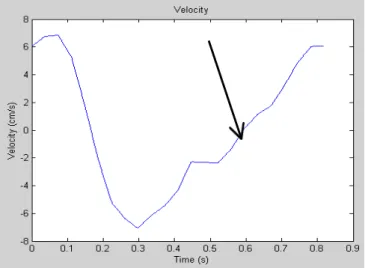
Cine phase-contrast (PC) MRI has been proven to become an effective tool in the quantification of the hydrodynamics of cerebrospinal fluid (CSF) through the aqueduct of Sylvius (1). Aqueductal CSF movement can be regarded as being to-and-fro due to the relatively straight and uniform hour-glass shape of the aqueductal of Sylvius (2-3). When the CSF flow velocity is depicted as a function of time in one cardiac cycle, the flow profile shows a “V” shape with average velocity close to zero (3). The purpose of this project was to further investigate the behavior of a higher frequency harmonic showing up as a “motch” in the CSF flow profile (arrow in Fig.1). In addition, in this preliminary report we examined the presence of notch with respect to other hydrodynamic parameters derived from the entire velocity-time curve. Possible meanings of the notch regarding intracranial pressure transmission conditions will be discussed.
A total of 91 subjects were included in this study. These consist of 38 patients with normal pressure hydrocephalus (NPH), 15 with communicating hydrocephalus (CH) and 38 healthy volunteers. The NPH group was further divided into Lo-NPH (n=21) and Hi-NPH (n=17), according to presence of
the cerebral aqueduct using phase contrast imaging technique with retrospective ECG gating (VENC=20cm/sec, FOV=16~10cm,
TR=45ms, 256x256, 18~30 cardiac phases interpolated from 54~64 phases).
Semi-automatic image segmentation was applied to select the entire aqueduct cross section as region of interest (ROI) on magnitude images to avoid errors caused by phase fluctuations, whereas the numerical integration and calculation of the hydrodynamic parameters was performed on the velocity-time curve using information from the velocity maps. The CSF hydrodynamic parameters analyzed included the total stroke volume (TSV) and net stroke volume (NSV), according to definitions reported previously (1). The presence of notch in the velocity-time curve was scored using a three-level scale: 0, 1, and 2, to represent significant, mild, non-existing presence of the notch (Figs.1 & 2), respectively. The association between presence of notch and hydrodynamic parameters were examined separately for different pathological conditions, and the Pearson’s correlation coefficients were reported.
三、結果與討論
From analysis the outcome of notch, one found out that Hi-NPH and CH lack of notch, but normal and Lo-NPH have notch wavelet. Figures 1 and 2 showed the velocity-time curves for a healthy subject with notch and a CH patient without evident notch, respectively. Lo-NPH patient showed an averaged score
6
notch scores with TSV/NSV was shown in Table 1. The total stroke volume correlated strongly with presence of notch in Lo-NPH and healthy subjects but not in the other groups, while the net stroke stroke volume did not show significance as high as TSV.
A pulsatile pressure wave could be divided into a forward transmission and a backward reflection. In situations where the reflection wave is of sufficient amplitude, reflection superimposed on the transmission wave could result in the notch pattern seen in the velocity-time curve. A larger impedance gradient should be accompanied by more wave reflection and thus a more significant presence of notch pattern. Patients with Hi-NPH and CH are known to exhibit larger cerebral tissue compliance, leading to hyperdynamic CSF flow (1). At the present stage, therefore, we inferred the physiological meanings of the notch pattern to be a pressure reflection caused by mismatched impedance at the end of the aqueduct of Sylvius, although the evidence from the preliminary results is admittedly limited. Clearly, further investigations are needed to answer these questions in more details, which will be the major portion in our project for the subsequent year. In the future we shall also consider Fourier analysis as an alternative means to objectively quantify the presence of the notch pattern.
四、計畫成果自評
Our efforts spent in the CSF-related project have so far created fairly satisfactory products. The preliminary results have been
presented in the annual meeting of the
International Society for Magnetic Resonance in Medicine. One paper
discussing CSF-motion-related effects on the FLAIR MR imaging technique has been published in the prestigious American Journal of Neuroradiology (4). Another
slightly unrelated project on cerebral glioma, with graduate students receiving financial support from this project, has also resulted in one journal article published in the same journal (5). One article prepared for journal paper has been submitted to Radiology (6).
Although the results are still preliminary and the methodology is admittedly somewhat difficult, current achievements from this project may have potential in assisting understanding of the CSF hydrodynamics related to intracranial pathology, such as increase of the intracranial pressure.
五、參考文獻
1. Bradley WG Jr, Scalzo D, Queralt J, Nitz WN, Atkinson DJ, Wong P. Normal-pressure hydrocephalus: evaluation with cerebrospinal fluid flow measurements at MR imaging. Radiology
1996;198:523-529.
2. Gideon P, Stahlberg F, Thomsen C, Gjerris F, Sorensen PS, Henriksen O. Cerebrospinal fluid flow and production in patients with normal pressure hydrocephalus studied by MRI.
Neuroradiology 1994;36:210-215.
3. Nilsson C, Stahlberg F, Thomsen C, Henriksen O, Herning M, Owman C. Circadian variation in human cerebrospinal fluid production measured by magnetic resonance imaging.
American Journal of Physiology
1992;262:R20-R24.
4. Wu HM, Yousem DM, Chung HW, Guo WY, Chang CY, Chen CY. Influence of scanning parameters on high-intensity CSF artifacts in fast-FLAIR imaging.
2002;23:393-399.
5. Wu WC, Chen CY, Chung HW, Juan CJ, Hsueh CJ, Gao HW. Discrepant MR spectroscopic and perfusion imaging results in a case of malignant transformation of cerebral glioma.
American Journal of Neuroradiology,
2002;23:1775-1778.
6. Huang TY, Chung HW, Chen MY, Giiang LH, Chin SC, Lee CS, Chen CY, Liu YJ. Quantification of CSF production rate in healthy adults: A high temporal and spatial resolution 2D cine
phase-contrast MRI study, submitted to
Radiology.
Abbreviations used in this report: TSV: total stroke volume.
NSV: net stroke volume (corresponding to CSF production rate).
Lo-NPH: patients with normal pressure hydrocephalus with low CSF flow (chronic NPH).
Hi-NPH: patients with normal pressure hydrocephalus with high CSF flow (acute NPH suitable for shunting surgery).
CH: communicating hydrocephalus.
六、圖表
Figure 1. Velocity of CSF flow in the aqueduct of Sylvius plotted versus time in one cardiac cycle for a healthy young subject (male, 26 yo) where the notch was obviously present.
8
Figure 2. Velocity of CSF flow in the aqueduct of Sylvius plotted versus time in one cardiac cycle for a patient with communicating hydrocephalus showing no evident presence of the notch.
Lo-NPH Normal Hi-NPH CH TSV 0.93 0.97 0.10 0.08 NSV 0.42 0.53 0.01 0.02
Table 1. Pearson’s correlation coefficients between the scores for the presence of notch in the CSF flow profile and two hydrodynamic parameters calculated from the CSF velocity-time curves as shown in Figure 1.