For Peer Review
Multifunctional Gentamicin Supplementation of Poly(γ-glutamic acid)-Based Hydrogels for Wound Dressing
Application
Journal: Journal of Applied Polymer Science Manuscript ID: APP-2010-04-1162.R2
Wiley - Manuscript type: Research Article
Keywords: hydrogels, crosslinking, biomaterials, biodegradable
For Peer Review
Multifunctional Gentamicin Supplementation of Poly(γ-glutamic acid)–Based
Hydrogels for Wound Dressing Application
Yu-Hsin Lin1*, Jui-Hsiang Lin2, Shu-Fen Peng1, Chia-Lin Yeh1, Wen-Chen Chen1, Tsai-Luan Chang3, Ming-Ju Liu3, Chih-Ho Lai4
1
Department of Biological Science and Technology, Center for Inflammation Research, China Medical University, Taichung, Taiwan, ROC.
2
Department of Materials Science and Engineering, Feng Chia University, Taichung, Taiwan, ROC.
3
Department of Applied Cosmetology and Graduate Institute of Cosmetic Science Hungkuang University, Taichung, Taiwan, ROC.
4
Department of Microbiology, School of Medicine, China Medical University, Taichung, Taiwan, ROC.
*
Correspondence to:
Yu-Hsin Lin, PhD Assistant Professor
Department of Biological Science and Technology China Medical University,
Taichung, Taiwan, 40402 Fax: 886-4-2207-1507
For Peer Review
ABSTRACT
The process of wound healing is composed of coagulation, inflammation, fibroplasia, collagenation, epithelization and wound contraction. The wound dressing should protect the wound from bacterial infection, maintain a moist healing environment, and promote cell migration to reconstruct damaged tissue, and be easy to apply and remove to improve patient comfort. The purpose of our study was developed multifunctional hydrogels composed of genipin–crosslinking biodegradable biomaterials of poly(γ-glutamic acid) and gelatin, encapsulating gentamicin to accelerate wound healing. The results of swelling ratio measurements clearly indicate that hydrogel composition of poly(γ-glutamic acid)–gelatin had a higher swelling ratio and lower peel adhesion properties than gelatin hydrogel alone. In an in vitro study, the gentamicin incorporated in prepared hydrogels effectively inhibited target microorganisms, and produced a higher expression of type I collagen in fibroblast cells. Confocal laser scanning microscopy revealed that the fibroblast cells cultured in the hydrogel membranes produced fibroblast cell migration, and showed a continuous lined cytoskeletal distributing status. In the in vivo study, it was found that the gentamicin incorporated in genipin–crosslinked γ-PGA–gelatin wound dressing demonstrate the potential of such biologically functionalized dressing to accelerate wound closure and hence its potential clinical usefulness.
For Peer Review
INTRODUCTION
Treatment of wounds remains an important surgical objective. Wound healing is a dynamic process in which the response to injury is aimed at reconstructing damaged tissue, and requires precise coordination of connective tissue repair, re–epithelialization, and angiogenesis.1 To generate new tissue and heal the wound, fibroblasts not only proliferate to increase cell numbers, but also produce several extracellular matrix proteins and growth factors.1 Collagen plays an important role in connective tissue healing by providing tissue strength. The type I collagen is a key function for cell adhesion and migration within connective tissues.2,3 Another challenge facing caregivers is the growing incidence of infection by antibiotic–resistant bacterial strains in surgery wounds.4,5 Gentamicin is the most common antibiotic agent used for local application in surgery.6 Junge et al. have demonstrated that the polyvinylidenfluoride mesh with gentamicin could significantly increase type I collagen protein expression in tissue repair process.7
The ideal dressing needs to ensure that the wound remains moist with exudates and proper adherence to the wound surface, and must be easy to apply and remove, to improve patient compliance and comfort.8,9 To accomplish ideal wound healing dressing, wound dressings should be changed from traditional passive materials to active and functional materials.10,11 As a hydrophilic and natural compound, poly(γ-glutamic acid) (γ-PGA) is produced either as a capsular substance or as slime by members of the genus Bacillus.12,13 It was previously reported that poly-glutamic acid showed high cell adhesion and enhanced cell migration properties.14,15 Furthermore, γ-PGA has been reported recently to have the ability to prevent post-surgical tissue adhesion and wound healing, probably because the hydrophilicity of γ-PGA prevents the shrinkage of the membrane and enables it to act as an appropriate barrier at the wound site.16
Nevertheless, γ-PGA dissolves rather rapidly in aqueous environments. This adverse aspect requires the use of crosslinking procedures by a crosslinking agent, reducing polymer dissolution at body temperature by the formation of non–soluble networks in the membranes.17,18 Various crosslinking agents, such as formaldehyde and glutaraldehyde have been used to chemically modify biomaterial for biomedical applications; in particular,
For Peer Review
glutaraldehyde is used widely.19,20 The disadvantage of these agents is that the crosslinkers are built into the biomaterial, and that these reactive, or even toxic compounds, may impair the biocompatibility of the crosslinked products.21,22 In an attempt to overcome this problem, a naturally occurring crosslinking agent (genipin) was used to crosslink amine–group–containing biomaterials.23 Genipin and its related iridoid glucosides extracted from the fruits of Gardenia jasminoides Ellis have been widely used as an antiphlogistic and cholagogue in herbal medicine.24 Genipin has been used to crosslink natural tissues and native polymers with very minimal cytotoxic effects, as compared to the crosslinking agent glutaraldehyde.22,25
The γ-PGA is a naturally edible polypeptide in which glutamate is polymerized via γ-amide linkages, each molecule of γ-PGA has only a single free amine group.26 According to our results, the genipin crosslinked with only γ-PGA cannot form a complete structure due to the relatively weak interaction between the amine group on γ-PGA and the aldehyde group on genipin. Lien et al. demonstrated that increasing the amine groups of gelatin, enhances the mechanical strength of genipin–crosslinked gelatin scaffolds.27 Thus, to overcome this problem, a complex composed of type A gelatin blended with γ-PGA was prepared to form a fulfilling consummation structure. Gelatin is a natural and biodegradable polymer obtained by extraction from collagen by acidic and alkaline pretreatment followed by thermal denaturation.21,28 Depending on the processing method, two types of gelatin can be produced: type A and B, with different bloom numbers.29 Type A gelatin is derived from acid–processed collagen, while type B gelatin is obtained by alkaline treatment of collagen, resulting in a difference in isoelectric point, 7–9 for type A gelatin and 4–5 for type B gelatin.30 Investigators have evaluated the triple–helix content and increases with its bloom number, leading to an increase in the stress of gelatin films.31
We prepared multifunctional hydrogel of γ-PGA and gelatin in genipin–crosslinked membranes to create an ideal wound dressing, and examined its physicochemical characteristics, namely mechanical probability, and peel adhesion force. The antimicrobial property, skin fibroblast cells migration assay, and type I collagen generation by fibroblasts, as well as cell viability were demonstrated. Additionally, an in vivo experiment was performed
For Peer Review
that compared wound healing area in the rat model with the prepared gentamicin incorporated in genipin–γ-PGA–gelatin wound dressing versus gauze (control).
EXPERIMENTAL Materials
Gelatin was obtained from Sigma-Aldrich (St Louis, MO, USA). γ-PGA was purchased from Vedan Co. Ltd. (Taichung, Taiwan). Genipin was purchased from Wako Pure Chemical Industries, Ltd. (Osaka, Japan). Oregon Green 514 phalloidin was from Molecular Probes (Eugene, OR, USA). Collagenase, 3-(4,5-dimethyl-thiazol-yl)-2,5-diphenyltetrazolium bromide (MTT), acetic acid, 4′,6-diamidino-2-phenylindole (DAPI), Triton X-100, phosphate-buffered saline (PBS), and paraformaldehyde were purchased from Sigma-Aldrich (St Louis, MO, USA). Eagle’s minimal essential medium (MEM), fetal bovine serum (FBS), penicillin, streptomycin, gentamicin sulfate, and trypsin-EDTA were from Gibco (Grand Island, NY, USA). All other chemicals and reagents were of analytical grade.
Preparation of the hydrogel membranes
Membranes gelatin membranes or γ-PGA–gelatin membranes of different compositions were prepared following crosslinking with genipin. First, a stock solution of gelatin (20%, w/v) in PBS was prepared by dissolving 4 g of gelatin in 20 mL PBS and stirring for 3 h at 50˚C. The genipin–crosslinked gelatin membranes were prepared by blending various amounts of aqueous gelatin solution (5.00%, 7.50%, 10.00%, or 12.50% by w/v) with an aqueous genipin solution in a concentration of 0.0250%, 0.0375%, 0.0500%, or 0.0750% (w/v), and then stirring at room temperature for 10 min, before sonication to remove the trapped air bubbles. Following thorough stirring, 10 mL of each air bubble–free mixed solution was poured into a polystyrene Petri–dish (with a diameter of 8.5 cm) for crosslinking for 12 h. The cast membranes were then dried in an oven at 37˚C. After drying, the gelatin membranes were cut into small dumbbell specimens for mechanical measurements.
The genipin–γ-PGA–gelatin membranes with particular γ-PGA–gelatin compositions (0%:10.00%, 1.25%:10.00%, 2.50%:10.00%, 3.75%:10.00%, and 5.00%:10.00% by w/v)
For Peer Review
were prepared by blending with a 0.05% (w/v) aqueous genipin solution. The γ-PGA was
dissolved in distilled water and desalted by dialysis against distilled water for 12 h, with water exchanges several times, and finally was lyophilized to obtain pure γ-PGA. The purified γ-PGA was dissolved in PBS at 4˚C for 3 h to prepare 10% (w/v) solutions. The genipin–γ-PGA–gelatin membranes composed of γ-PGA and gelatin after genipin crosslinking were prepared by a series of aqueous γ-PGA–gelatin solutions with 10.00% (w/v) gelatin, and 0%, 1.25%, 2.50%, 3.75% and 5.00% (w/v) γ-PGA. Fourier transformed infrared spectroscopy (FT–IR, Perkin–Elmer Spectrum RX1 FT–IR System, Buckinghamshire, England) was used to examine the peak variation of the carboxylic groups of γ-PGA and amine groups of gelatin on the prepared hydrogels. The sample powder then was mixed with KBr (1:100) and pressed into a disk and analysis was performed on an FT–IR spectrometer from 400 to 4000 cm-1. The crystalline forms of γ-PGA, gelatin and the γ-PGA–gelatin membrane were determined by X–ray diffraction (MXP18, MAC Science, Japan) under the operating conditions of 40 kV and 20 mA. The scanning rate was 4º/min and the diffraction
patterns were determined over a range of diffraction angles (2θ), from 5º to 50º.32
Characterization of the prepared hydrogel membranes
The mechanical measurement was carried out in a sample extension on dumbbell specimens (gauge length 30.0 mm, gauge width 5.0 mm, and thickness 0.2 mm). The maximum tensile force at breakage was determined by uni–axial measurements using a HUNGTA/HT–9102 operating system at a constant speed of 10 mm/min. Measurements were made five times for each sample and averages were reported. The degree of crosslinking, determined by the ninhydin assay, was defined as the percentage of free amine groups in gelatin that had reacted with genipin subsequent to crosslinking. In the ninhydrin assay, the test sample first was lyophilized for 24 h and then weighed. Subsequently, the lyophilized sample was heated with a ninhydrin solution for 20 min. The optical absorbance of the solution was recorded with a microplate spectrofluorometer Molecular Devices SpectraMax M2e (Sunnyvale, CA, USA), using glycine at various known concentrations as the standard. To assess biodegradation, each test sample was placed in a PBS solution (pH 7.4) with 16
For Peer Review
units/mL collagenase, and incubated at 37ºC. After the determined time, the test samples were removed from the collagenase solution, and lyophilized for 24 h and then weighed. The remaining weight of the test samples was calculated as described elsewhere.9
Water swelling ratio
To measure the water swelling ratio of the hydrogel membranes, a pre–weighed dry sample was immersed in PBS at 37˚C. The weight of the swollen membrane was determined subsequently by sandwiching the membrane between two paper towels to remove excess water on the surface, and weighed immediately.33 The moisture permeability of the test sample was determined by measuring the water vapor transmission rate. Each test membrane was mounted on the mouth of a bottle (diameter 25 mm) containing 10 mL of water and kept at 37˚C and 35% relative humidity in an incubator. The water vapor transmission rate was calculated by using the following formula:9
where the water vapor transmission rate is expressed in g/m2/day, A is the area of the bottle mouth (mm2), and Wi and Wt are the weights of the bottle containing water before and after
permeation of water in an incubator, respectively.
Morphology
The surface morphologies of the prepared membranes were examined using a scanning electron microscope (SEM, S3000H, Hitachi, Japan). All of the test samples were mounted on metal grids using double–sided carbon adhesive tape, and the SEM was used at an accelerating voltage of 8 kV. To examine the peeling strength of the prepared membranes composed of γ-PGA and gelatin, an Instron peel device was used to determine the adhesive strength of the system. The test samples were tightly clipped by the clamping apparatus. They were placed on a flat surface with the test side facing up. An electromechanical test frame configured with a load cell to peel samples at 90˚ angles between the peel and the substrate, with a constant speed of 50 mm/min. The peeling strength was evaluated by using the following formula:34
For Peer Review
Cytotoxicity and type I collagen test for gentamicin
Normal human skin fibroblast (NHF) cells were obtained from the American Type Culture Collection and used between passages 15 and 30. The cells were initially grown in 25 cm2 tissue culture flasks with MEM medium supplemented with 0.1 mM non-essential amino acids, 1.0 mM sodium pyruvate, 10% FBS, penicillin (100 U/mL), and streptomycin (100 µg/mL), and were kept in an incubator at 37 °C, 95% humidity, and 5% CO2. The cells were
harvested for subculture every three days with 0.25% trypsin plus 0.05% EDTA solution and were used for the cell migration experiments. The cytotoxicity of the gentamicin was evaluated in vitro with the MTT assay. The assay is based on mitochondrial dehydrogenase activity as an indicator of cell viability. Briefly, MTT was dissolved in PBS to a concentration of 5 mg/mL as a stock MTT solution and filtered for sterilization. The NHF cells were seeded
at 5 × 10⁴ cells/well in 96–well plates for overnight, and then replaced by the growth medium
containing various concentrations (0, 0.12, 0.25, 0.50, or 0.75 mg/mL) of gentamicin sulfate
for 4 days.
To determine type I collagen production by fibroblasts following treatment with gentamicin, the cells were collected in 0.5 mL 0.05 M acetic acid (pH 3.0) and measured using a human type I collagen detection kit (Chondrex, WA, USA).35 In brief, a 100 µL sample was added to each well, and type I collagen was detected by specific antibodies. The color reaction was stopped by the addition of an acid solution and the intensity of the color was read at 490 nm on a microplate spectrofluorometer. The protein concentration was determined by interpolation from a standard curve using known concentrations of type I collagen standards as supplied.
Cellular compatibility of gentamicin incorporated in hydrogel membranes
Gentamicin was incorporated in membranes to prepare the wound dressing. The gentamicin stock solution A was 10 mg/mL gentamicin in PBS. A 0.5 mL volume of stock solution A was mixed with 10 mL of a solution of aqueous γ-PGA–gelatin, with stirring at
For Peer Review
37˚C as described for the preparation of hydrogel membranes. To observe the cellular compatibility of the prepared hydrogel membranes, test samples (diameter 16 mm) cut from the membranes, were glued to the bottoms of 6 cm diameter Petri plates. Subsequently, NHF cells were seeded at 1 × 10⁶ cells/mL in each dish and maintained in a humidified incubator at 37˚C for 4 days. The samples were drew out, and the viability of cells was evaluated qualitatively according to a live/dead assay using calcein-AM and ethidium homodimer.36
To examine the morphology of the cells on the test membranes following incubation, the membranes were washed three times with PBS before they were fixed in 3.7% paraformaldehyde. The membranes were washed again three times with PBS and permeabilized with 0.2% Triton X-100 for 15 min at 37°C. The washes were repeated, and the cells were stained with Alexa Fluor® 488 phalloidin and DAPI, which specifically stain the F-actin and the nucleus, respectively. The stained cells were examined with excitation at 340 and 488 nm, under a confocal laser scanning microscopy (CLSM). The images were superimposed with the LCS Lite software (version 2.0).
Antimicrobial activity
The antibacterial properties of gentamicin incorporated in γ-PGA–gelatin membranes were tested on agar plates inoculated with Escherichia coli (ATCC 25922) and Pseudomonas aeruginosa (ATCC 27853), measuring the zone of inhibition. Gentamicin was released from membranes into inoculated medium to inhibit the growth of the two bacterial strains. After 24 h of incubation at 37˚C, inhibition zones around the gentamicin–incorporated membranes were observed and compared with that of a blank membrane without gentamicin.
Animal study
Animal care and use complied the 1996 revision of the ‘‘Guide for the Care and Use of Laboratory Animals’’ prepared by the Institute of Laboratory Animal Resources, National Research Council, and published by the National Academy Press. Male 8–week–old Sprague–Dawley rats weighing 250–300 g were housed individually in polycarbonate cages maintained at constant temperature (22 ± 2℃) and humidity (55%). The rats had free access to
For Peer Review
food and water and were subjected to a 12:12–hour light–dark cycle. Average weight and behavior did not change significantly during the experiment. Rats were anesthetized with an intraperitoneal injection of ketamine (90 mg/kg) with xylazine (10 mg/kg).37 The skin over the dorsal area was shaved, application fields were outlined with marking pen just before skin excision, and surgical area was disinfected with 70% ethanol. On the back of each rat, a full–thickness wound with dimensions 1×1 cm was created on each side of the spine by dermoepidermic excision. The wounds were covered immediately after photography with gentamicin incorporated in genipin–γ-PGA–gelatin membrane on one side and gauze (as a control) on the other. The samples were placed alternately on the right or left following the order of the animals. Each dressing was covered with a sterile compress secured by a hypoallergenic elastic adhesive bandage. Animals were caged individually following identification. The dressings were removed on days 2, 4, 8 and 12 post–surgery, and the wounds were examined and photographed to measure wound size reduction. The wound size measurements taken at the time during surgery and biopsy were used to calculate the percentage size reduction using equation.38
Where A0 and At denote initial wound area and wound area after time interval ‘t’,
respectively. Wound area was measured from photographs using the Image–Pro Plus (Media Cybernetics, Silver Spring, MD, USA) following calibration.
Statistical analysis
Statistical analysis of the differences in the measured properties of the groups was performed with one–way analysis of variance and the determination of confidence intervals, with the statistical package Statistical Analysis System, version 6.08 (SAS Institute Inc., Cary, NC). All data are presented as means and standard deviations, indicated as “mean ± SD”. Differences were considered to be statistically significant when the P values were less than 0.05.
For Peer Review
RESULTS AND DISCUSSION
Caracterization of the prepared membranes
The characteristics of the prepared membranes after genipin crosslinking were investigated and compared (Fig. 1). After drying, the diameter of the genipin–crosslinked γ-PGA membrane was 0.12 ± 0.01 mm in thickness and had a rough surface with large wrinkles. On the contrast, when adding gelatin into γ-PGA to form a genipin–crosslinked γ-PGA–gelatin membrane, it was observed that the membrane turned dark bluish. The dark bluish color observed in the genipin–crosslinked gelatin was a result of the reaction of genipin with the amine groups on gelatin. It is known that genipin may form blue pigments upon spontaneous reaction with amine groups. It was reported by Touyama et al. that only primary amines, rather than secondary or tertiary amines, can react with genipin.39 Additionally, it was observed that the γ-PGA and gelatin membranes crosslinked with genipin were swollen in PBS at 37˚C [Fig. 1(a)]. This result revealed that γ-PGA membrane had a faster dissolution in PBS as compared to its γ-PGA–gelatin membrane counterpart.
To choice the optimum gelatin concentration of the prepared membrane for increasing its firm structure, we evaluated the influence of various gelatin concentrations (5.00%, 7.50%, 10.00% and 12.50% by w/v) on the tensile mechanical property of genipin–crosslinked gelatin.
As shown in Table Ⅰ, the tensile strengths of genipin–crosslinked gelatin membrane at
breaking were 5.96 ± 0.61 MPa (for a gelatin concentration of 5.00%), 7.21 ± 1.25 MPa (for
7.50%), 11.53 ± 0.95 MPa (for 10.00%), and 11.81 ± 0.66 MPa (for 12.50%). It was found
that the gelatin concentration of 12.50% (w/v), which is even more brittle than a gelatin concentration of 10.00%, obviously increases the brittleness of the prepared gelatin membranes, leading to a corresponding effect in their tensile strength. We propose that a genipin amine group monomer is formed through nucleophilic attack by the amine–group–containing compounds, such as gelatin, on the third carbon of genipin. This is followed by the opening of the genipin ring and the formation of an aldehyde group. The resulting aldehyde group is attacked subsequently by the attached secondary amine group.18 Dimerization occurs at the second stage, perhaps by radical reaction. Therefore, genipin may form intramolecularly and intermolecularly crosslinked products with a heterocyclic structure.
For Peer Review
The degree of crosslinking of test membranes with a gelatin content of 10.00% but with different genipin concentrations (0.0250%, 0.0375%, 0.0500% and 0.0750% by w/v) are analyzed (Table Ⅱ). In this study, the crosslinking degree refers to the percentage of free amine groups in gelatin reacted with genipin subsequent to crosslinking.40 According to a previous study, genipin crosslinks gelatin through a nucleophilic attack by amine groups on lysine and arginine residues on gelatin to fabricate a wound dressing.22,41
The various concentrations of genipin yielded degrees of crosslinking of the membranes of 17.31 ± 2.91% (at 0.0250% genipin, n = 5), 24.51 ± 3.92% (at 0.0375%), 33.51 ± 2.02% (at 0.0500%), and 35.42 ± 2.43% (at 0.0750%). The degradation behavior was examined as a function of the degree of crosslinking using a series of samples with increasing crosslinking densities [Fig. 1(b)]. When the genipin content was at a low concentration, such as 0.0250% (w/v), the test membrane showed almost completely biodegraded in a period of 72 h. When the genipin content was in the range 0.050% to 0.075% (w/v), the biodegradation rate was slow. This indicates that the higher degree of crosslinking in gelatin membranes causes more resistance to degradation in PBS collagenase solutions. It is known that bacterial collagenase is generally capable of cleaving peptide bonds within the triple helical structure of collagen and has specificity for the Pro–X–Gly–Pro–Y region, splitting between X and Gly.42 Based on these result, the crosslinking agent genipin was used at a concentration of 0.050% (w/v) in the prepared gelatin membranes for the rest of the study.
Swelling characteristics of genipin–crosslinked γ-PGA–gelatin hydrogel
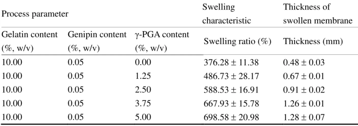
The swelling characteristics of genipin–γ-PGA–gelatin membranes prepared at distinct compositions are shown in Figure 2. γ-PGA, produced by certain Bacillus species, is a naturally occurring anionic homo–polyamide that is made of L-glutamic acid units connected by amide linkages between α-amine and γ-carboxylic acid groups.12,43 This biomaterial has received renewed interest owing to its excellent properties, including its water–solubility, biodegradability, biocompatibility, high capacity to absorb water and non–toxicity towards humans and the environment.44 The swelling of the test membranes reached a stable state at a
For Peer Review
376.28 ± 11.38% (for a γ-PGA–gelatin composition of 0%:10.00%), 486.73% ± 28.17% (1.25%:10.00%), 588.53 ± 16.91% (2.50%:10.00%), 667.93% ± 15.78% (3.75%:10.00%), and 698.58 ± 20.98% (5.00%:10.00%) (p < 0.05), because the electrostatic repulsion between the
ionized acid groups (–COO–) promoted swelling [Fig. 2(b)]. Micrographs of these swollen and
dried test membranes are shown in Figure 2(a). The dried membranes prepared in the study appeared to be about 0.2 mm in thickness. When the hydrogel membranes were allowed to swell for 120 min, it had a smooth surface and a different from swollen membranes deep level appearance. As shown in Table Ⅲ, the thickness of the swollen membranes were 0.48 ± 0.03 mm (for the γ-PGA–gelatin composition of 0%:10.00%), 0.67 ± 0.01 mm (for 1.25%:10.00%), 0.91 ± 0.02 mm (for 2.50%:10.00%), 1.26 ± 0.01 mm (for 3.75%:10.00%), and 1.28 ± 0.07 mm (for 5.00%:10.00%) with increasing amounts of γ-PGA. Because the combination of γ-PGA and gelatin at 3.75% and 10.00% (w/v) produced the best physicochemical characteristics among all the studied groups, this concentration was used in the rest of the study.
Morphology
Analysis of the morphologies of γ-PGA–gelatin and gelatin membrane by SEM at the different magnifications indicates that the surface of the prepared membranes exhibited genipin crosslinking. As displayed in Figure 3, the morphology of the surface of the pure gelatin membrane was rough, and small wrinkles were visible at a magnification of 3000. The
surface of the γ-PGA–gelatin membrane was smoother than that of the pure gelatin membrane.
Gelatin is a derived protein with good biocompatibility, obtained by the partial hydrolysis of collagen.45 The gelatin A that was used in this study was produced by acid hydrolysis with an isoelectric point of approximately 8.0.46 The hydrogel membranes were prepared at pH 7.4, at which type A gelatin still exhibits a positive net charge. γ-PGA, an anionic peptide, is a natural compound that is formed as a capsular substance or as slime by members of the genus Bacillus.12,13 Accordingly, in the preparation of genipin–γ-PGA–gelatin hydrogels with positively charged type A gelatin, an oppositely charged γ-PGA interacts ionically to form a complex, as revealed by the FT–IR spectra and X–ray diffractograms of γ-PGA, gelatin and
For Peer Review
γ-PGA–gelatin complexes (Fig. 4 and Fig. 5).Figure 4 shows the FT–IR spectra of the hydrogels prepared with γ-PGA and gelatin. The characteristic peaks at 1618 cm–1 and 1539 cm–1 represented carboxylate ions (–COO–) on γ-PGA and protonated amine groups (–NH3+) on gelatin. As shown, in the case of
γ-PGA–gelatin complexes the characteristic peak at 1618 cm–1 for carboxylate ions on γ-PGA disappeared was replaced by a new peak at 1607 cm–1, while the characteristic peak of protonated amine groups on gelatin at 1539 cm–1 shifted to 1528 cm–1. The ionized γ-PGA and gelatin therefore were able to form polyelectrolyte complexes via electrostatic interactions.
The incorporation of gelatin and γ-PGA was evident in X–ray diffraction date (Fig. 5). Gelatin has an obvious peak at 2θ = 7º and a large broad amorphous peak at 2θ = 22º. In γ-PGA–gelatin complex, it may be seen that the diffraction peak of gelatin decreased and became broad. This phenomenon may be attributed to the electrostatic interactions between gelatin and γ-PGA that has disrupted the regular crystallites of gelatin molecules. Thus, in the preparation of the genipin–crosslinked γ-PGA–gelatin hydrogel, γ-PGA entangled through the gelatin network (Fig. 5).
A wound dressing with a smooth surface structure could effectively counteract adhesion and would be easy to peel off the wound, thus being easily removable and comfortable to wear.47,48 Peeling–testing is a well established methodology in industrial applications involving membrane adhesives and has been used to a limited extent in the biomaterials field.49 Thus, according to the peel adhesion force test on the genipin–crosslinked gelatin and γ-PGA–gelatin membranes, the mean peel strength (N/m) was 168.3 ± 15.1 for 10.00% gelatin , and 109.8 ± 9.4 for γ-PGA:gelatin in a ratio of 3.75%:10.00%.
Cytotoxicity and type I collagen generation by gentamicin
Cytotoxicity of NHF cells was assayed as the percentage of cells surviving after treatment with various concentrations of gentamicin, using an MTT assay [Fig. 6(a)]. Cell viability was generally not affected by gentamicin sulfate at concentrations below 0.50 mg/mL but declined slightly when exposed to 0.75 mg/mL. The expression of type I collagen by NHF cells treated with various concentrations of gentamicin was measured with the human type I collagen
For Peer Review
detection kit. It was reported that gentamicin sulfate modification of polyvinylidenfluoride mesh induces a higher expression of type I collagen mRNA, and, consequently, an increased collagen type I ratio at the mesh/host tissue interface, with a decreased expression of collagenases (MMP-8 and MMP-13).7 As shown in Figure 6(a), the type I collagen content of NHF cells were 562.5 ± 29.8 ng/mL (control), 591.7 ± 41.8 ng/mL (at 0.12 mg/mL gentamicin), 711.7 ± 50.3 ng/mL (at 0.25 mg/mL gentamicin), 891.6 ± 81.7 ng/mL (at 0.50 mg/mL gentamicin), and 879.9 ± 47.8 ng/mL (at 0.75 mg/mL gentamicin). Because it was found that since 0.50 mg/mL gentamicin induced a significantly higher expression of type I collagen as compared to control (p < 0.05), and without damaging the cultured cells, this concentration was used in the rest of the study.
In vitro antibacterial study
E. coli and P. aeruginosa are Gram–negative aerobic rods and can be environmental contaminants of burn wounds and of other trauma, through dressing fluids or other sources, thereby causing sepsis.50 Gentamicin is a broad–spectrum aminoglycoside antibiotic and useful primarily in infections involving E. coli and P. aeruginosa.51 Aminoglycoside antibiotics that bind to the ribosome site cause misreading of the genetic code and inhibit translocation.52 The antibacterial ability of the composite membranes loaded with gentamicin (0.50 mg/mL) was examined using the Kirby–Bauer disk diffusion test. Zones of no growth (inhibition zones) were clearly seen around the discs of gentamicin–incorporated membranes in culture plates inoculated with either E. coli or P. aeruginosa [Fig. 6(b), right]. Conversely, control samples composed of the same formulation without the drug, did not reveal any antibactericidal effect [Fig. 6(b), left], thus indicating that the active antibacterial agent, gentamicin, could be immobilized in the composite membrane and subsequently released, thereby effectively inhibiting target microorganisms.
Cellular compatibility of gentamicin incorporated in hydrogel membrane
The cellular compatibility of the gentamicin incorporated in membranes was observed qualitatively by a live/dead cell viability assay [Fig. 7(a)]. The results indicated that the
For Peer Review
viability of the cells was not affected by the treatment with the gentamicin incorporated–membranes for 2 or 4 days. Live/dead staining was performed to show the living cells stained with calcein-AM (green color) versus the dead cells stained with ethidium homodimer (Ethd-1) (red color) by CLSM. Almost all the NHF cells in the test membranes were stained a green color. Phase contrast microscopy and CLSM observation also confirmed that the gentamicin membranes were not cytotoxic. Figure 7(b) shows that the cell growth in membrane has the same morphology of cell growth characteristic as the control (Petri–dish alone). To examine whether the use of the membrane was associated with redistribution of actin, F-actin staining with FITC–labelled phalloidin was carried out in the study. Figure 7(b) shows the staining pattern of fluorescence images of NHF cells grown on membranes and the control (Petri–dish). A continuous lined pattern of a green fluorescent cytoskeleton was observed, together with the blue nuclei. These results indicate that wound dressing membrane developed in this study was cytocompatible.
In vivo wound healing effects
Obvious differences in wound closure were observed among 2, 4, 8, and 12 days post–treatment (Fig. 8). The wounds treated with gentamicin incorporated in genipin–γ-PGA–gelatin membrane were considerably smaller than those treated with gauze (control) after 4 days. Because of gentamicin is a broad–spectrum aminoglycoside antibiotic and the most common locally applied antibiotic agent used in wound healing.51,53 Furthermore, subcutaneous aspect appeared grossly normal for the test membrane (gentamicin incorporated in genipin–γ-PGA–gelatin hydrogel) and evidence of infection of the wound site appeared uninfected [Fig. 8(a)]. Previous evidence showed that wounds covered with gauze alone were hemorrhagic and the wound exhibited scab.25 All test wounds appeared healed after 12 days for test membrane and gauze (control), whereas control wounds were incompletely healed and displayed less scab. Reduction in wound defect area was calculated by measuring the wound area at different times [Fig. 8(b)]. The results show a slight difference between the control group (wound closure of 39.32 ± 5.69%) and prepared membrane group (wound closure of 56.08 ± 5.71%) after 4 days (p < 0.05). This difference grew after 8 and 12 days, as wounds
For Peer Review
treated with prepared membrane showed 76.96 ± 5.73% and 89.79 ± 2.99% closure, compared to 60.51 ± 4.15% and 77.06 ± 6.13%, respectively, for wounds in the control group. The γ-PGA has been reported to be able to prevent post–surgical tissue adhesion and induce wound healing.16 Thus, the results of this investigation on gentamicin incorporated in genipin–γ-PGA–gelatin wound dressing demonstrate the potential of such biologically functionalized dressing to accelerate wound closure and hence its potential clinical usefulness.
CONCLUSIONS
The hydrogel membranes composed of γ-PGA and gelatin, encapsulating gentamicin, was developed as an ideal wound dressing system. Our results indicate that the γ-PGA–gelatin hydrogel had a higher swelling ratio and lower peel adhesion properties than the gelatin hydrogel alone. In an in vitro study, the gentamicn incorporated in hydrogels could effectively inhibit target microorganisms, elevate the skin fibroblast cells type I collagen expression, and produced fibroblast cell growth in the membrane, indicating that the developed wound dressing was cytocompatible for wound healing. In vivo experiment result on gentamicin incorporated in genipin–γ-PGA–gelatin wound dressing demonstrate the potential of such biologically functionalized dressing to accelerate wound closure.
ACKNOWLEDGEMENTS
This work was supported by grants from the National Science Council (NSC 97-2320-B-039-002-MY3 and NSC 98-2815-C-039-006-E) and the Center for Inflammation Research, China Medical University (CMU 98-CT-12 and CMU 98-N1-08), Republic of China. The confocal microscopy SP2 experiment and Ziess microscope apparatus supported by the Medical Research Core Facilities center, Office of Research & Development, China Medical University were gratefully acknowledged.
For Peer Review
References
1. Singer, A. J.; Clark, R. A. N Engl J Med 1999, 341, 738.
2. Jettanacheawchankit, S.; Sasithanasate, S.; Sangvanich, P.; Banlunara, W.; Thunyakitpisal, P. J Pharmacol Sci 2009, 109, 525.
3. San Antonio, J. D.; Lander, A. D.; Karnovsky, M. J.; Slayter, H. S. J Cell Biol 1994, 125, 1179.
4. Ong, S. Y.; Wu, J.; Moochhala, S. M.; Tan, M. H.; Lu, J. Biomaterials 2008, 29, 4323. 5. Murray, C. K.; Roop, S. A.; Hospenthal, D. R.; Dooley, D. P.; Wenner, K.; Hammock, J.;
Taufen, N.; Gourdine, E. Mil Med 2006, 171, 826.
6. Gilbert, A. I.; Felton, L. L. Surg Gynecol Obstet 1993, 177, 126.
7. Junge, K.; Klinge, U.; Rosch, R.; Lynen, P.; Binnebösel, M.; Conze, J.; Mertens, P. R.; Schwab, R.; Schumpelick, V. Langenbecks Arch Surg 2007, 392, 465.
8. Yücedag, F.; Atalay-Oral, C.; Erkal, S.; Sirkecioglu, A.; Karasartova, D.; Sahin, F.; Tantekin-Ersolmaz, S. B.; Güner, F. S. J Appl Polym Sci 2010, 115, 1347.
9. Yoo, H. J.; Kim, H. D. J Biomed Mater Res B Appl Biomater 2008, 85, 326. 10. Purna, S. K.; Babu, M. Burns 2000, 26, 54.
11. Choate, C. S. J Am Podiatr Med Assoc 1994, 8, 463.
12. Richard, A.; Margaritis, A. Crit Rev Biotechnol 2001, 21, 219.
13. Park, C.; Choi, J. C.; Choi, Y. H.; Nakamura, H.; Shimanouchi, K.; Horiuchi, T.; Misono, H.; Sewaki, T.; Soda, K.; Ashiuchi, M.; Sung, M. H. J Mol Cat B: Enzymatic 2005, 35, 128.
14. Matsusaki, M.; Yoshida, H.; Akashi, M. Biomaterials 2007, 28, 2729.
15. Guan, H.; McGuire, M. J.; Li, S.; Brown, K. C. Bioconjug Chem 2008, 19, 1813.
16. Izumi, Y.; Yamamoto, M.; Kawamura, M.; Adachi, T.; Kobayashi, K. Surgery 2007, 141, 678.
17. Leo, E.; Vandelli, M. A.; Cameroni, R.; Forni, F. Int J Pharm 1997, 155, 75.
18. Yan, C. H.; Li, X. W.; Chen, X. L.; Wang, D. Q.; Zhong, D. C.; Tan, T. Z.; Kitano, H. Biomaterials 1991, 12, 640.
For Peer Review
20. Neumann, P. M.; Zur, B.; Ehrenreich, Y. J Biomed Mater Res 1981, 15, 9.
21. Kuijpers, A. J.; Engbers, G. H.; Krijgsveld, J.; Zaat, S. A.; Dankert, J.; Feijen, J. J Biomater Sci Polym Ed 2000, 11, 225.
22. Chang, W. H.; Chang, Y.; Lai, P. H.; Sung, H. W. J Biomater Sci Polym Ed 2003, 14, 481. 23. Akao, T.; Kobashi, K.; Aburada, M. Biol Pharm Bull 1994, 17, 1573.
24. Sung, H. W.; Huang, D. M.; Chang, W. H.; Huang, R. N.; Hsu, J. C. Biomed Mater Res 1999, 46, 520.
25. Balakrishnan, B.; Mohanty, M.; Umashankar, P. R.; Jayakrishnan, A. Biomaterials 2005, 26, 6335.
26. Bae, S. R.; Park, C.; Choi, J. C.; Poo, H.; Kim, C. J.; Sung, M. H. J Microbiol Biotechnol 2010, 20, 803.
27. Lien, S. M.; Li, W. T.; Huang, T. J. Mater Sci Eng C 2008, 28, 36.
28. Liu, Y.; Liu, X.; Wang, X.; Yang, J.; Yang, X. J.; Lu, L. J Appl Polym Sci 2010, 116, 2617.
29. Peng, H. T.; Martineau, L.; Shek, P. N. J Mater Sci Mater Med 2008, 19, 997. 30. Vandervoort, J.; Ludwig, A. Eur J Pharm Biopharm 2004, 57, 251.
31. Bigi, A.; Panzavolta, S.; Rubini, K. Biomaterials 2004, 25, 5675. 32. Qu, X.; Wirsén, A.; Albertsson, A. C. Polymer 2000, 41, 4589. 33. Dong, Z.; Wang, Q.; Du, Y. J Memb Sci 2006, 280, 37.
34. Dong, C.; Mead, E.; Skalak, R.; Fung, Y. C.; Debes, J. C.; Zapata-Sirvent, R. L.; Andree, C.; Greenleaf, G.; Cooper, M.; Hansbrough, J. F. Ann Biomed Eng 1993, 21, 51.
35. Meng, L.; Ye, X.; Fan, M.; Xiong, X.; Von den Hoff, J. W.; Bian, Z. Arch Oral Biol 2008, 53, 1050.
36. Chen, M. C.; Tsai, H. W.; Liu, C. T.; Peng, S. F.; Lai, W. Y.; Chen, S. J.; Chang, Y.; Sung H. W. Biomaterials 2009, 30, 2102.
37. Lee, W. R.; Park, J. H.; Kim, K. H.; Kim, S. J.; Park, D. H.; Chae, M. H.; Suh, S. H.; Jeong, S. W.; Park, K. K. Wound Repair Regen 2009, 17, 505.
For Peer Review
38. Balakrishnan, B.; Mohanty, M.; Fernandez, A. C.; Mohanan, P. V.; Jayakrishnan, A. Biomaterials 2006, 27, 1355.
39. Touyama, R.; Takeda, Y.; Inoue, K.; Kawamura, I.; Yatsuzuka, M.; Ikumoto, T.; Shingu, T.; Yokoi, T.; Inouye, H. Chem Pharm Bull 1994, 42, 668.
40. Silvestro, L.; Viano, I.; Macario, M.; Colangelo, D.; Montrucchio, G.; Panico, S.; Fantozzi, R. Semin Thromb Hemost 1994, 20, 254.
41. Mi, F. L. Biomacromolecules 2005, 6, 975.
42. Harper, E.; Berger, A.; Katchalski, E. Biopolymers 1972, 11, 1607.
43. Liang, H. F.; Chen, C. T.; Chen, S. C.; Kulkarni, A. R.; Chiu, Y. L.; Chen, M. C.; Sung, H. W. Biomaterials 2006, 27, 2051.
44. Chang, K. Y.; Lin, C. C.; Ho, G. H.; Huang, Y. P.; Lee, Y. D. Polymer, 2009, 50, 1755. 45. Zhang, Y.; Wang, Z.; Wang, Y.; Zhao, J.; Wu, C. Polymer, 2007, 48, 5639.
46. Tapia, C.; Ormazabal, V.; Costa, E.; Yazdani-Pedram, M. Drug Dev Ind Pharm 2007, 33, 585.
47. Pei, H. N.; Chen, X. G.; Li, Y.; Zhou, H. Y. J Biomed Mater Res A 2008, 85, 566.
48. Wokovich, A. M.; Brown, S. A.; McMaster, F. J.; Doub, W. H.; Cai, B.; Sadrieh, N.; Chen, M. L.; Machado, S.; Shen, M.; Buhse, L. F. J Biomed Mater Res B Appl Biomater 2008, 87, 105.
49. Bundy, K.; Schlegel, U.; Rahn, B.; Geret, V.; Perren, S. J Mater Sci Mater Med 2000, 11, 517
50. Matsushita, A.; Iwase, M.; Kato, Y.; Ichihara, S.; Ichihara, G.; Kimata, H.; Hayashi, K.; Hashimoto, K.; Yokoi, T.; Noda, A.; Koike, Y.; Yokota, M.; Nagata, K. Exp Anim 2007, 56, 339.
51. Bryan, L. E.; Van Den Elzen, H. M. Antimicrob Agents Chemother 1977, 12, 163. 52. Yoshizawa, S.; Fourmy, D.; Puglisi, J. D. EMBO J 1998, 17, 6437.
53. Rosen, H. R.; Marczell, A. P.; Czerwenka, E.; Stierer, M. O.; Spoula, H.; Wasl, H. Am J Surg 1991, 162, 438.
For Peer Review
Figure Captions
Figure 1. (a) Micrographs of the genipin–crosslinked γ-PGA and γ-PGA–gelatin membranes were taken under a microscope; (b) The remaining weight of genipin–crosslinked gelatin hydrogel membranes by enzyme biodegradation (pH 7.4, PBS with 16 units/mL collagenase).
Figure 2. (a) Micrographs of genipin–γ-PGA–gelatin hydrogel membranes prepared at different compositions (by w/v) by swelling for 120 min in a solution at pH 7.4 at 37˚C were taken under a microscope; (b) Swelling characteristics of genipin–γ-PGA–gelatin hydrogel membranes prepared at different compositions (by w/v) determined by immersing dried test samples to swell for 360 min in a solution at pH 7.4 at 37˚C. γ-PGA: poly-γ-glutamic acid.
Figure 3. SEM micrographs of the γ-PGA–gelatin and only gelatin membranes prepared with genipin crosslinking. SEM: scanning electron microscope; γ-PGA: poly-γ-glutamic acid.
Figure 4. FT–IR spectra of γ-PGA, gelatin and γ-PGA–gelatin complexes. γ-PGA: poly-γ-glutamic acid. FT–IR: fourier transformed infrared spectroscopy.
Figure 5. X–ray diffractograms of γ-PGA, gelatin and γ-PGA–gelatin complexes. γ-PGA: poly-γ-glutamic acid.
Figure 6. (a) Cell viability and type I collagen levels after treatment with various concentrations of gentamicin, determined by the MTT assay and the human type I collagen detection kit; (b) Results of the antibacterial–activity study of the genipin–γ-PGA–gelatin membranes incorporating gentamicin. MTT: 3-(4,5-dimethylthiazol-2-yl)-2,5-diphenyltetrazolium bromide. γ-PGA: poly-γ-glutamic acid. *Statistical significance at a level of p < 0.05. Date expressed as mean ± SD.
Figure 7. Results of the activity assay of the gentamicin incorporated in genipin–γ-PGA–gelatin membranes. (a) CLSM images showing the viability of NHF cells following treatment with experimental membranes for distinct time periods; (b) Micrographs and fluorescent images of NHF cells fluorescently stained for F-actin and
For Peer Review
nuclei, following incubation with an experimental membrane for 4 days. NHF: normal human skin fibroblast; γ-PGA: poly-γ-glutamic acid; CLSM: confocal laser scanning microscope.
Figure 8. (a) Representative photographs of the wound on days 2, 4, 8, and 12, taken postoperatively following full–thickness skin excision; (b) Percentage wound closure following distinct time intervals. Data points are the average of n = 5 and error bars represent ± SD.
For Peer Review
TablesTable Ⅰ. The tensile strength at breaking at room temperature of the genipin-crosslinked gelatin membranes (n = 5).
Table Ⅱ. The degree of crosslinking of the genipin-crosslinked gelatin membranes (n = 5).
Table Ⅲ. The swelling characteristic and thickness of genipin–crosslinked γ-PGA–gelatin membranes (γ-PGA: poly-γ-glutamic acid) (n = 5).
Process parameter Swelling
characteristic Thickness of swollen membrane Gelatin content (%, w/v) Genipin content (%, w/v) γ-PGA content
(%, w/v) Swelling ratio (%) Thickness (mm)
10.00 0.05 0.00 376.28 ± 11.38 0.48 ± 0.03
10.00 0.05 1.25 486.73 ± 28.17 0.67 ± 0.01
10.00 0.05 2.50 588.53 ± 16.91 0.91 ± 0.02
10.00 0.05 3.75 667.93 ± 15.78 1.26 ± 0.01
10.00 0.05 5.00 698.58 ± 20.98 1.28 ± 0.07
Process parameter Tensile strength
Gelatin content (%, w/v) Genipin content (%, w/v) Strength at the break point (MPa)
5.00 0.05 5.96 ± 0.61
7.50 0.05 7.21 ± 1.25
10.00 0.05 11.53 ± 0.95
12.50 0.05 11.81 ± 0.66
Process parameter Extent of crosslinking
Gelatin content (%, w/v) Genipin content (%, w/v) Crosslinking degree (%)
10.00 0.0250 17.31 ± 2.91
10.00 0.0375 24.51 ± 3.92
10.00 0.0500 33.51 ± 2.02
For Peer Review
Figure 1a
For Peer Review
Figure 1b
For Peer Review
Figure 2a
For Peer Review
Figure 2b
For Peer Review
Figure 3
For Peer Review
Figure 4
For Peer Review
Figure 5
For Peer Review
Figure 6
For Peer Review
Figure 7
For Peer Review
Figure 8