Synthesis and Luminescent Properties of Strong Blue
Light-Emitting Al
2O
3ÕZnO Nanocables
Chi-Sheng Hsiao,a Wan-Lin Kuo,a San-Yuan Chen,a,zJi-Lin Shen,b Chin-Ching Lin,cand Syh-Yuh Chengc
a
Department of Materials Sciences and Engineering, National Chiao Tung University, Hsinchu, Taiwan b
Physics Department, Chung Yuan Christian University, Chung-Li, Taiwan c
Materials Research Laboratories, Industrial Technology Research Institution, Chutung, Taiwan
Photoelectronic characteristics are investigated in well-aligned aluminum-coated ZnO nanorods共Al2O3/ZnO nanocables兲 grown
on Si substrates buffered with ZnO film at a low temperature. Photoluminescence measurement indicates that a strong blue emission peak at⬃450 nm appears at 400 and 600°C in O2and N2atmospheres, respectively. A 30⫻ enhancement of the relative
intensity ratio of blue emission 共IB兲 to ultraviolet emission 共IUV兲 has been observed for the Al2O3/ZnO nanocables.
High-resolution transmission electron microscopy and X-ray photoelectron spectroscopy analyses reveal that the origin of the strong blue emission can be attributed to the structure transition and cleavage of the oxygen–hydrogen bond共OH兲 of the Al2O3/ZnO
nanocable, which leads to the formation of singly ionized oxygen vacancies共Al–O•兲.
© 2008 The Electrochemical Society. 关DOI: 10.1149/1.2890289兴 All rights reserved.
Manuscript submitted August 16, 2007; revised manuscript received January 2, 2008. Available electronically March 18, 2008.
Al2O3has been used for capacitor dielectrics and gate oxides in memory devices due to its high dielectric constant, very low perme-ability, and high thermal conductivity.1However, the photolumines-cence共PL兲 property of alumina film or nanoparticles has not been studied in detail. Yoldas et al. studied alumina–silica powders and stated that the presence of pentahedrally coordinated aluminum ap-pears to be strongly correlated with the occurrence of PL.2Suga et al. studied alumina gel from an inorganic salt and alkoxide, and mentioned that the PL is closely related to oxygen defects and the development of the AlVsite.3However, no data exist for determin-ing the dependence of the properties of a defect center on the struc-ture of its coordinate sites and the presence of luminescence. Re-cently, Li et al. reported that a broad band located around 422 nm could be detected from nanosized␥-Al2O3powder.4It is suggested that the produced defect level could induce␥-Al2O3nanopowder to emit blue PL bands. However, most of those studies have been fo-cused on nanopowders or gel films. In past years, ZnO/Al2O3core/ shell nanofibers have been prepared from the Al2O3deposition of ZnO nanowires with an atomic layer deposition technique, but no PL properties have been reported.5These findings indicate that so far, the PL property of nanoscale alumina film has not been investi-gated. In addition, our previous study found that, when an alumina film is deposited on a ZnO-coated silicon substrate by a wet chemi-cal process, the alumina film not only emits blue, but the blue emis-sion can also be much enhanced compared to that of coatings on pure silicon. This indicates that ZnO plays an important role in the PL properties of nanosized alumina films.
Wet chemical approaches are widely used for the fabrication of large oriented arrays of ZnO nanorods on Si or polymer organic substrates. Furthermore, zinc oxide is an important photoelectronic material because of its wide direct bandgap of 3.37 eV and a rela-tively large exciton binding energy of 60 meV,6which makes ZnO a promising material for light-emitting diodes or diode lasers. In ad-dition, depending on the processing methods, a variety of defects can be easily generated from nanostructured ZnO. Therefore, it is possible to develop the Al2O3/ZnO nanostructure into white phos-phor if the blue emission from alumina can be incorporated into the ZnO nanostructure. So far, to the best of our knowledge, there have been no further systematic investigations on the light-emitting prop-erties of alumina-coated ZnO nanorods grown in aqueous solutions at lower temperatures. Furthermore, it has been challenging to de-velop Al2O3/ZnO or ZnO-based nanostructures with a strong light emission by simple wet chemical processing. Therefore, in this work, a simple method of combining the aqueous solution process
with a thermal treatment is proposed to develop Al2O3/ZnO nano-cables with a strong blue emission. The structure and optical prop-erties of the nanocables also will be discussed. In addition, the ap-plication of this advanced structure to modify luminescent properties demonstrates a way to develop nanostructures for optoelectronic ap-plications.
Experimental
Previous experiments have shown that highly arrayed ZnO nano-rods can be developed on ZnO film-coated Si共ZnOf/Si兲 substrates.7
Following the sol–gel porous alumina templating method proposed by Martin et al.,85 M ammonia was added to an aqueous alumina nitrate solution 共0.4 M兲 at room temperature. After the hydrated precipitate was formed, it was centrifugally separated and washed several times with distilled water. The precipitates were then pep-tized with nitric acid to obtain a translucent, homogeneous, and stable sol. After that, the ZnO nanorods were exposed to surface modification by oxygen plasma treatment, and were subsequently immersed in the precursor sol of Al+3to grow an alumina shell on the ZnO nanorods at 80°C for 1 h. The thickness of the alumina shell on the ZnO nanorods can be modulated by controlling the reaction parameters. After washing with distilled water, rapid ther-mal annealing was performed for aluminum sol-coated ZnO nano-rods at 200–600°C in nitrogen and oxygen atmospheres. Micro-structure observation of Al2O3/ZnO nanocables was performed by a transmission electron microscope 共TEM, JEOL 2010兲 operated at 200 keV. Room-temperature PL measurement was performed on the nanorod samples, which were excited by a 325 nm He–Cd laser with an excitation power of 25 mW. The emitted luminescence light was collected through a 0.32 m spectrometer with a charge-coupled de-vice detector. The focused spot size of the He–Cd laser was esti-mated to be about 200 m in diameter. X-ray photoelectron spec-troscopy 共XPS兲 was used to evaluate the Al–O chemical binding states. The 27Al magic angle spinning nuclear magnetic resonance 共MAS NMR兲 was used to examine the chemical shift of alumina-coated ZnO nanrods scraped from the Si substrate using a Bruker Dsx400wc NMR spectrometer. The XPS analyses were performed on a Kratos AXIS Ultra with a monochromatic Mg X-ray source at 150 W.
Results and Discussion
Figure 1a shows the scanning electron microscope共SEM兲 sur-face image of the arrayed as-synthesized Al2O3/ZnO nanocables grown vertically on ZnOf/Si substrates. It was found that the
nano-rods have a well-defined hexagonal plane with a diameter of approximately 20–30 nm. Further analysis on as-synthesized Al2O3/ZnO nanocables is shown in the TEM image of Fig. 1b, z
E-mail: [email protected]
Journal of The Electrochemical Society, 155共5兲 K96-K99 共2008兲
0013-4651/2008/155共5兲/K96/4/$23.00 © The Electrochemical Society
K96
) unless CC License in place (see abstract). ecsdl.org/site/terms_use
address. Redistribution subject to ECS terms of use (see 140.113.38.11
indicating that alumina was conformably deposited on the ZnO na-norods and that the nana-norods remained in their original shape. Energy-dispersive spectroscopy 共EDS兲 analysis further confirmed that only strong Zn and O signals without Al were identified in the core nanorods, but that a weak peak corresponding to Al could be detected from the shell, indicating that an alumina shell was formed on the ZnO nanorods at low temperature.
The high-resolution共HR兲 TEM image in Fig. 2a illustrates that alumina was conformably deposited on the ZnO nanorods to be-come a core/shell Al2O3/ZnO nanocable structure. From the se-lected area electron diffraction in the inset of Fig. 2a, two diffraction patterns共rings and spot patterns兲 are observed. The spot pattern is from the single-crystal ZnO structure, while the ring pattern can be attributed to an amorphous alumina structure that may be a
pseudo-boehmite phase developed in aqueous solution according to the re-port of Ishizaka.9Upon annealing at 200°C, a short-range ordered structure seems to develop in the alumina shell, as shown in the HRTEM image of Fig. 2b. It is believed that rapid thermal treatment in an O2 atmosphere is favorable for the cleavage of the oxygen– hydrogen bond 共OH兲, causing the departure of H ions from the pseudoboehmite and initiating the occurrence of the phase transfor-mation of amorphous pseudoboehmite into a␥-phase alumina struc-ture. This structure can be thought of as a cubic spinel, with some suggestion of a tetragonal共tetrahedrally coordinated aluminum兲 dis-tortion in boehmite-derived␥-Al2O3in the I41/amd space group.10 When annealed at 400°C in an O2atmosphere, a long-range ordered structure can be clearly observed from the HRTEM image in Fig. 2c. According to the calculation of the lattice fringe共about 0.2 nm兲, it was indexed as a共400兲 plane, indicating a ␥-phase alumina structure 共d = 0.198 nm兲. This suggests that with increasing the temperature to 400°C in an O2atmosphere, the ␥-phase was developed in the alumina shell. As annealed at 600°C, two kinds of lattice fringes are clearly observed for annealed Al2O3/ZnO nanocables as shown in Fig. 2d, indicating that, in addition to single-crystalline ZnO nano-rods, a crystalline␥-alumina structure has been completely formed in the alumina shell at 600°C.
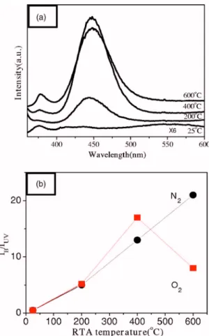
Figure 3a shows the room-temperature PL property of Al2O3/ZnO nanocables at different temperatures. Three important emission peaks can be observed from the PL spectrum of as-synthesized alumina-coated ZnO nanocables. Both ultraviolet共UV兲 emission共378 nm兲 and green–yellow 共visible兲 emission 共525–575 nm兲 bands are attributed to ZnO nanorods, and the other broad blue emission共425–475 nm兲 is possibly from the alumina shell. The near bandedge共UV emission兲 is generally attributed to free-exciton re-Figure 1.共Color online兲 SEM images of 共a兲 side view of ZnO nanorods. 共b兲
TEM image and EDS analysis of Al2O3/ZnO nanocables.
Figure 2. HRTEM of Al2O3/ZnO nanocables 共a兲 as-synthesized and
an-nealed at共b兲 200°C; 共c兲 400°C and 共d兲 600°C in oxygen ambient.
Figure 3. 共Color online兲 共a兲 Room-temperature PL spectra of Al2O3/ZnO
nanocables after rapid thermal anneal at various temperatures in O2
atmo-sphere.共b兲 The relative PL ratios 共IB/IUV兲 of blue emission to UV emission
on annealing temperature as a function of O2and N2atmospheres. K97
Journal of The Electrochemical Society, 155共5兲 K96-K99 共2008兲 K97
) unless CC License in place (see abstract). ecsdl.org/site/terms_use
address. Redistribution subject to ECS terms of use (see 140.113.38.11
combination, but the green–yellow emission is produced from the oxygen defect of the pure ZnO nanorods fabricated by chemical solution methods.11 In fact, it was found that the UV emission in-tensity of the as-grown nanocables increases with the annealing tem-perature at 200–600°C in an oxygen ambient, but the visible emis-sion in postannealed samples tends to disappear, suggesting that the native defects can be reduced by postannealing treatment as shown in Fig. 3a. It was noted that when the Al2O3/ZnO nanocables are annealed at 200°C in an O2atmosphere, an obvious blue emission at around 450 nm, which was primarily contributed from the alumina shell, can be clearly identified. According to the report of Lippens et al.12and TEM analysis 共Fig. 1兲, an increase of the annealing tem-perature may cause the cleavage of the oxygen–hydrogen bond 共OH兲 in pseudoboehmite structure, and consequently, both Al–O–O• and Al–O•defects would probably be produced in the alumina shell and vary with annealing temperature and atmospheres. At a lower temperature, i.e., 200°C, Al–O–O• defects are much more easily generated than Al–O•. However, according to Ishizaka’s report, the distance between the aluminum atom and the electron spin in Al–O–O• is long enough to neglect the hyperfine interaction with Al, indicating that Al–O–O• would not be a luminescent center.13 With the increase of the annealing temperature, the OH bond in the pseudoboehmite structure tends to be broken and Al–O• would be formed during the phase transformation from the pseudoboehmite phase共octahedrally coordinated structure兲 to the ␥-phase aluminum oxide共tetrahedrallly coordinated structure兲, favoring pentahedrally coordinated aluminum and the occurrence of singly ionized oxygen vacancies which are regarded as the F+center.14
With increasing the temperature to 400°C, dehydration proceeds and the structure evolves through a series of disordered states during the phase transformation from pseudoboehmite to␥-phase as dem-onstrated in Fig. 2c. In this condition, pentahedrally coordinated aluminum is easily formed, indicating that more singly ionized oxy-gen vacancies 共F+ center兲 defects can be generated. Therefore, a strong blue emission can be observed. At a higher temperature, i.e., 600°C, although the positions of the blue emission remain un-changed, it is found that the peak intensity is weakened compared to the sample annealed at 400°C. This indicates that the defect density of the main defects共F+centers兲 responsible for the blue emission is decreased with increasing the annealing temperature up to 600°C. Consequently, the density of the F+centers is reduced due to com-pensation in the O2atmosphere.
As reported, ZnO material usually generates a strong UV emission.15However, it can be seen in the figure that UV emis-sion decreases while blue light emisemis-sion increases. It can be inferred that UV emission from the ZnO band-edge recombination can excite the blue light of aluminum oxide. This may result in different inten-sity ratios of blue light to UV in the PL spectra. To further investi-gate the effect of heat treatment on the F+centers and pentahedrally coordinated aluminum, the as-synthesized Al2O3/ZnO nanocable samples were subjected to annealing at different temperatures in N2 and O2atmospheres. The relative PL ratios共IB/IUV兲 of blue emis-sion to UV emisemis-sion on annealing temperature as a function of O2 and N2atmospheres is presented in Fig. 3b. It is observed that the strongest blue emission of the nanocables appears at 600°C in a nitrogen ambient instead of 400°C in an oxygen atmosphere. Fur-thermore, the blue emission of postannealed Al2O3/ZnO nanocables is dominated not only by the annealing temperature but also the annealing atmosphere because the PL emission is very sensitive to defect type.16 As dehydration proceeds, the structure evolves through a series of disordered states, probably from octahedral to pentahedral and then tetrahedral. Ishizaka et al. reported that the luminescence is maximized for alumina treated at 600°C, at which temperature ␥-alumina is formed.13 Yoldas et al. studied calcium aluminate glass and suggested that the pentahedrally coordinated aluminum共AlV兲 may be strongly correlated with the occurrence of luminescence.2 Therefore, 27Al NMR analysis was performed to characterize such complexes.17Figure 4 shows the27Al MAS NMR
spectra of these samples as a function of the annealing temperature. Besides the resonance lines attributable to tetrahedrally共AlIV兲 and octahedrally共AlVI兲 coordinated aluminum, a line corresponding to pentahedrally共AlV兲 coordinated aluminum has also been observed. It was found that the concentration of pentahedrally coordinated 共AlV兲 aluminum is dependent on the thermochemical environment within the samples. Furthermore, the AlVsignal increases with the firing temperature, reaches a maximum intensity at about 600°C, and then decreases at a higher temperature in both O2and N2 an-nealing atmospheres. When comparing Fig. 3b, it was noted that, although the concentration of pentahedrally coordinated aluminum is higher at 600°C, the relative peak intensity 共IB/IUV兲 of the O2-annealed nanocables is decreased. This may imply that with the increase of annealing temperature, the OH bond in the pseudoboeh-mite structure tends to be broken and produce more pentahedrally coordinated aluminum and singly ionized oxygen vacancies 共F+兲. However, the F+ centers would be compensated in the case of an-nealing in oxygen at a higher anan-nealing temperature, i.e., 600°C. Therefore, in this condition F+ centers decrease, but the pentahe-drally coordinated aluminum still exists as indicated from the NMR result.
Because the equilibrium structure during the transformation of boehmite to ␥-alumina phase is determined by the number of the OH groups,18 the surface composition of Al2O3/ZnO nanocables was further examined by XPS for samples annealed at 400°C in N2 and O2 atmospheres as shown in Fig. 5. XPS spectra of Al 2p in annealed Al2O3/ZnO nanocables are shown in Fig. 5a. The peak position for the samples with nearly stoichiometric composition an-nealed in a N2atmosphere is located at a binding energy of 74.3 eV, indicating that the Al–O binding state is dominated by the boehmite/ ␥-alumina phase. In contrast, the Al 2p position of the samples annealed in an O2atmosphere is located at a binding energy of 74.6 eV, indicating that␥-alumina is easily formed for the sample treated in O2 gas. The asymmetric spectrum of O 1s in Fig. 5b and c contains three peaks at 529.6, 530.8, and共broadly兲 532.6 eV. The peaks around 529.6 and 530.8 eV can be assigned to O2−ions in the Zn–O bonds and Al–O bonds, respectively. The peak related to the highest binding energy around 532.6 eV is attributed to OH bonds. As a result, a strong OH–bond peak was observed in a N2 atmo-Figure 4. 27Al MAS NMR spectra for the alumina-coated ZnO nanorods
annealed in O2.
K98 Journal of The Electrochemical Society, 155共5兲 K96-K99 共2008兲
K98
) unless CC License in place (see abstract). ecsdl.org/site/terms_use
address. Redistribution subject to ECS terms of use (see 140.113.38.11
sphere compared to that in an O2atmosphere. This again demon-strates that more F+centers are produced by the rapid thermal treat-ment of 400°C in an O2 atmosphere, resulting in a stronger blue emission.
However, with a further increase of the annealing temperature up to 600°C, it was found that the IB/IUV decreases in an O2 atmo-sphere but increases in a N2atmosphere. The F+ centers would be diminished due to compensation in O2 atmosphere. In contrast, IB/IUVcontinuously increases and reaches the maximum intensity at 600°C in a N2atmosphere. Therefore, the relative IB/IUVintensity of the Al2O3/ZnO nanocables annealed in a N2atmosphere is stron-ger than that in an O2atmosphere at 600°C, indicating the difficulty of compensating F+centers in a nitrogen ambient. Therefore, in this work, it can be concluded that the blue emission is primarily domi-nated by F+centers that are accompanied by the occurrence of pen-tahedrally coordinated aluminum during the phase evolution of the Al2O3shell on the ZnO nanorods.
Conclusion
In summary, we have developed well-aligned arrays of Al2O3/ZnO nanocables on ZnOf/Si substrates buffered with a ZnO
film by combining a simple chemical solution with a low-temperature treatment. The PL measurement indicates that a strong blue emission peak at⬃450 nm appears at 400 and 600°C in O2 and N2atmospheres, respectively. The phenomenon is strongly re-lated to OH bond cleavage and a phase transition from amorphous to ␥-phase alumina, as evidenced by XPS and NMR analyses. The results provide an effective method for constructing nanostructures with a strong blue light emission.
National Chiao Tung University assisted in meeting the publication costs of this article.
References
1. E. P. Gusev, M. Copel, E. Cartier, I. J. R. Baumvol, C. Krug, and M. A. Gribelyuk,
Appl. Phys. Lett., 76, 176共2000兲.
2. B. E. Yoldas, J. Non-Cryst. Solids, 147–148, 614共1992兲.
3. Y. Kurokawa, T. Suga, S. Nakata, T. Ikoma, and S. Tero-Kubota, J. Mater. Sci.
Lett., 17, 275共1998兲.
4. Z. Q. Yu, D. Chang, and C. Li, J. Mater. Res., 16, 1890共2001兲.
5. J. Hwang, B. Min, J. S. Lee, K. Keem, K. Cho, M. Y. Sung, M. S. Lee, and S. Kim,
Adv. Mater., 16, 422共2004兲.
6. D. B. Laks, Appl. Phys. Lett., 63, 1375共1993兲.
7. S. C. Liou, C. S. Hsiao, and S. Y. Chen, J. Cryst. Growth, 274, 438共2005兲. 8. B. Cheng and E. T. Samulski, J. Mater. Chem., 11, 2901共2001兲. 9. T. Ishizaka and S. T. Kubota, J. Phys. Chem. Solids, 64, 801共2003兲. 10. G. Paglia, S. Božin, and J. L. Billinge, Chem. Mater., 18, 3242共2006兲. 11. D. Li, Y. H. Leung, A. B. Djurisic, Z. T. Liu, M. H. Xie, S. L. Shi, S. J. Xu, and W.
K. Chan, Appl. Phys. Lett., 85, 1601共2004兲.
12. B. C. Lippens and J. H. de Boer, Acta Crystallogr., 17, 1312共1964兲. 13. T. Ishizaka and Y. Kurokawa, J. Appl. Phys., 90, 2257共2001兲.
14. Y. Du, W. L. Cai, C. M. Mo, J. Chen, L. D. Zhang, and X. G. Zhu, Appl. Phys.
Lett., 74, 2951共1999兲.
15. N. E. Hsu, W. K. Hung, and Y. F. Chen, J. Appl. Phys., 96, 4671共2004兲. 16. X. L. Wu, G. G. Siu, C. L. Fu, and H. C. Ong, Appl. Phys. Lett., 78, 2285共2001兲. 17. T. Ishizaka and S. T. Kubota, J. Phys. Chem. Solids, 64, 801共2003兲.
18. M. Nguefack, A. F. Popa, S. Rossignol, and C. Kappenstein, Phys. Chem. Chem.
Phys., 5, 4279共2003兲. Figure 5.共Color online兲 XPS spectra for the Al2O3/ZnO nanocables of 共a兲
Al 2p and O 1s annealed in共b兲 N2and共c兲 O2at 400°C.
K99
Journal of The Electrochemical Society, 155共5兲 K96-K99 共2008兲 K99
) unless CC License in place (see abstract). ecsdl.org/site/terms_use
address. Redistribution subject to ECS terms of use (see 140.113.38.11