行政院國家科學委員會專題研究計畫 期中進度報告
快速穩定態磁振造影及其臨床應用之進階研究(1/3)
計畫類別: 個別型計畫 計畫編號: NSC93-2213-E-002-135- 執行期間: 93 年 08 月 01 日至 94 年 07 月 31 日 執行單位: 國立臺灣大學電機工程學系暨研究所 計畫主持人: 鍾孝文 共同主持人: 陳震宇 計畫參與人員: 吳明龍、王福年、林益如、蔡尚岳、莊子肇、施逸優、趙梓程 報告類型: 精簡報告 報告附件: 出席國際會議研究心得報告及發表論文 處理方式: 本計畫可公開查詢中 華 民 國 94 年 5 月 25 日
行政院國家科學委員會專題研究計畫進度報告
快速穩定態磁振造影及其臨床應用之進階研究(1/3)
Advanced investigations on rapid steady-state MR imaging and its
clinical applications (1/3)
計畫編號:NSC93-2213-E-002-135
執行期限:93 年 8 月 1 日至 94 年 7 月 31 日
主持人:鍾孝文教授 台大電機系
[email protected]
一、中文摘要 本計畫第一年特定目標,係探討三維 穩定態自由旋進成像法中,偏折角之選取 對於腦脊髓液與腦實質對比的影響,同時 檢查切面方向外來假影的嚴重程度。理論 推導顯示偏折角減小有利於減輕切面方向 外來假影,但相對犧牲腦脊髓液與周邊腦 實質的對比。六位健康受試者與四位病患 的實驗結果指出,在常用的 700 偏折角 下,外來假影可影響將近多達三分之一的 切面而減至 400之後,外來假影減少 60%、 僅犧牲 20%的影像對比。實驗結果與理論 預測高度相符。因此在臨床可接受的假影 範圍內,我們建議三維穩定態自由旋進技 術做腦室腦池影像擷取時,應採用 400的偏 折角。 關鍵詞:磁振造影、腦室腦池影像、腦脊 髓液、穩定態自由旋進、切面方向外來假 影。 AbstractThe specific aim for the first fiscal year of this project is to investigate the effects of flip angle setting in 3D balanced steady-state free precession (SSFP) imaging on the image contrast between cerebrospinal fluid (CSF) and its surrounding cerebral parenchyma, at the same time to examine the presence and
extent of aliasing artifacts along the slice selection direction. Theoretical derivations of the signal behavior in the steady state regime indicated that the extent of slice aliasing artifacts decreased as the flip angle was lowered, at the expense of a sacrifice in CSF-parenchyma contrast. Experimental data from six healthy subjects and four patients showed that prominent slice aliasing artifacts were found to affect nearly 30% of the imaging slices when using the commonly employed 700 flip angle. The extent of slice aliasing artifacts and CSF-parenchyma contrast agreed well with theoretical predictions. In particular, a designated flip angle of 400 showed nearly 60% reduction in the extent of slice aliasing with only 20% decrease in CSF-parenchyma contrast, as compared with the use of 700 flip angle. A flip angle of about 400 is thus recommended for 3D balanced SSFP MR ventriculo- cisternography with sufficient CSF- parenchyma contrast at a clinically acceptable level of slice aliasing artifacts.
Keywords: Magnetic resonance imaging, ventriculocisternography, cerebrospinal fluid, steady-state free precession, slice aliasing artifacts.
二、計畫緣由與目的
precession technique (SSFP; alternatively termed TrueFISP, FIESTA, or balanced FFE by different manufacturers) has recently evolved as an attractive means for magnetic resonance ventriculocisternography (1). Besides being able to provide strong T2/T1 contrast that highlights the cerebrospinal fluid (CSF), the balanced SSFP imaging is known to be signal-to-noise ratio efficient, directly three-dimensionally compatible, and inherently flow compensated, all being highly advantageous. Previous reports have documented the potential of 3D SSFP imaging in MR ventriculocisternography, however, detailed issues such as the optimization of imaging parameters are lacking (1,2). Therefore, in this study we investigate the influence of the RF excitation flip angle in 3D SSFP MR ventriculocisternography, one of the very few parameters in SSFP imaging that could be flexibly selected by the operators. Specifically, we hypothesize that the use of a large flip angle around 700 or so (1,2) could lead to increased aliasing artifacts along the slab-selection direction, whereas lowering of the flip angle is able to reduce the artifacts at the expense of decreased contrast between CSF and the brain parenchyma.
The slab profile in 3D SSFP imaging as a function of the designated flip angle (i.e., flip angle at the slab center) was investigated first using computer calculations. The excitation RF pulse was assumed to be a sinc-shaped pulse with no side lobes, lasting for 0.9 msec in time duration with Hamming window smoothing. The profile of the flip angle distribution was then estimated using Fourier transformation. The ideal designated slab thickness was defined as the full-width-at-half-maximum (FWHM) of the flip angle profile. Subsequently, the profile of the SSFP signal distribution along the slice direction was computed for CSF. The designated flip angle was varied from 100 to 800 and the SSFP signal profiles plotted, with the actual slab thickness defined as FWHM of the SSFP signal profile. The percentage
slice aliasing was derived as the difference between the actual and the ideal slab thickness, divided by ideal slab thickness. In addition, the contrast between CSF and brain parenchyma was also calculated as a function of flip angle. Since the gray matter and white matter show relatively poor contrast in SSFP imaging (3), we simply used the white matter signals to represent the signals from brain parenchyma.
Experimental validation of the theoretical calculations was carried out on six healthy subjects (23-48 years of age, all males) who volunteered participation in this study, and on four patients (33-84 years of age, all males) who were referred to the MR suites for an examination of possible CSF-related diseases. Imaging was performed on a 1.5 Tesla MR system using a 3D SSFP sequence. The patients were scanned with TR = 5.9 msec, TE = 1.8 msec (fractional echo), designated flip angle = 700, FOV = 150 to 180 mm, matrix = 320x320, slice thickness = 0.8 mm zero-filled to 0.4 mm, 84 slices within one single 3D slab after zero-filling interpolation, and 4 signal averages. For the healthy subjects, five coronal scans were performed, with the designated flip angle varied from 300 to 700 at 100 increments. The imaging parameters were TR ~ 5.5 msec (varied slightly depending on the choice of designated flip angle), TE ~ 1.7 msec (minimum value with fractional echo), FOV = 180 mm, matrix = 256x256, slice thickness = 2 mm, 36 slices within one single 3D slab, and 3 signal averages. The images were visually examined for the extent of the slice aliasing artifacts by recording the numbers of slices showing wrap-around of the lateral ventricles from other regions. In addition, the signal intensities of CSF and white matter were measured from regions-of-interest and the CSF-parenchyma contrast subsequently derived.
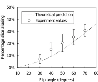
The slab profiles at two designated flip angles 400 and 700 show that at larger designated flip angles, the actual slab profiles for the SSFP signals tend to become wider. Figure 1 plots the percentage slice aliasing as a function of the designated flip angle. Note that at 700 flip angle as chosen in previous studies (1,2), the percentage slice aliasing is as large as nearly 30%, meaning that about one third of the slices would be subject to interference from CSF outside the image slab. Indeed, prominent slice aliasing was clearly visible even in the 21st slice out of an 84-slice slab (Fig.2). Experimental measurements from our subjects agreed fairly well with the theoretical deductions (Fig.1).
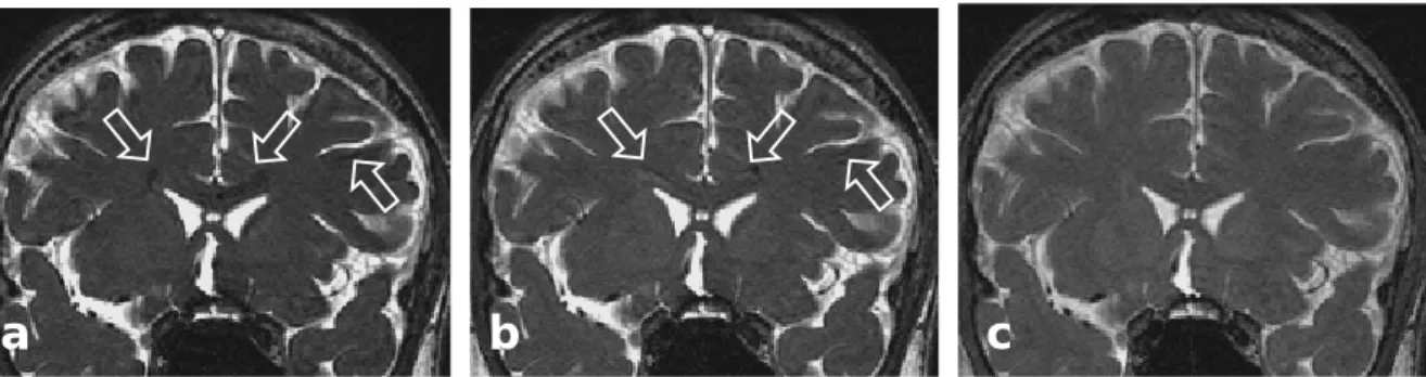
It is seen from Fig.1 that by lowering the designated flip angle, the percentage extent of slice aliasing could be reduced. However, this is achieved at the expense of decreased CSF-parenchyma contrast, as is shown in Fig.3 which plots the CSF-white matter contrast. Experimental values again showed good agreement with theoretical predictions. Note that as one decreases the designated flip angle to reduce slice aliasing, contrast between CSF and white matter becomes lowered. Figures 4a to 4c demonstrate this situation of trade-off. An inner slice (the 8th anterior slice in a 36-slice volume) exhibits aliased CSF signals from the lateral ventricles when the images were acquired using 700 flip angle. By lowering the flip angle to 500 (Fig.4b) and 300 (Fig.4c), the slice aliasing artifacts reduce and become invisible, respectively, but the contrast between CSF and the brain parenchyma gets lower as predicted.
The 3D balanced SSFP imaging technique is attractive for MR ventriculocisternography because it is capable of providing sub-millimeter voxel resolution along all three directions (1,2). Unfortunately, the current literature is in lack of comprehensive documentation on the optimization of imaging parameters in SSFP imaging, especially for neural imaging.
Results from our study indicate that the flip angle effect alone is an important factor that presents an obvious trade-off between image contrast and aliasing artifacts in 3D SSFP MR ventriculocisternography. The typical designated flip angle of 700 (1,2), aiming at maximizing fluid signals in SSFP imaging, is shown in our study to cause prominent slice aliasing artifacts affecting nearly one third of the imaging slices. The presence of the artifacts, if superimposed on the pathology, could possibly hamper accurate diagnostic interpretation. Therefore, it would be desirable to reduce the extent of aliasing in 3D balanced SSFP imaging, even though the artifacts have not yet led to misleading diagnosis in our institute.
As shown both theoretically and experimentally in this study, a simple means to reduce the slice aliasing artifacts is to use a smaller designated flip angle for SSFP imaging. By choosing a designated flip angle such that the SSFP signals vary more steeply with the actual flip angle (such as 400), the slice aliasing artifacts are shown to decrease. In our experience, we found that the designated flip angle of 400 reaches a good compromise between artifacts and contrast, showing nearly 60% reduction in the extent of slice aliasing with only about 20% decrease in CSF-parenchyma contrast, as compared with the use of 700 flip angle. We therefore conclude that SSFP imaging at a flip angle of about 400 could serve as an effective method suitable for 3D MR ventriculocisternography in clinical practice. 四、 計畫成果自評
Our efforts spent in this project have created results substantially greater than that mentioned in this brief report, which is an excerpted version of a recently published 2005 paper in the American Journal of
Neuroradiology (4). Overall, the project
has generated five conference papers, all presented in the 2005 Annual Meeting of the International Society of Magnetic Resonance
in Medicine. In addition, two journal articles (5,6) has been accepted, plus some other ones under active preparation. The achievements from this project have already influenced the routine protocols at Taipei and Kaohsiung Veterans General Hospitals. In short, we have confidence that a successful execution of the second year of this project will result in better utilization of the MR systems in routine diagnosis.
五、參考文獻
1. Schmitz B, Hagen T, Reith W. Three-dimensional true FISP for high-resolution imaging of the whole brain. Eur Radiol 2003;13:1577-1582. 2. Tsuchiya K, Aoki C, Hachiya J.
Evaluation of MR cisternography of the cerebellopontine angle using a balanced fast-field-echo sequence: preliminary findings. Eur Radiol 2004;14:239-242. 3. Huang TY, Huang IJ, Chen CY, Scheffler
K, Chung HW, Cheng HC. Are TrueFISP images T2/T1-weighted? Magnetic
Resonance in Medicine 2002;48:684-688.
4. Wu ML, Ko CW, Chen TY, Wu MT, Chung HW, Huang TY, Lin YR. MR ventriculocisternography by using 3D balanced steady-state free precession imaging: technical note. American
Journal of Neuroradiology, 2005;26:
1170-1173.
5. Huang TY, Chung HW, Wang FN, Ko CW, Chen CY. Fat and water separation in balanced steady-state free precession using the Dixon method. Magnetic
Resonance in Medicine, 2004;51:243-
247.
6. Chuang KH, Wu MT, Lin YR, Wu ML, Chung HW. Application of model-free analysis in the MR assessment of pulmonary perfusion dynamics. Magnetic
Resonance in Medicine, 2005, accepted.
六、圖表 0% 10% 20% 30% 40% 50% 10 20 30 40 50 60 70 80 Flip angle (degrees)
Per c ent age s lic e ali a s in g Theoretical prediction Experiment values
Figure 1. Percentage slice aliasing plotted as a function of the flip angle (dotted line). Experimental measurements (circles) agreed well with theoretical deductions. Note that at 700 flip angle, the percentage slice aliasing is as large as nearly 30%. By lowering the flip angle to 300 or 400, the percentage slice aliasing decreases to less than 20%.
Figure 2. Increased slice aliasing artifacts in 3D steady-state free precession imaging on a 33-year male subject. The 21st anterior slice in a 84-slice volume acquired with a designated flip angle of 700 is shown to exhibit prominent aliased CSF signals (arrows) from the lateral ventricles actually located at a posterior region. The posterior slice also shows evidence of slice aliasing from anterior region (image not shown).
六、圖表 0 0.1 0.2 0.3 0.4 10 20 30 40 50 60 70 80
Flip angle (degrees)
C S F -W M c o n tra s t (a rb it ra ry u n it s ) Theoretical prediction Experiment values
Figure 3. Contrast between CSF and white matter calculated as a function of the designated flip angle (dotted line), superimposed by experimental values (circles) measured from the images obtained from the healthy subjects recruited in our study. The measurements showed that the CSF-parenchyma contrast reduces as flip angle decreases, in good agreement with theoretical derivations.
Figure 4. Images acquired from a 23-year-old male healthy subject showing comparison of slice aliasing artifacts in 3D steady-state free precession imaging, as a function of the designated flip angle. The 8th anterior slice in a 36-slice volume acquired with a designated flip angle of 700 (a), exhibits prominent aliased signals from CSF (arrows). By lowering the flip angle to 500 (b) and 300 (c), the slice aliasing decreases (arrows) and becomes invisible, respectively. Also note the continuous reduction in the contrast between CSF and brain parenchyma from (a) to (c).