國立交通大學
材料科學與工程學系
博士論文
設計與製備具顯影與磁場刺激藥物釋放之
多功能型奈米藥物輸送載體元件
Design and Fabricate Multifunctional Nanocapsules for Imaging
and Magnetically-triggered Drug Release
研究生: 胡尚秀
指導教授:陳三元 博士
i
設計與製備具顯影與磁場刺激藥物釋放之
多功能型奈米藥物輸送載體元件
研究生:胡尚秀 指導老師:陳三元
國立交通大學材料科學與工程學系
摘要
在控制藥物釋放的系統中,尤其在是在及外力場刺激下作用之智慧型藥物釋 放系統,在全球已經引起各學界與業界人士的注意。在本論文中,利用材料與藥 物的結合,設計與製備一種具多功能性的磁性藥物奈米載體,除了具有利用外部 磁場控制釋放藥物之功能,並可以同步監測藥物釋放的情形。然而,傳統的藥物 釋放,僅利用藥物載體的特性與所在的環境變化來運作,在人體中並未真正達到 完善地控制釋放的目標。故本論文根據此創新的研究構想,使用"磁場"的非接觸 力特性,達到遙控藥物載體快速藥物釋放的效果。首先,用奈米級氧化鐵與明膠 /幾丁聚醣製備複合材料水膠,該水膠可以利用外加高頻磁場驅動水膠內部氧化 鐵生熱與轉動,進而使該溫度敏感型水膠高分子結構鬆散,達到快速藥物釋放的 效果。為達到可應用於體內之藥物載體,因此將藥物載體奈米化,提出利用新穎 製程控制,先將 poly-(N-vinyl-2-pyrrolidone) (PVP)做為奈米核之結構,其表面具 有螯合鐵離子的特性,再利用共沈法,將單晶奈米氧化鐵殼層成長於表面上,發 展具有磁敏感性的奈米核殼膠囊結構,將藥物完美的包覆於該奈米膠囊當中,在 未加磁場的狀態下,該奈米膠囊藥物自然釋放量趨近於零,如此可大幅降低高毒 性藥物在體內的副作用,然而當此奈米膠囊抵達治療位置時,可以利用外部的磁 場刺激磁奈米載體,控制藥物快速的局部釋放,有效的控制藥物釋放於定點。此 外,利用奈米晶體成長技術,架接具光學特性的量子點於奈米膠囊的表面上,此ii 具光學特性量子點,可以利用其螢光顯影,來追蹤該奈米膠囊於體內的位置,並 且搭配磁場的控制下,可監測藥物於定點釋放的情形,可用來評估釋放量對於疾 病的影響。並且奈米膠囊的表面經由化學表面改質,可帶有標靶癌細胞的分子, 在體內中大量提升奈米膠囊進入癌細胞的效率。此奈米複合膠囊搭配外加磁場的 控制的結構元件,期望在未來可達到快速有效率的局部釋藥於腫瘤細胞,並同時 於體內偵測藥物釋放情形。 同時,為了達到包覆油相藥物與提升包藥率,設計另一種多功能性的磁性奈 米膠囊藥物載體,尺度約為 100 奈米,將油相藥物與氧化鐵作為奈米核,並在載 體外部包覆奈米二氧化矽殼層,達到大幅降低藥物自然釋放的效果;並藉由螢光 染劑標定二氧化矽殼層,可使該載體可同時具有控制釋放藥物與顯影之功能。未 加磁場的狀態下,由於二氧化矽為堅固緊密之結構,此奈米膠囊藥物利用奈米殼 層,將藥物包覆於載體當中,可大幅降低載體漏藥性與高毒性抗癌藥物在體內的 副作用,然而當此奈米膠囊抵達需要特定位置時,再施加外部的磁場刺激磁奈米 載體,控制藥物快速的局部釋放,將大量的藥物釋放於治療之位置,達到治療的 效果。此外,磁性奈米粒子還能利用 MRI 來作為細胞追蹤的標的,在磁性奈米 粒子表面接枝特定抗體,使此特殊架接的奈米粒子可以與特定細胞之抗原結合, 以達到標定式之藥物釋放。本研究後期將利用靜脈注射的方式,將奈米膠囊藥物 載體注射入老鼠的血管內,來作一些體內之標定式藥物釋放之測試,並觀察體內 腫瘤細胞的治療效果,期望達到新一代的治療效果。 關鍵字:藥物控制釋放,磁性奈米粒子,藥物載體,影像顯影
iii
Design and Fabricate Multifunctional Nanocapsules for Imaging and
Magnetically-triggered Drug Release
Student: Shang-Hsiu Hu Advisor: San-Yuan Chen
Department of Materials Science and Engineering
Nation Chiao Tung University
Abstract
Controlled drug release has been received greatest attention worldwide, especially stimuli-responsive drug-delivery systems. In order to accelerate the response of an adaptive nanocarrier to stimuli, the use of the magnetic-sensitive nanomedical platfoems as a new type of actuator has been developed in this thesis. In first part, ferrogels composed of thermosensitive polymers, gelatin and chitosan, and iron oxide nanoparticles were triggered by a high frequency magnetic field to achieve pulsatile drug release. Under cyclic exposures to the high frequency magnetic stimuli, a highly controllable and repeatable burst release were exhibited with desirable precision from the ferrogels. In second part, the drug carriers were designed and narrowed down to nanosize. A novel core-shell nanosphere which was constructed by a poly-(N-vinyl-2-pyrrolidone) (PVP)-modified silica core with an outer layer of single-crystal iron oxide shell was fabricated. This drug delivery nano-device capable of providing controlled release of precise dose of therapeutic molecules when body needs and in particular, zero release when no need in the host, is critically important for clinical practices. The nanosphere showed outstanding release-and-zero-release characteristic via the addition and removal of an external manipulation of high-frequency magnetic field, respectively. Upon magnetic stimulus, the single
iv
crystal iron oxide shell with a thickness in a few nanometers demonstrated atomic re-arrangement, forming lattice of varying orientations, where inter-crystal boundary were developed, allowing drug eluted or no released in a reversible behavior. Further stimulation causes rupturing, i.e., permanent damage, of the shell, where drug eluted rapidly and can hardly be ceased, even after removal of the stimulus. Such novel core-shell nanospheres showed a fast cancerous cell (human cervical cancer cell line) uptake behavior, which implies a highly efficient potential to achieve anti-cancer therapy when anti-cancer drug is delivered. On the other hand, constructing with other functional nanoparticles like quantum dots can form the multifunctional magnetic nanocarriers that could be applicable to a variety of fields as a new driving mechanism. Combining with fluorescent quantum dots, the nanocarriers can be tracked in the human body. The optical properties also can be tunable while applying the magnetic field for sensing the drug release. Drug release rate at on-off operation of AC magnetic fields (hyperthermia effects) and the conditions of killing-tumor cells also would be investigated.
Finally, the magnetic nanocapsules capable of carrying hydrophobic drug molecules were synthesized as the bifunctional magnetic vectors that can be triggered for control release of therapeutic agent by an external magnetic field. The drug release profiles of cpsules can be well-regulated through an ultra thin layer of silica shell. Remote control of drug release from the nanocapsules was successfully achieved using an external magnetic field where the core phase being structurally disintegrated to a certain extent while subjecting to magnetic stimulus, resulting in a burst release of the encapsulated drug. However, a relatively slow and linear release restored immediately right after removal of the stimulus. More than surprisingly, the nanocapsules demonstrated a relatively high uptake efficiency by HeLa cell line. In addition, the magnetic nanoparticles still provides some advantages, such as magnetic
v
resonance image (MRI) for cell labeling and grafted probe-proteins (such as biotin or antibody) onto magnetic nanoparticles for ―target‖ drug delivery. Therefore, the multifunctional drug-carriers is able to be triggered by thermal and magnetic changes, and especially put emphasis on ―target‖ drug-delivery using MRI, cell labeling or others detecting technique.
vi
致謝
經過四年的奮鬥與努力,終於在 2010 年拿到了博士學位,雖然一路上跌跌 撞撞,遇到了很多挫折與阻礙,過程難免有許多的失落、灰心與現實的壓力,不 過我總是相信這是自己選擇的路,一定要堅固自己的意志將學位完成,總是不斷 的提醒自己,雖然自己不是最聰明的學生,要做的最認真的學生。取得學位時, 那心中的激動與喜悅,真的難以言喻。 首先要先感謝我的指導教授陳三元老師,在研究上,總是很有耐心的與學生 討論,分析每項實驗的新穎性與創新性,找出好的研究方向,給學生很大的想像 空間與彈性,並誘導學生的創造力,不斷激發學生的潛能,訓練鋼鐵般的意志, 在生活上,也給學生許多的協助與鼓勵,我相信,我能遇到這麼優秀的指導老師, 真的是上輩子修來的福氣。我也要感謝另一位老師劉典謨教授,嚴格上來說,他 也是學生的指導教授,從碩士到博士班,指導學生論文的研究題目與方向,他的 創新思考模式與積極研究的態度,深深的感染了學生,讓學生對於研究的思維有 更深入的想法,在論文撰寫部分,也給予學生很多的指導,不厭其凡的幫助學生 修改論文,學生相當的感動,會永遠銘記在心。此外,學生也要感謝在美國華盛 頓大學的 Xiaohu Gao 教授,在學生於美國執行千里馬研究計畫時,給予學生很 大的協助與資源,學習美國在研究上活躍與開放式的思維,訓練學生的快速思考 模式,見識美國頂尖研究環境的訓練模式,雖然只在該實驗室待一年,對於研究 能力與創造性的開發,有相當的助益。在此也感謝交大電控所的陳右穎老師,在 核詞共振顯影與奈米載體的開發,給學生許多相當有用的建議,並給予學生相當 多研究資源。也感謝來幫學生口試的宋信文教授、林峰輝教授、鐘次文教授、林 志生教授、邱信程教授,給予學生許多各領域的專業建議,深入研究探討,讓學 生受益良多。最後要感謝台灣國科會,給予研究經費上支持,充裕的研究經費, 對於研究的速度與執行度有正面的幫助。 特別感謝劉定與學長,從碩士班就開始帶學生進入磁性材料與藥物釋放的領 域,學習許多實驗的技巧與方法。感謝劉澤英學長於藥物釋放分析上的協助與指 導。特別感謝蕭繼聖學長,在電子顯微鏡上分析的協助,沒有好的電鏡分析人才, 我相信也做不出好的研究,學長總是很有耐心的協助我完成各項樣品分析,很懷 念在路途遙遠的奈米中心一同打拼的日子。此外,感謝董維琳學妹,在細胞研究 上的協助,他總是很認真負責的完成許多任務,日以繼夜的完成工作量相當大的 細胞實驗,建構細胞實驗系統,讓研究順利進行。也要特別感謝同組的蔡佳惠、 黃信揚、陳柏溶、廖邦傑,在研究上的討論於協助,一同抱著冒險的精神往未知 的方向前進,一同解決許多難題,有了你們,研究才能順利的推進。此外,也感 謝實驗室其他的成員,昆和、彥妤、冠廷、泓洲、振富、志欣、翔銓、又維、薇 蓁、大方、彥博、純瑜、偉銘、俞君、博學、孟軒、衍人、簪華、輔宣、佩鈴、vii 岱容、立傑、嘉偉、吟芝,在研究與生活上的幫忙與協助,幫遇到打擊時,總能 適時的拉我一把,讓我又有了新的力量,有了你們,研究所生活變得更加的多采 多姿。 感謝我的家人,爺爺、奶奶、爸爸、媽媽、弟弟與姑姑們,在一路上無怨無 悔的支持與鼓勵,在我感到迷惘疲累時,督促我必頇努力勇敢的往前,給我很大 的關心,此刻終於畢業了,希望與他們一同分享這份喜悅,這個學位也是屬於他 們的。感謝毓瑄,陪伴我度過的日子,讓我感覺生命更有他的價值,更用心體會 這個世界。感謝我的機車與電腦,陪我完成十幾篇的 SCI 論文與國科會計畫,我 想我跟他們相處的時間是最長的。一個結束,也是另一個開始,我會秉持著博士 班所學到的態度與精神,繼續面對未來的挑戰。 2010 年 7 月 22 日 尚秀
viii
Contents
摘要... i Abstract ... iii 致謝...vi Chapter 1 Introduction ... 1Chapter 2 Literature Review and Theory ... 6
2.1 Introduction of controlled drug delivery and release ... 6
2.2 Responsive polymers for drug delivery ... 7
2.2.1 Temperature-responsive polymer ... 8
2.2.2 Responsive polymer incorporated magnetic nanoparticles ... 11
2.3 Nanotechnology on drug delivery system... 13
2.3.1 Nanotechnology for nanoplatforms ... 14
2.3.2 Nano-polymer drug delivery vehicles ... 14
2.4 Magnetic nanoparticles for biomedical applications ... 16
2.4.1 Synthesis of magnetic nanoparticles ... 19
2.4.2 Biocompatibility of magnetic nanoparticles ... 21
2.4.3 Magnetic nanoparticles for high performance magnetic resonance imaging ... 23
2.4.4 Magnetic nanoparticles for in vivo molecular imaging ... 26
2.4.5 Surface modification of magnetic nanoparticles ... 28
2.5 Magnetic Nano-Carriers for drug delivery and therapy ... 31
2.5.1 Amphiphilic micellar coatings ... 33
2.5.2 Magnetic nanoparticles encapsulated in polymer for small molecules drug delivery ... 34
2.5.3 Magnetic nanoparticles encapsulated in silica for drug delivery ... 36
2.5.4 Magnetic nanoparticles for other therapy ... 38
2.5.5 Magnetically triggered release for composite membrane ... 40
2.5.6 Magnetic-vortex microdiscs for targeted cancer-cell destruction ... 42
Chapter 3 Experimental Procedures ... 44
3.1 Experiment overviews ... 44
3.2 Synthesis of magnetic nanoparticles ... 45
3.3 Characterization ... 46
3.4 High frequency magnetic field (HFMF) ... 46
3.5 Drug loading efficiency and release... 48
3.6 Release kinetics ... 49
3.7 Cell culture ... 50
ix
4.1 Introduction ... 52
4.2 Fabrication of ferrogels ... 53
4.3 Characteristics of ferrogels ... 54
4.4 Drug release triggered by magnetic field from ferrogels ... 56
4.5 Cyclic drug release rates of ferrogels ... 59
Chapter 5 Core/Single-Crystal-Shell Nanospheres for Controlled Drug Release via a Magnetically Triggered Rupturing Mechanism ... 62
5.1 Introduction ... 62
5.2 Preparation of core/single-crystal-shell nanospheres... 64
5.3 Nanostructure of core-single crystal shell nanospheres ... 66
5.4 Triggered release of fluorescence dye from nanoparticles ... 67
5.5 BET analysis of core-shell nanpspheres ... 69
5.6 Nanostructural evolution ... 71
5.7 in vitro controlled release ... 74
5.8 Summary ... 85
Chapter 6 A Multifunctional Nanodevice capable of Imaging, Magnetically Controlling, and In-situ Monitoring Drug Release ... 86
6.1 Introduction ... 86
6.2 Experimental section of multifuntional nanodevices ... 88
6.3 Structures of multifunctional nanodevices ... 89
6.4 Drug release and in-situ monitoring abilities of the nanodevices ... 94
6.6 Operation mechanism of the nanodevice ... 101
6.7 In-situ-monitoring of drug release in cancerous cells ... 102
6.8 Summary ... 106
Chapter 7 PVA-Iron Oxide /Silica Core-Shell Nanocarriers for Magnetically Controlled Drug Release and Cancer Cell Uptake Efficiency ... 109
7.1 Introduction ... 109
7.2 Experimental section ... 110
7.3 Preparation of SAIO@SiO2 nanocarriers ... 114
7.4 Characterization of SAIO@SiO2 nanocarriers ... 117
7.5 Drug release of SAIO and SAIO@SiO2 nanocarriers ... 118
7.6 Magnetic-sensitive drug release behavior ... 120
7.7 Nanostructure evolution under magnetic treatment ... 122
7.8 Cell Uptake ... 126
7.9 Summary ... 129
Chapter 8 Thermosensitive Yolk/Shell Capsules for Magnetically-Triggered Drug Release ... 131
x
8.2 Experimental section ... 133
8.3 Preparation of thermosensitive yolk/shell capsules ... 136
8.4 Characterization of yolk/shell capsules ... 139
8.5 Drug release of yolk/shell capsules and F68/PVA nanocomposites ... 141
8.6 Thermosensitive behavior of the yolk/shell capsules ... 142
8.7 Magnetically-triggered drug release from yolk/shell capsules ... 145
8.8 Nanostructure evolution under magnetic treatment ... 147
8.9 Cell uptake and MR imaging ... 149
8.10 Summary ... 151
Chapter 9 Conclution ... 153
9.1 Ferrogels for magnetically-triggered drug release ... 153
9.2 Core/single-crystal iron oxide shell nanospheres for drug delivery ... 153
9.3 Self-assemble iron oxide/silica (SAIO) core-shell carriers ... 154
9.4 Thermosensitive yolk/shell capsules ... 155
Reference ... 156
xi
Figure Captions
Figure 2.1 (a) Drug concentration at site of therapeutic action under a controlled drug release
system (solid line) and a conventional injection (dash line). (b) An ideal distribution of drug concentration from a controlled drug release system. ... 7
Figure 2.2 Thermosensitive behaviors of poly (ethylene oxide)-poly (propylene oxide)-poly
(ethylene oxide) (PEO-PPO-PEO) tri-block polymer. ... 9
Figure 2.3 Gelatin possesses a triple helix structure at room temperature and forms a stable
gels (middle). While increasing above the breaking temperature of hydrogen bonds, the gelatin becomes solution states (right). ... 10
Figure 2.4 Magnetic Targeted Carriers (MTCs) has been designed which could adsorb
pharmaceutical agents through application of an externally magnetic field for site-specific targeting and sustained release of drugs. ... 12
Figure 2.5 Representative nanoparticles that can serve as nanoplatforms and polymeric
vehicles for targeted molecular imaging in living subjects. ... 16
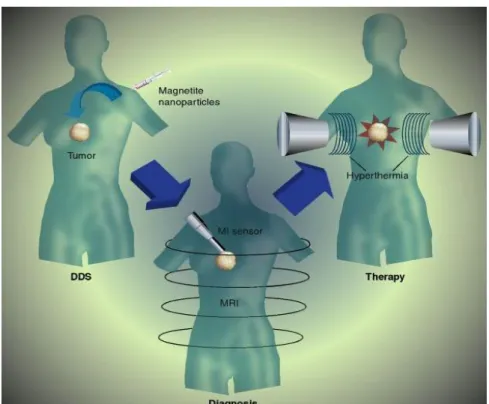
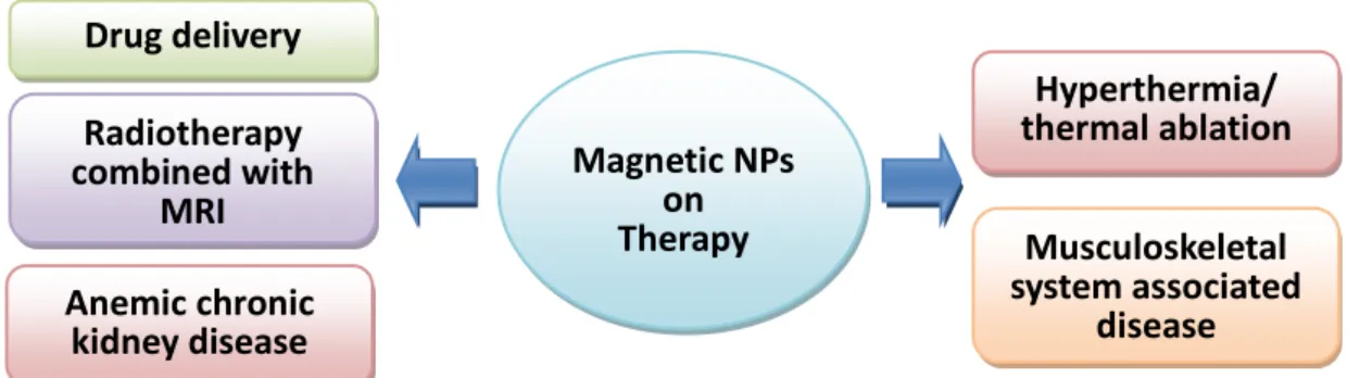
Figure 2.6 Magnetic nanoparticles for biomedical applications: (a) drug delivery, (b)
diagnosis, and (c) therapy by hyperthermia. ... 18
Figure 2.7 TEM micrographs of iron oxide NPs with diameters of (a) 6 nm, (b) 7 nm, (c) 8
nm, (d) 9 nm, (e) 10 nm, (f) 11 nm, (g) 12 nm, (h) 13 nm. The organic phase high-temperature synthetic route enables precise control of NP size. ... 21
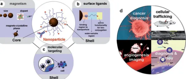
Figure 2.8 Functionalized MNPs for molecular and cellular magnetic resonance imaging
(MRI). a) Controlling the magnetism of the nanoparticle core, b) tailoring the surface ligands of the nanoparticle shell, and c) the molecular targeting capability of biomolecule-conjugated nanoparticles. d) High performance utilizations of nanoparticles for molecular and cellular MRI. ... 25
Figure 2.9 (a) Surface spin canting effect of a nanoparticle upon magnetization (M magnetic
moment, H external magnetic field). (b–e) Nanoscale size effects of Fe3O4 nanoparticles
on magnetism and MR contrast effects. (b) Transmission electron microscopic (TEM) images of 4, 6, 9, and 12 nm of iron oxide nanoparticles. (c) Mass magnetization values, d) T2-weighted MR images (top: black and white, bottom: color), and (e) relaxivity coefficient r2 of the nanoparticles presented in (a). (f) FeCo magnetism-controlled metal-alloy MNPs. These FeCo MNPs have an exceptionally high magnetization value of 215 emu per gram metal ... 26
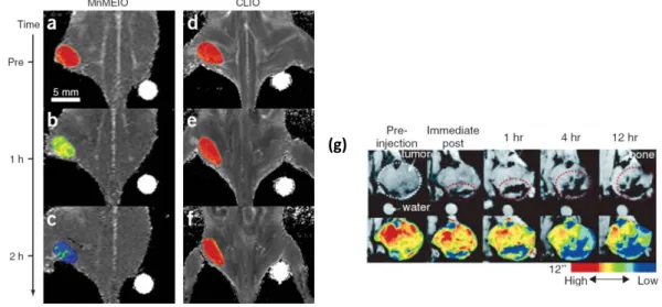
Figure 2.10 (a-f) Color maps of T2-weighted MR images of a mouse implanted with the
cancer cell at different time points after injection of MnFe2O4–Herceptin conjugates or
CLIO–Herceptin conjugates. In (a–c), gradual color changes at the tumor site, from red
(low R2) to blue (high R2), indicate progressive targeting by MnFe2O4–Herceptin
xii
model imaged at 9.4 T. Color-coded MR images to further delineate MR signal changes. ... 27
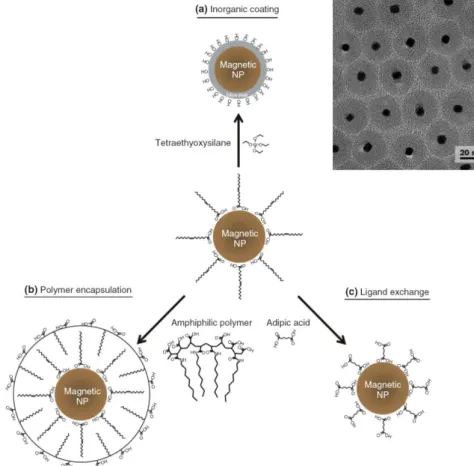
Figure 2.11 Three types of surface modification schemes for magnetic nanoparticles. (a)
Inorganic surface coating with an amorphous silica shell. (b) Amphiphilic polymer coating on the magnetic nanopartiels. (c) Ligand exchange is to replace native surface ligands. These routes exhibit polar or charged functional groups onto the outer surface of the NP for water solubility. ... 30
Figure 2.12 Biomedical application of magnetic nanoparticles. ... 32 Figure 2.13 (a) Synthesis scheme and TEM image of polystyrene–poly(acrylic acid) (PSPAA)
block copolymer micelles encapsulating several MNPs. (b) Schematic and TEM image of poly(d,l-lactide)–PEG block copolymer micelles encapsulating several MNPs and a therapeutic agent. ... 34
Figure 2.14 (a) Synthetic procedure for the multifunctional polymer nanoparticles. TEM
images of nanoparticles. (b) CLSM of DOXO fluorescence and optical images in KB cells treated with a) naked PLGA(MNP/DOXO), b) PLGA(MNP/DOXO)-PEG, c)
PLGA(MNP/DOXO)-FOL nanoparticles, and d) PLGA(MNP/DOXO)-FOL
nanoparticles under an external magnetic field, respectively. ... 36
Figure 2.15 (a) Schematic illustration of multifunctional nanoparticles showing iron oxide
nanocrystals encapsulated within mesoporous silica, hydrophobic anticancer drugs stored inside the pores, and surface modifications with phosphonate and folic acid targeting ligands. SEM and TEM images of the iron oxide incorporated within the mesoporous silica NPs. (b) Fluorescence microscopy images showing the effect of folic acid modification on the NPs (green fluorescence). The cell nuclei were stained with DAPI (blue fluorescence), and the membranes were stained with WGA (red fluorescence).. ... 38
Figure 2.16 (a) Therapeutic applications of hybrid FePt@CoS2 metal-alloy MNPs. Left: the
lethal effect of the hybrid MNPs on cells. Middle: optical detection of changes in cell morphology of untreated HeLa cells (top) and the MNP-treated HeLa cells (bottom). Right: dose-dependent cell viability, b) hybrid iron oxide nanoparticles consisting of SiO2@Fe3O4@Au and anti-HER2/neuantibody for simultaneous MRI diagnosis and laser-assisted hyperthermia therapy. ... 39
Figure 2.17 Stimulus-responsive membrane triggering in vitro: (a) temperature-triggering,
comparison of nanogel particle size in suspension (blue data, right y-axis) and differential flux of sodium fluorescein through the nanogel-loaded membranes (red data, left y-axis) as a function of temperature; (b) magnetic triggering, temperature profile in the sample chamber and differential flux of sodium fluorescein out of membrane-capped devices as a function of time over four successive on/off cycles of the external magnetic field. ... 41
xiii
Figure 2.18 (a) The concept of targeted magnetomechanical cancer-cell destruction using
disc-shaped magnetic particles possessing a spin-vortex ground state. The microdiscs are biofunctionalized with anti-human-IL13 2R antibody, specifically targeting human glioblastoma cells. When an alternating magnetic field is applied, the magnetic discs oscillate, compromising membrane integrity and initiating spin-vortex-mediated programmed cell death. (b,d) and MD–mAb-functionalized cells subjected to 20 Hz–90 Oe a.c. fields for 10 min and TUNEL stained 4 h after the magnetic-field exposure (c,e). The control cells with well-organized chromatin structures have a blue fluorescence, whereas the treated cells are stained with a dark orange–brown dye owing to chromatin fragmentation—an indication of apoptosis. ... 43
Figure 3.1 (a) Intrinsic (Néel) and (B) extrinsic (Brown) remagnetization mechanism
(schematic). Equipment of high frequency magnetic field (HFMF). ... 47
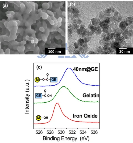
Figure 4.1 (a) SEM, (b) TEM and (c) XPS analyses of ferrogels composed by iron oxide
nanoparticles and gelatin matrix. ... 55
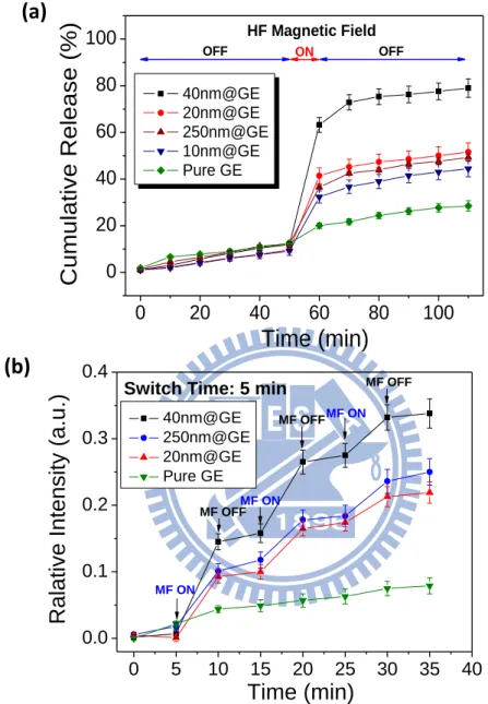
Figure 4.2 (a) Drug release behaviors of the feoogels under the 10 minutes exposing of high
frequency magnetic field (HFMF) and (b) The on-off switch operations of high frequency magnetic field (HFMF) manipulated the cumulative drug release of the ferrogels with different sizes of the nano-magnets. ... 57
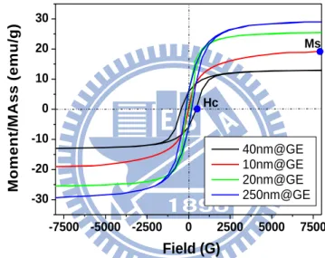
Figure 4.3 Vibrating sample magnetometry measurements for the ferrogels with various iron
oxide nanoparticles sizes. ... 58
Figure 4.4 Schematic drawing of structures of ferrogel which exhibited a triple-helix structure
to restrict the drug molecules to release. While applying the high frequency magnetic field (HFMF), the magnetic nanoparticles provides the heat energy to loose the structures, and twist and shake the polymer molecular chains to effectively accelerate drug release rate. ... 58
Figure 4.5 Cyclic drug release rates of 40nm@GE ferrogels under a 5-min period of HFMF
stimuli and a long 180-minute switching duration, where a longer switching time period ensures the drug to reach a kinetically favorable distribution in the ferrogel for a subsequent burst release. ... 60
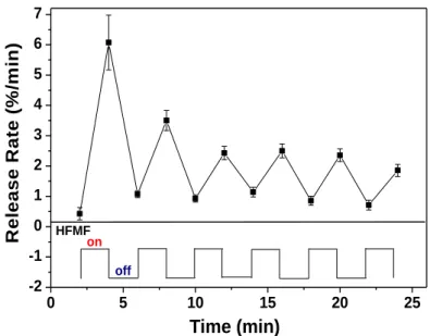
Figure 4.6 Cyclic drug release rates of 40nm@GE ferrogels (a) under a 2-minute period of
HFMF stimuli and a short 2-min switching duration. ... 61
Figure 5.1 (a) TEM images and (b) HR-TEM of PVP-modified Silica/Fe3O4 core-shell
nanospheres. Local Fourier transfer patterns indicate that the crystallographic structure is extremely uniform and homogenous through the shell. Some facet {1 1 0} planes are observed inside the nanosphere, and the Fourier transform pattern indicates that the shell is oriented at z = [0 0 1]. (c) Local Fourier transfer patterns indicate that the crystalline is uniform and homogenous through the particle. (d) X-ray diffractometer verified the
xiv
according to JCPDS [85-1436]). ... 65
Figure 5.2 (a) Photographs of the cuvettes with fluorescence-loaded PVP-modified
Silica/Fe3O4 core-shell nanospheres dispersed in water solution. Before HFMF exposure,
fluorescence-loaded PVP-modified Silica/Fe3O4 core-shell nanospheres displayed no
sign of fluorescence under the UV light (left); after exposure, green fluorescence
released from the PVP-modified Silica/Fe3O4 core-shell nanospheres was clearly
detected (right). (b) Emission spectra of PVP-modified Silica/Fe3O4 core-shell
nanospheres (15 mg per 10 mL water) for applying HFMF from 30 s to 180 s. (c)
Emission spectra of PVP-modified Silica/Fe3O4 core-shell nanospheres. Negligibly small
amount of the dye molecule being further released from the nanospheres for a time period of 120 seconds in the absence of the stimulus. ... 68
Figure 5.3 Nitrogen-adsorption spectra for the nanospheres before and after a 20-min HFMF
exposure. BET measurements of silica nanospheres treated with HFMF for 0 sec, 60 sec and 20 minute. ... 69
Figure 5.4 Schematic illustration of the thin shell with a proposed mechanism for control
release of the fluorescence dye: (I) while applying HFMF, vibration enlarges the dimension of the nano-faults, making dye molecules easy to release out and the change in the dimension of the nano-faults is physically reversible upon a short-term field exposure. (II) under long-term exposure, the nano-faults received sufficient amount of the energy, vigorous energy (although not specifically identified yet in this communication) ruptures the thin shell permanently. ... 72
Figure 5.5 TEM images of long-term HFMF exposure, the nano-faults received sufficient
amount of the energy, vigorous energy (although not specifically identified yet in this communication) ruptures the thin shell permanently. ... 73
Figure 5.6 Fluorescence micrographs of HeLa cells after 10 hours incubation with
fluorescence-loaded PVP-modified Silica/Fe3O4 core-shell nanospheres. (a) without
HFMF treatment; (b) after 30 seconds of HFMF stimulus, green fluorescence was clearly observed within the cell bodies of these HeLa cells upon excitation at 494 nm. ... 75
Figure 5.7 (a)Temperature curve of nanocapsules during application of HFMFs. (b) Drug
release behavoirs of nanocapsules under HFMFs, and their release rates (k). ... 76
Figure 5.8 A near-linear relationship between the rate constant k and SAR values, with
correlation coefficient as high as 0.98 can be obtained. ... 79
Figure 5.9 SAR effects on the different manetic field regions. Drug release behaviors in cells
are highly relative with the strength of SAR. ... 81
Figure 5.10 The TEM electron beam is focus on the nanocapsule for different time durations.
While increasing the heating time to 40 minutes, the obvious deformation of melting was observed. ... 82
xv
for different time of HFMF treatment. ... 83
Figure 6.1 (a) Schematic of the stimuli-response nanodevice delivery system where quantum
dots were deposited on the shell of the PVP/Fe3O4 core-shell nanospheres. Attachment of
the ZCIS QDs on the surface of the nanospheres acts not only as a strong fluorescence-emitting agent, but also as a sensor to monitor the drug release in a real-time basis under magnetic induction. (b) TEM image and (c) HRTEM image of the
PVP-Fe3O4 core-shell nanospheres. (d) HRTEM image of the ZCIS-doped nanospheres.
After incorporation of the ZCIS QDs on the core-shell nanospheres, the suspension displayed a fluorescence character under the UV light (inset picture). ... 90
Figure 6.2 Local Fourier transfer patterns of single-crystal iron oxide shell indicate that the
shell exhibits uniform and homogenous crystalline orderliness along the surface of the core phase. ... 91
Figure 6.3 (a) The TEM image and local Fourier transfer patterns of ZCIS QD-Single crystal
Fe3O4 shell nanoplatform. The local Fourier transfer pattern also demonstrated a high
crystallinity of the ZCIS QD. (b) The TEM image of CIS QD-Single crystal Fe3O4 shell
nanoplatform. The EDS investigated the Fe3O4 shell and ZCIS QD, corresponding to the
regions 1 and 2 in the HRTEM image, respectively. HRTEM image shows the solid nanoparticle attaching the ring-like shell region being ZCIS QDs in the heterodimer. The energy dispersive X-ray spectrometer (EDS) analysis confirms that the ring-like region mainly consists of Fe and the small solid particle consists of Cu and Se. ... 91
Figure 6.4 Field-dependent magnetization curve of nanodevices with and without quantum
dots... 92
Figure 6.5 Schematic drawing showing multi-functionalities of each compartment from
as-designed nanodevices for nanoimaging, controlled drug release, and in-situ motoring of drug release. ... 93
Figure 6.6 Cellular uptake of the nanodevice was evaluated using HeLa cells, by incubating
the cell line with both mercaptoundecanoic acid-modified nanodevice (MUA-NDs) and folic acid-modified nanodevice (FA-NDs). Both the MUA-NDs and FA-NDs were uptaken by the cells in 4 hours of period, probably through endocytosis, with different degrees of efficiency. In comparison, the majority of the FA-NDs can be clearly observed in the cytoplasm region of the cell, but only a few MUA-NDs was uptaken by HeLa cells, indicating that the folic acid-modified version promotes a stronger cell-specific intake by the HeLa cell line than that of the mercaptoundecanoic acid-modified version. The folic-acid-modified nanodevices showed excellent cell-specific uptake efficiency, through possibly endocytosis. ... 93
Figure 6.7 (a) Emission spectra of dye-loaded nanodevices (30 mg per 10 mL water) under
HFMF treatment over a time period from 0 s to 100 s. Before HFMF exposure, the dye-loaded nanodevices displayed no sign of dye release, which causes green
xvi
fluorescence at an emission wavelength of 517 nm, as determined by fluorescence spectrophotometer. However, a strong emission signal from the QDs after exposure, red fluorescence at an emission wavelength of 581-614 nm, show that dye was released from the nanodevices. A degenerative green florescence appeared concurrently. (b) Model drug intensity versus quantum dot intensity curves originate from both the dye and ZCIS emitting spectra and show a near symmetrical profile under different magnetic field strengths. ... 95
Figure 6.8 Temperature curve of nanodevices during application of HFMFs of different
strengths. ... 97
Figure 6.9 Binding energy of S 2p of the nanodevices with and without exposure to a high
frequency magnetic field (HFMF) for a time period of 180 seconds. XPS results indicate the binding energy shift of the S 2p line from 162.3 eV - 166.2 eV before the stimulus to a single, strong binding-energy peak at 167 eV after the magnetic exposure. This could
be attributed to S2- from ZnS, CuS, In2S3, or SO4
2-, SO2. The binding energy at 167 eV on
the ZCIS surface is strongly associated with the presence of oxide groups including SO4
2-,
SO2, etc on the surface. This demonstrates a rapid oxidation of the ZCIS QDs after a
short period of magnetic induction. ... 99
Figure 6.10 PL emission spectrum at various time intervals of nanodevice incubation in PBS
solution. ... 100
Figure 6.11 (a) Schematic illustration of the nanodevice with a proposed mechanism for
controlled release of the dye molecules, as well as the degeneration of fluorescence intensity of the ZCIS QDs (b) Shell vibration causing enlargement of the dimensions of nano-crevices along the deformed single-crystal shell, rendering dye release upon short-term magnetic stimulation. (c) After long-term exposure, the deformed shell has received a sufficient amount of the energy to cause a final, permanent, mechanical rupture. Meanwhile, a rapid surface oxidation altered the surface structure of the QDs, leading to substantial degeneration of the fluorescence intensity. ... 102
Figure 6.12 Cell viability of HeLa cells after 12 to 48 hours of incubation with increasing
amounts of folic acid modified nanodevices (FA-NDs). Cell viability was measured using an MTT assay. ... 103
Figure 6.13 Fluorescent combination of the HeLa cells with the dye-loaded nanodevices after
12 hours incubation. With increasing duration of HFMF treatment, both the controlled release of the dye molecules (green channel intensity increased) and the associated real-time, in-situ monitoring capability of the doped ZCIS QDs (red channel intensity decreased) can be manipulated simultaneously to single-cell resolution. This implies that dye release can be precisely monitored by the variation of the ZCIS QDs from the
nanodevices. Gsum/Bsum represents the ratio of the green channel intensity to blue channel
xvii
Rsum/Bsum is then defined as the relative intensities of the nanodevices in each cell... 105
Figure 6.14 The ratio of Gsum/Bsum and Rsum/Bsum versus the duration of magnetic stimulus in
the cells gives rise to two curves. The results demonstrate the relative drug concentration,
represented by Gsum/Bsum, in the cells with increasing duration of stimulus. The
fluorescence intensity originating from the ZCIS QDS, represented by Rsum/Bsum,
decreased in proportion at the same time... 106
Figure 6.15 Fluorescent morphologies of HeLa cells after 12 hours incubation with drug
loaded nanodevices. With increasing the time duration of HFMF treatment, the control of drug release associated with a real-time self monitoring of the ZCIS QDs from the nanodevices were manipulated simultaneously. The digital analysis software (Nikon, Japan) was use to analyze the fluorescence intensities of model drug and nanodevices. The conditions of the exposure are the same for each color channels. The analyzed areas were determined by the software which defined the fluorescence intensity from 1 to 255. The ranges of the fluorescence intensities: Blue channel (60-255), Green channel (40-255), and Red channel (30-255). ... 107
Figure 7.1 Schematic illustration of the synthesis and structure of the self-assemble iron oxide
/silica core-shell (SAIO@SiO2) nanocarriers for magnetically controlled drug release.115
Figure 7.2 TEM images of (a) iron oxide nanoparticles, (b) self-assemble iron oxide (SAIO)
nanospheres, and (c, d) self-assemble iron oxide /silica core-shell (SAIO@SiO2)
nanocarriers. The thickness of silica shells coated on the SAIO nanoparticle is 4-5 nm. The inside pictures displayed the solution color for different particles. (e-g) The size
distributions of iron oxide nanoparticles, SAIO nanoparticles and SAIO@SiO2
nanocarriers were measured by dynamic light scattering (DLS). ... 116
Figure 7.3 The SEM images of (a) SAIO and (b) SAIO@SiO2 nanospheres. (c)After 4
minutes, the SAIO@ SiO2 nanospheres did not show obviously cracks under the SEM
analysis. ... 116
Figure 7.4 (a) X-ray diffraction patterns of iron oxide nanoparticles, SAIO and SAIO@SiO2.
(b) Field-dependent magnetization curve of iron oxide nanoparticles, SAIO and
SAIO@SiO2 nanocarriers. The inside pictures: the SAIO@SiO2 nanocarriers were
attracted by an external magnet. ... 118
Figure 7.5 Cumulative drug release of SAIO and SAIO@SiO2 nanocarriers. Coated with
silica shells, the SAIO@SiO2 nanocarriers exhibited relatively smaller amount of
cumulative drug release than SAIO nanoparticles. ... 120
Figure 7.6 Cumulative drug release profiles of ibuprofen (IBU) from (a) SAIO and (b)
SAIO@SiO2 nanocarriers were triggered by 1 to 4 minutes of high frequency magnetic
field. ... 121
Figure 7.7 TEM images of nanostructures of (a, b) SAIO@SiO2 nanocarriers, and (c) to (f)
xviii
displayed no obviously crack after HFMF treatment. (c-f) SAIO without silica shells showed obvious cracks or deformation after applying HFMF. ... 123
Figure 7.8 Time-course PL microscopy images of HeLa cells labeled with FITC- SAIO@SiO2
nanocarriers, the cell skeleton wsa stained with rhodamine phalloidin (red), and cell
nucleus with DAPI (purple). Cells were incubated with FITC- SAIO@SiO2 nanocarriers
for (a) 30 min, (b) 1 h, (c) 2 h, and (d) 4 hour. ... 127
Figure 7.9 (a) Cell viability of HeLa cells after 12 to 48 hours of incubation with increasing
amounts of SAIO and SAIO@SiO2 nanospheres. Cell viability was measured using an
MTT assay. (b) Flow cytometry analysis for the FITC- SAIO@SiO2 nanocarriers
accumulated in HeLa cells for incubation of 30 min and 2 hours. ... 128
Figure 7.10 After the HeLa cells took up the SAIO@SiO2 nanospheres, the HFMF subjected
to these cells for 30 seconds to 4 minutes. The cytotoxicity with short time of HFMF treatment is small, indicating that the heat produced by nanospheres could not kill most of the cancer cells. ... 130
Figure 8.1 (a) Schematic illustration of the synthesis and structure of the thermosensitive
yolk/shell capsules for magnetically-triggered controlled drug release. TEM images of (b) yolk/shell-1, (e) yolk/shell-2, (f) yolk/shell-3, and (g) yolk/shell-4. (c) The SEM images of yolk/shell capsules. (d) The TEM image of lattice structure of iron oxide nanoparticles in the capsules. The yolk/shell capsules with an average diameter about 76 nm possessed an ultra-thin layer of dense silica shell of about 7 nm in thickness. ... 137
Figure 8.2 TEM images of yolk/shell capsules without PVA. Without PVA as a stabilizer, the
nanocapsules displayed a un-uniform morphologies. Some magnetic nanoparticles were not encapsulated in the nanocapsules. ... 138
Figure 8.3 SEM images of (a) yolk/shell-1, (b) yolk/shell-2, (c) yolk/shell-3, and (d)
yolk/shell-4. ... 138
Figure 8.4 (a) Field-dependent magnetization curves of yolk/shell-1 to yolk/shell-4, showing
the capsules are superparamagnetic. (b) Cumulative drug release of F68/PVA nanocomposites (NCs) and yolk/shell capsules. Coated with silica shells, the yolk/shell capsules showed relatively smaller amount of cumulative drug release than F68/PVA nanocomposites (NCs). ... 140
Figure 8.5 (a) Diameter of F68/PVA nanocomposites (NCs) measured by DLS decreased
abruptly at about critically micellization temperature (CMT). (b) DSC cooling scans of F68 and F68/PVA nanocomposites (NCs). ... 144
Figure 8.6 (a) Cumulative drug release profiles of ibuprofen (IBU) from yolk/shell capsules
were triggered by 1 minute of high frequency magnetic field (HFMF) at forth minute. Schematic illustration as shown in inside picture with a proposed mechanism for controlled drug release under HFMF. After the exposure to a magnetic field, the
xix
volume/hydrophobicity transition of cores produced a strong inner stress, making the capsules collapse or rupture as demonstrated in (b) and (c) TEM images. ... 145
Figure 8.7 Time-course confocal images of ARPE-19 cells incubated with 10 FITC-labeled
yolk/shell capsules (green dots) for (a) 4 and (b) 24 hours. The cells were stained with rhodamine phalloidin (red), and cell nucleus with DAPI (blue). (c) The cross section images of cells viewed by laser-scanning confocal microscope exhibited that the capsules are localized in cells. (d) ARPE-19 cell viability under the incubation of capsules for 24 hours with and without 1-minute of magnetic field treatment. (e) Magnetic resonance images of rat brain before and after the intravenous injection of yolk/shell capsules. The local hyperintensity generated by capsules was visualized using a 3 T small animal MR. Image was acquired pre-injection (Left) and 2 hr post-injection (Right). ... 150
1
Chapter 1
Introduction
Control release of therapeutic agents from nanometric carriers has been received increasingly interest because it provides numerous advantages, such as high delivery efficiency and site-specific therapy, compared to traditional dosing techniques. Owing to these advantages, many researches proposed to integrate active drug molecules with host materials, aimed at manipulating drug release profile. It is desirable that drug release behavior can be optimized with either a slow, zero-order release pattern or a burst fashion mimicking natural release of biological molecules, such as hormones like insulin or thyroxine formed in endocrine glands, in the body.[1] Therefore, many studies have been reported in response to specific stimuli, such as temperature,[2,3] pH, [4] electric field,[5] ultrasound,[6,7] and magnetic field[8-11] to deliver drugs in a therapeutically desirable manner. Many traditional stimuli such as electric signal, mechanical force, pH, etc, were usually needed a physical contact with the drug carriers in order to trigger drug release, however, such a manner of triggering drug release may not be practically applicable in human body. Furthermore, real-time release upon a short-time stimulus is also hard to achieve for traditional stimuli-responsive polymeric materials, which is especially critical for a certain clinical complications. Therefore, a practical development of a desired drug carrier should possess real-time responsive to the stimuli when an urgent need is required for disease control and/or slow, sustained release to meet different clinical complications.
A major challenge in designing drug release systems involves the precise control of drug release, both spatially and temporally. Optimizing drug-release behavior to enable pulsatile release mimicking the natural release of biomolecules is also desirable. Many focal diseases require local drug release systems that can act at a
2
specific site, thus reducing the side effects of a toxic drug molecule or enhancing a drug‘s therapeutic efficiency. Functional nanomaterials have recently been used as an energy-induced media to trigger drug release for cancer therapy.[12,13] For example, the photothermal effects of gold nanostructures, nanocages and carbon nanotubes can be activated by irradiation with near-infrared (NIR) light for tumor therapy.[14-18] These materials are effective at increasing the local temperature using NIR light, but the short penetration depth of NIR lasers may require the injection of an invasive optical-fiber into the tumor tissue to treat deep-seated cancer cells. Using a magnetic field in place of these NIR systems would allow for remote management to trigger drug release from the magnetic drug carriers.[19, 20]
A high frequency magnetic field can cause an increase in the temperature of magnetic nanoparticles, which could be useful for creating local hyperthermia and as an approach to cancer therapy. So far, to our best knowledge, little investigation has been addressed on the controlled drug release precisely from nanosized carriers under an external magnetic stimulation. However, nano-carriers with controllable drug release property is highly desirable because such small carriers can be designed to deliver drug to a specific site of disease, and then, drop the therapeutic molecules in a right position at a right time with a therapeutically effective dose. For the purpose to control release by magnetic field, this thesis was mainly designed and constructed by preparing a self-assembly iron oxide nanoparticles with drug molecules embedded in an ultra-thin nanoshell. A structurally dense shell can be acted as a physical barrier to eliminate undesirable drug release before reaching the target sites, which appears to be practically desired.
First, in Chapter 4, in order to investigate the ability of high frequency magnetic field (HFMF) to trigger drug release from magnetic composites, we synthesized smart ferrogels fabricated by gelatin/chitosan polymer in the presence of iron oxide particles
3
averaging from 10nm to 250 nm in diameter. Iron oxide particles embedded in the ferrogel exhibit a good affinity with carboxylic acid groups of gelatin. An external HFMF of 50k-100k Hz was applied to rotate the embedded iron oxide particles, which subsequently disturbed the polymer chains and dramatically increase the drug diffusion rate from ferrogels. Pulses of HFMF were also applied to the nanocarriers to examine the programmed drug release. The interaction of iron oxide particles and gelatin were estimated by the ESCA and SEM analysis.
In Chapter 5, we shifted our focus on nanosized magnetic drug-carriers which were designed and prepared by a poly-(N-vinyl-2-pyrrolidone) (PVP)-modified silica core covered with a thin, single-crystal iron oxide shell. The iron shell is used for outstanding on-off characteristic of controlled release of model molecule. The fluorescence dye as a model drug molecule was be encapsulated in the cores and released under a stimulus of high-frequency magnetic field (HFMF). While applying HFMF, the thin oxide shells may subject to developing crevices along the spherical shell, which may be formed the ―nano-faults‖, where magnetically-induced vibration enlarges the dimension of the nano-faults, making drug molecules easy to diffuse out and the change in the dimension of the nano-fault is reversible upon a short-term magnetic field exposure.
To be continued, in Chapter 6, based on the core-iron oxide shell nanocarriers, the multifunctional nanodevice were fabricated by integrating nano-imaging, targeting, and controlled drug delivery. The nanodevice is composed of a polymer core/single-crystal iron oxide shell nanostructure bonded to a quantum dot. It shows outstanding release and retention characteristics via an external on/off manipulation of a high-frequency magnetic field. This allows a variation between retention and slow release of the drug. Further stimulation causes permanent rupturing of the shell, causing release of the drug in a burst-like manner. The quantum dot bonded to the
4
nanodevice provides optical information for in-situ monitoring of the drug release through use of a magnetic field.
In addition, in Chapter 7, to encapsulate hydrophobic drug molecules and enhance the encapsulation efficiency, self-assemble iron oxide/silica core-shell (SAIO@SiO2) nanocarriers were synthesized as the bifunctional magnetic vectors that
can be used for control release of therapeutic agent by an external magnetic field and drug release profiles can be well-regulated through an ultra thin layer of silica shell. The hydrophobic drug molecules were encapsulated within the iron oxide-PVA core and then further covered with a thin-layer silica shell to regulate release pattern. The nanostructural evolution of the nanocarriers upon the stimulus was examined and the mechanism of control release of drug is proposed for such a core-shell nanocarrier. The surfactant-free SAIO@SiO2 nanocarriers were incubated with HeLa cell line to
investigate the uptake efficiency.
Chapter 8 expanding our study from SAIO@SiO2 nanocarriers in Chapter 7
investigate synthesis of thermosensitive yolk/shell capsules containing cores with a stimulus-responsive volume and hydrophobicity and an ultra-thin silica shell. The core of these yolk/shell capsules is composed of thermosensitive PEO-PPO-PEO copolymers with iron oxide nanoparticles and hydrophobic drug molecules incorporated via a mini-emulsion process. The core is also coated with a thin-layer silica shell to regulate the release from the capsule before application of an external magnetic field. When the magnetic field induces temperatures approaching 47 C, the thermosensitive cores exhibit a significant shrinkage in size resulting in a greater than 10-fold decrease in diameter. The dramatic volume/hydrophobicity transition destroys the solid silica shells and causes them to collapse, leading to rapid drug release. These yolk/shell capsules can also be used to enhance magnetic resonance imaging (MRI) as demonstrated in vivo in rat brains.
5
We make a conclusion in Chapter 9 for each parts of our drug carriers with controllable functionalities. These carriers are simultaneously being multifunctional, compact in size, and sensitive to environmental stimulation, which are not available for conventional drug carriers. Future development of this new class of multifunctional carriers includes targeted imaging and therapy in vitro and in vivo, and we envision this enabling technology will open exciting opportunities in nanomedicine, electronics, spintronics, and catalysis.
6
Chapter 2
Literature Review and Theory
2.1 Introduction of controlled drug delivery and release
Controlled drug delivery and release is one of the most rapidly advancing areas of science in which material and chemical researchers are contributing to human health care. Drug delivery and release have typically focused on optimizing drug compounds, enhancing their effects, and reducing their side effect and toxicity. The role now includes diagnosis, site-specific targeting, and rapid dosing to high therapeutic concentration. The technique can also enhance the screening and evaluating new compounds. The key concept of controlled drug delivery and release is defined as "Making old drugs new."[21] Generally, developing new drug compounds costs a lot of money ,and at least, spend several years for animal studies and clinical trials. Designing functional molecules or materials conjugated with well-developed drug molecules offers rapid and efficient way to achieve the concepts. By these wide advantages of the drug delivery and release system, a number of researches have been successfully proposed to integrate active drug molecules and host materials, where to manipulate drug release desirably. For example, through conventional bolus injection, drug concentrations at site of therapeutic actions are only a portion of the treatment period in the therapeutic window. By contrast, drug delivery from the controlled polymeric systems can maintain drug concentrations within the therapeutic window for prolonged time.[22] As shown in Figure 2.1a, the concentrations of drug at the site of activity are compared 4 injections per six hours and a controlled drug release system. Drug concentrations fluctuate via the bolus injections during the 24 hours, and the drug concentrations reach the therapeutic window for only portion of
7 0 6 12 18 24 Drug concentration at site of action hours Controlled release system Injection
Injection administered every 6 hours
Therapeutic window
Toxic drug level
Sub-therapeutic drug level 0 6 12 18 24 Hours Drug at therapeutic site Systemic drug concentration Drug concentration at site of action
Systemic concentration at which side-effect occur
Therapeutic window
Systemic window
treatment period. The controlled drug release system matches the rate of drug elimination maintaining the drug concentration within the therapeutic window for the vast majority of the 24 hours periods. In distribution control, the drug release systems aim to release the drug to the targeting site of activity, in which the drug concentrations reaching the therapeutic window as well as reducing the side-effect production in Figure 2.1b. This literature reviews exhibit the smart drug delivery and release systems including environment-sensitive hydrogels, micro- and nano- carriers, in which the functional materials such as quantum dots, iron oxide, or gold nanoparticles incorporating for imaging, diagnosis, energy absorbing or triggering drug release. In the later sections of this review, we focus on the current applications of nanotechnology inducing and triggering the drug delivery systems, especially for magnetic field and magnetic nanoparticles.
Figure 2.1 (a) Drug concentration at site of therapeutic action under a controlled drug release system (solid line) and a conventional injection (dash line). (b) An ideal distribution of drug concentration from a controlled drug release system.[22] Copyright 1999, Chemical Reviews.
2.2 Responsive polymers for drug delivery
Responsive hydrogels provide enormous advantages in various applications. Some environmental variables, such as low pH and raised temperature, are occurred in the human body. By tailoring their molecular structure, polymer chains can be
8
modified to interact with their environment in a preprogrammed and intelligent manner. The types of responsive hydrogels are also called 'intelligent' or 'smart' hydrogels. Many physical or chemical stimuli have been used to induce the various responses of the smart hydrogel systems. Such smart hydrogels possess such ‗sensing‘ properties which allow to change in swelling behaviors, permeability, and elasticity upon only minute alternations in the environmental conditions. Many physical and chemical stimuli have been applied to induce various responses in response to change in temperature,[2,3] pH,[4] electric field[5] and magnetic field,[8] for the smart hydrogels which administer drug release considerably and can be potentially used in extended field. These smart hydrogels have widely been used in diverse applications, such as in artificial muscles, chemical valves, immobilization of enzymes, and concentrating dilute solutions in bioseparation. Owing to these advantages, responsive hydrogels are ideal candidates for developing self-regulating drug delivery systems.
2.2.1 Temperature-responsive polymer
Temperature-sensitive hydrogels are one of the most commonly studied class of environmentally responsive polymer systems in controlled drug release. Many polymers show a thermodynamic structures possessing a temperature-responsive phase transition property. The changes are categorized by the phase diagrams. For example, polymers are one-phase in that solution may thermodynamically separate into two distinct phases at another temperature, which is causing the presence of hydrophobic groups, such as methyl, ethyl and propyl groups. The responsive polymer is related to polymer phase separation as the temperature is raised to a critical value so-called the lower critical solution temperature (LCST), above which the solution partitions into two phases: water and polymer rich phase. The polymer chains showing a lower critical miscibility temperature tend to collapse or shrink while the
9
temperature increasing above the LCST. In contrast, the polymers swell upon the temperature below the LCST. Among the homopolymers that exhibit LCST, the most studied is poly(N-isopropylacrylamide) (PNIPAAm) and its derivatives. PNIPAAm showing a LCST around 33 oC has been widely studies for variety of applications, including controlled drug release and tissue engineering.[23-25]
Figure 2.2 Thermosensitive behaviors of poly (ethylene oxide)-poly (propylene oxide)-poly
(ethylene oxide) (PEO-PPO-PEO) tri-block polymer. Copyright 1999, American Chemical
Society.[26]
Another nontoxic temperature-sensitive hydrogels is tri-block copolymers, poly (ethylene oxide)-poly (propylene oxide)- poly (ethylene oxide) (PEO-PPO-PEO) which also exhibits the reversible solution transition behaviors in aqueous solution. Commercially known as Pluronic (BASF) or poloxamers (ICI), this amphiphilic polymer is non-ionic surfactant composed of the hydrophilic PEO segments and hydrophobic PPO segments. They are approved by the US Food and Drug Administration (FDA) for clinical purposes. In figure 2.2, inter-chain aggregation of polymer occurs while the temperature above LCST, forming alternating PEO and PPO arranged into micelles, cylinders or other supermolecular structures.[27,28] On the other word, the LCST also means the critical micellization temperature (CMT).[29,30] Therefore, in this thesis, this thermosensitive polymer is proposed to the formation of nanocapsules to optimize the operating temperature of the magnetic field inducing
10
triggered release. Under the suitable temperature, the drug molecules or biomolecules can maintain their activities. Recently, Choi et al. reported that the nanocapsules synthesized by PEO-PPO-PEO copolymer is biocompatible (commercially known as pluronic) and manifest a range of critical micellization temperature for volume/hydrophobicity transition, showing a reversible (1000×) volume transition when cycled between 25 and 37 oC.[31]
Figure 2.3Gelatin possesses a triple helix structure at room temperature and forms a stable gels (middle). While increasing above the breaking temperature of hydrogen bonds, the
gelatin becomes solution states (right). Copyright 2000, American Chemical Society.[32]
Natural biomolucules such as protein or poly-peptides generally show structural transitions at increasing temperature. For example, the conformation of single strand peptides can be changed from a helix to a coil under the characteristic temperature. Such changes of secondary and tertiary structures of biopolymers demonstrate a significant effect on the biological functionalities. This conformation changes are caused by the formation or de-formation of the hydrogen bonds between amino acids and relatively immune to the entropy-dominated influences of hydrophobility. For these biomolecules, the temperature sensitive behaviors is like the 'melting' of the hydrogen bonds. For example as shown in Figure 2.3, gelatin, a protein, possesses a triple helix structure at room temperature and forms a stable gels.[32] While increasing above the breaking temperature of hydrogen bonds, the gelatin becomes solution states. Cooling the solution below the gelation temperature, the random coils of the peptides self-assemble again into triple-helix structure. In this situation, both
11
hydrogen bonds and hydrophobic molecular aggregation contribute to gelation.
2.2.2 Responsive polymer incorporated magnetic nanoparticles
Such smart hydrogels possess such ‗sensing‘ properties which allow to change in swelling behaviors, permeability, and elasticity upon only minute alternations in the environmental conditions. So far, many kinds of magnetic sensitive hydrogels (ferrogels) have been developed and studied with regard to biomedical materials. This magnetic field sensitive gel, also known as ferrogel, finely distributed colloidal superparamagnetic magnetic nanoparticles are incorporated into the swollen network. A macroscopic change in the shapes of the resulting ferrogels in response to external magnetic stimuli can be easily manipulated, which permit these ferrogels to be employed as muscle-like soft linear actuators and drug delivery systems.[33-35] On the other hand, the magnetic nanoparticles couple the shape of the ferrogel to external magnetic field. Shape distortion can be induced instantaneously and disappears abruptly while removing the magnetic field. A discontinuous elongation and contraction in response to infinitesimal change can be controlled by an external magnetic field. Such gels has been reported with regard to their applications to several boimedical and industrial fields. For example, magnetic-field-sensitive gelatin microspheres were reported for pulsed release of insulin via an oscillating magnetic field[36] and the release rate of insulin in the alginate microspheres with magnetic particles is much faster than that in absence of an external magnetic field. Although magnetic nanoparticles (MNPs) were widely used for magnetic resonance contrast enhancement, tissue repair, immunoassay, hyperthermia, drug targeting and delivery and in cell separation,[37,38] to the best of our knowledge, there has been little investigation on drug delivery under a direct current (DC) magnetic field through the use of magnetic nanoparticles in the ferrogels. Drug delivery from the magnetic
12
sensitive ferrogels can be triggered by a non-contact force (an external magnetic field), which is superior to the traditional stimuli response polymers, such as pH or temperature sensitive polymer. By this concept, a Magnetic Targeted Carriers (MTCs) has been designed which could adsorb pharmaceutical agents through application of an externally magnetic field for site-specific targeting and sustained release of drugs[39] as shown in Figure 2.4. In addition, according to our previous study, it was demonstrated that a direct current (DC) magnetic field can be used to manipulate the drug release behaviors from a smart magnetic hydrogel through an on-off switch of a magnetic field.[40] Zhang et al. reported that macroporous temperature-sensitive hydrogels exhibited a tremendously faster response to the external temperature changes due to their unique macroporous structures.[41] In addition, pHEMA sponges were developed to achieve rapid and reliable delivery of bioactive substances for long-term implantable drug delivery devices,[42] and plasmid DNA with a sustained release from polymeric scaffolds was investigated for tissue regeneration.[43]
Figure 2.4 Magnetic Targeted Carriers (MTCs) has been designed which could adsorb pharmaceutical agents through application of an externally magnetic field for site-specific
13
2.3 Nanotechnology on drug delivery system
The application of nanotechnology to drug delivery received a lot of attention because it is widely expected to change the landscape of pharmaceutical and biomedical industries.[44] The development of nanotechnology products may play an important role in adding a new function of therapeutics. Appling the nanotechnology to drug delivery system, it may be possible to achieve (1) improved delivery of poorly water-soluble drugs; (2) targeted delivery of drug in a cell- or tissue-specific manner; (3) delivery of large macromolecule drugs to intracellular sites of action; (4) co-delivery of two or more drugs or therapeutic modality for combination therapy; and (5) visualization of sites of drug delivery by combining therapeutic agents with imaging modalities. Additionally, the fabricating complexity of nanotechnology therapeutics may also display a significant hurdle for generic companies to develop equivalent therapeutics readily. These are just a few of the many compelling reasons that nanotechnology holds enormous promise for drug delivery. There are a number of parameters that are important for successful development and manufacturing of drug delivery vehicles. For example, the use of biocompatible materials with simple robust processes for biomaterial assembly, conjugation chemistry, and purification steps; and developing scalable unit operations amenable to manufacturing large quantities of targeted drug delivery system. It has been shown that the development of drug delivery vehicles by self-assembly of prefunctionalized biomaterials simplifies the optimization and the potential manufacturing of these systems.[44] The physical and chemical properties of vehicles, such as size, charge, and surface hydrophilicity, can all impact the circulating half-time of the particles and their distribution. Recently, surface properties of drug delivery nanosystem such as ordered striations of functional groups as well as their shape and size have been shown to enhance particle uptake.
14
2.3.1 Nanotechnology for nanoplatforms
Nanotechnology, an interdisciplinary research field involving chemistry, engineering, biology, medicine, and more, has great potential for early detection, accurate diagnosis, and personalized treatment of diseases. Nanoscale devices are typically smaller than several hundred nanometers and are comparable to the size of large biological molecules such as enzymes, receptors, and antibodies. With a size about 100 to 10000 times smaller than human cells, these nanoscale devices can offer unprecedented interactions with biomolecules both on the surface of and inside cells, which may revolutionize disease diagnosis and treatment. The most well-studied nanomaterials include quantum dots (QDs),[45,46] carbon nanotubes,[47,48] nanoshells,[49] paramagnetic nanoparticles,[50] and many others (Figure 2.5).[51,52]
One of the major applications of nanotechnology is in biomedicine. Nanoparticles can be engineered as nanoplatforms for effective and targeted delivery of drugs and imaging labels by overcoming the many biological, biophysical, and biomedical barriers. For in vitro and ex vivo applications, the advantages of state-of-the-art nanodevices (nanochips, nanosensors, and so on) over traditional assay methods are obvious.[52,53] Several barriers exist for in vivo applications in preclinical animal models and eventually clinical translation of nanotechnology, among which are the biocompatibility, in vivo kinetics, targeting efficacy, acute and chronic toxicity, and cost-effectiveness.
2.3.2 Nano-polymer drug delivery vehicles
Drugs entrapped in polymer matrices can be released via passive diffusion of the drug from a static polymer scaffold (e.g., through the pores in the polymer matrix or between polymer chains). Enhanced diffusion can be attained with swelling of the polymer matrix (via changes in pH, ionic strength, temperature, enzymatic conversion,