行政院國家科學委員會專題研究計畫 期中進度報告
快速穩定態磁振造影及其臨床應用之研究(1/2)
計畫類別: 個別型計畫 計畫編號: NSC91-2213-E-002-078-執行期間: 91 年 08 月 01 日至 92 年 07 月 31 日 執行單位: 國立臺灣大學電機工程學系暨研究所 計畫主持人: 鍾孝文 報告類型: 精簡報告 處理方式: 本計畫可公開查詢中
華
民
國 92 年 5 月 21 日
國科會工程處
九十一年度計畫
NSC91-2213-E-002-078
進度報告
計畫主持人:鍾孝文副教授 台大電機系
行政院國家科學委員會專題研究計畫進度報告
快速穩定態磁振造影及其臨床應用之研究(1/2)
Rapid steady-state MR imaging and its clinical applications
計畫編號:NSC91-2213-E-002-078
執行期限:91 年 8 月 1 日至 92 年 7 月 31 日
主持人:鍾孝文副教授 台大電機系
[email protected]
一、中文摘要 一般咸信由 TrueFISP 技術所取得之 磁振影像為 T2/T1 對比。本計畫中顯示, 若以常見的「半偏折角」準備方式,影像 擷取當中進入穩定態的時間將接近於全部 信號讀取時間,表示暫態響應將會主導影 像對比。本計畫取得志願受試者腦部之二 維影像,並且改變相位編碼的數目,以觀 察不同暫態響應的作用。隨著相位編碼數 的提高,腦部灰質與白質的對比由質子密 度逐漸進入 T2/T1 對比,並且與理論計算 吻合。結果顯示在一般 TrueFISP 影像中, 對比形式應為質子密度與 T2/T1 對比的綜 合體,其加權比重則由暫態響應時間長短 所決定。臨床判讀 TrueFISP 影像特性因此 需加以謹慎考量。believed to exhibit T2/T1-weighting. In this project, it is demonstrated that with the widely used half-flip-angle preparation scheme, approaching the steady state requires a time length comparable to the scan time such that the transient-state response may dominate the TrueFISP image contrast. Two-dimensional images of the human brain were obtained using various phase encoding matrices to investigate the transient-state signal behavior. Contrast between gray and
white matter was found to change
significantly from proton-density- to T2/T1-weighted as the phase encoding matrix size increased, which was in good agreement with theoretical predictions. It is concluded that TrueFISP images in general exhibit T2/T1-contrast, but should be more appropriately regarded as exhibiting a transient-state combination of proton-density
二、計畫緣由與目的
Rapid gradient-echo imaging with balanced gradient waveforms in all three gradient channels (SSFP or balanced FFE; referred to as TrueFISP for True Fast Imaging with Steady-state Precession in this article) (1) has recently raised significant attention in clinical practice. Combining the advantages of sub-second scan time per slice, high fluid-tissue contrast (1), three-dimensional imaging compatibility, and inherent flow compensation, the TrueFISP
technique has found wide clinical
applications (2). Image contrast in generic TrueFISP images is generally believed to be T2/T1-weighted. In the present study, however, we show that even with the (–α/2)-(TR/2) preparation scheme commonly used for rapid and smooth stabilization of the magnetization vector (3), the number of “preparatory” RF pulses needed to approach the steady state can exceed one hundred. As a result, the usual appearance of TrueFISP images depends on the number of RF pulses experienced by the magnetization prior to data acquisition near the center portion of the k-space.
Two-dimensional transaxial images of the brain were acquired from seven healthy volunteers on a 1.5T system (Siemens Vision+, Erlangen, Germany) using the TrueFISP technique (TR/TE = 6.4/3.0 msec, one signal average) with 1800 phase alternation of the excitation RF pulses. An initial RF pulse with –α/2 flip angle was placed at TR/2 before the imaging pulse train to facilitate smooth evolution to the steady state (3). The order of phase encoding was linear, hence the image contrast, as dominated by the central portion of the
whenever possible. The flip angles for the excitation RF pulse were varied from 300 to 900 at 100 increments to investigate flip-angle dependency. The theoretical behavior of MR signal intensity undergoing a continuous train of RF pulses, with (–α/2)-(TR/2) preparation, was calculated for the same scanning parameters as that described for the imaging experiments.
三、結果與討論
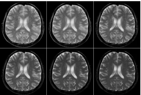
Figures 1a-1f show brain images
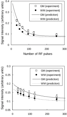
acquired at a 300 flip angle with 48, 64, 96, 128, 256, and 768 RF pulses preceding data acquisition around the center of the k-space, respectively. Note the decrease in contrast between gray and white matter from proton-density- to T2/T1-weighting. Figure 2 shows the theoretical calculation of signal intensity for gray and white matter at flip angles of 700 and 300, respectively, plotted as a function of the RF pulses preceding data acquisition around the center of the k-space. Approaching the steady state for gray (T1 ~ 1200 msec; T2 ~ 100 msec) and white matter (T1 ~ 900 msec; T2 ~ 80 msec) was faster than for CSF (T1 ~ 4500 msec; T2 ~ 2200 msec; data not shown), but still required about 150 preparatory RF pulses. For images at 256 phase encoding or lower, the image contrast is therefore a transient-state combination of proton-density and T2/T1, if using a linear phase encoding order. This transient-state proton-density weighting accounts for the clearer contrast between gray and white matter in Figs.1a-1c, which were acquired at low phase encoding values. These results depart from the expectation of poor contrast from steady-state T2/T1 weighting. The experimental data (open squares and filled circles for gray and white matter in Fig.2, respectively) taken from the
combining proton-density- and
T2/T1-weighting, if the (–α/2)-(TR/2)
preparation scheme is used with a linear phase encoding order. In this regard, TrueFISP images are not strictly T2/T1-weighted in general, although the transient-state and steady-state contrast between fluids and parenchymal tissues are basically similar. The actual image appearance strongly depends on the number of RF pulses that precede data acquisition around the center of the k -space. The speed in reaching steady state also depends on tissue T1 and T2.
For fluids with a long T1 and T2, such as CSF, approaching the steady state is relatively slow. The amount of change in signal intensity is thus
small during continuous data
acquisition, making the
transient-state behavior of fluids in TrueFISP images relatively unimportant. For parenchymal tissues, such as gray and white matter, the number of preparatory RF pulses needed to reach steady state is on the order of one hundred. Consequently, TrueFISP images acquired using different phase encoding values (e.g., 128 vs. 512, standing for 64 and 256 preparatory RF pulses, respectively, if using a linear phase encoding order) can show dramatic differences in contrast. This effect is particularly evident at large flip angles (Fig.2).
Therefore, when using a linear phase encoding order, particularly at large flip angles and depending on the phase encoding value utilized, it may be more appropriate to consider 2D TrueFISP
images as a transient-state
combination of proton-density and T2/T1 contrast. Since the parenchymal contrast differs significantly between
transient-state and steady-state
imaging, interpretation of 2D TrueFISP images in clinical practice requires particular caution.
四、計畫成果自評
Our efforts spent in this project have created results substantially greater than that mentioned in this brief report, which is an excerpted version of a recently published paper in the prestigious journal Magnetic Resonance in Medicine (4). Overall, the
project has generated five conference papers, including two presented in the Annual Meeting of the International Society of Magnetic Resonance in Medicine. In addition, one journal article (4) has been published, plus another one recently submitted (5). Achievements from this project are under investigation by Tri-Service General Hospital for potential use in the clinical practice. In short, we have confidence that a successful execution of the second year of this project will result in better utilization of the MR systems in routine diagnosis.
五、參考文獻
of the heart with segmented true fast imaging with steady-state precession.
Radiology 2001;219:828-834.
3. Deimling M, Heid O. Magnetization prepared true FISP imaging (abstr). Proc 2nd Annual Meeting SMR, San Francisco, CA, 1994. p.495.
4. Huang TY, Huang IJ, Chen CY, Scheffler K, Chung HW, Cheng HC. Are TrueFISP
images T2/T1-weighted? Magnetic
Resonance in Medicine 2002;48:684-688.
5. Huang TY, Chung HW, Wang FN, Ko CW, Chen CY. Fat and water separation in TrueFISP imaging using the Dixon
method, submitted to Magnetic
Resonance in Medicine.
六、圖表
Figure 1. Brain images acquired at a 300 flip angle with (a) 48, (b) 64, (c) 96, (d) 128, (e) 256, and (f) 768 RF pulses preceding data acquisition around the center of the k-space, respectively. Notice the decreasing contrast between gray and white matter from proton-density- to T2/T1-weighting as the number of preparatory RF pulses increases.
0 0.1 0.2 0.3 0.4 0 100 200 300 Number of RF pulses S ig n al I n ten s it y (ar bi tr a ry u n it s) GM (experiment) WM (experiment) GM (prediction) WM (prediction) 0.05 0.1 0.15 0.2 0.25 0.3 0 100 200 300 Number of RF pulses S ign al I n te n s it y ( ar bi tr ar y u n it s ) GM (experiment) WM (experiment) GM (prediction) WM (prediction)
Figure 2. Theoretical calculation of signal intensity for gray matter and white matter at flip angles of 700 (left) and 300 (right), respectively, shown as a decreasing function of the number of RF pulses preceding data acquisition around the center of the k-space. Approaching the steady state requires about 150 preparatory RF pulses. Experiment results are in good agreement with theoretical predictions.