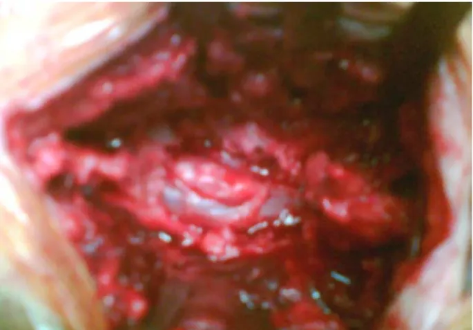
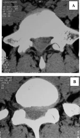
Posterior epidural migration of a sequestrated lumbar disc fragment: MRI findings.
全文
數據
相關文件
In 1942, Stafne described for the first time 35 asymptomatic, radiolucent cavities, unilaterally located in the posterior region of the mandible, between the mandibular angle and
The aggressiveness of the lesion and destructive growth noted in the present case, along with fascicular pattern of spindle cells in a mixed inflammatory infiltrate ruled out
expansion of the alveolar ridge near the left deciduous mandibular second molar -Panoramic findings:.. A well-defined unilocular radiolucent lesion located in the left
Henceforth, the aim of the study was to evaluate whether MRI findings of various degrees of disk displacement could be correlated with the presence or absence of clinical signs
Case Presentation: In this clinical case report, it is described a case of a 16-year-old male patient with an asymptomatic osteolytic lesion at first upper left molar apical level,
→ In the displaced coronal fragment of a tooth with a root fracture, pulp revascularization with subsequent pulp canal calcification will proceed on its own concurrently with
Periapical radiographs of the presenting case of intraosseous MPNST showed a unilocular periapical lesion with a defined border and external dental resorption in the lateral region
Dental variations like dens invaginatus in the upper left central incisor (Figure 5) and taurodontism in posterior molars (Figure 6) are associated with talon cusps.. On
