國 立 交 通 大 學
生物科技學系
碩 士 論 文
探討
乙醯膽鹼酯酶在固定化和游離狀態下之酵素動力學
Kinetics of Free and Immobilized Acetylcholinesterase
研究生: 李奇叡
指導教授: 楊裕雄 教授
探討
乙醯膽鹼酯酶在固定化和游離狀態下之酵素動力學
Kinetics of Free and Immobilized Acetylcholinesterase
研究生: 李奇叡
Student: Chi-Ruei Lee
指導教授: 楊裕雄 Advisor: Yuh-Shyong Yang
國 立 交 通 大 學
生 物 科 技 學 系
碩 士 論 文
A Thesis
Submitted to Department of Biological Science and Technology
National Chiao Tung University
in Partial Fulfillment of the Requirements
for the Degree of
Master of Science
in
Biological Science and Technology
May 2012
Hsinchu, Taiwan, Republic of China
i
探討
乙醯膽鹼酯酶在固定化和游離狀態下之酵素動力學
學生: 李奇叡 指導教授: 楊裕雄 教授 國立交通大學生物科技學系碩士班 摘要 在過去的文獻中,關於乙醯膽鹼酯酶(E.C. 3.1.1.7)的研究,大多是探討在游 離狀態下的酵素特性變化。但是在生物體中乙醯膽鹼酯脢並不是以游離的狀態存 在,而是以鑲嵌的方式固定在細胞膜上,因此酵素的特性可能在不同的生理條件 下而有差異。本研究將乙醯膽鹼酯酶固定在二氧化矽晶片上以模擬此酵素固定在 細胞膜上的環境。配合實驗室之前開發的量測固定化酵素平台,測量出被固定化 的乙醯膽鹼酯酶的酵素動力學參數 K*m 和 V*max,探討三維和二維的乙醯膽鹼 酯酶在兩種不同狀態下的酵素動力學。本研究利用LabVIEW 圖形化程式將固定 化酵素量測平台過於繁雜的分析過程,開發為自動化計算的人機介面。此程式縮 短了繁瑣的計算程序同時在短時間內就能得到固定化酵素的動力學參數 K*m 和 V*max。 整合此兩套系統將能夠快速得到研究所需要之固定化酵素動力學參數。 乙醯膽鹼酯酶在二維狀態下酵素動力學參數 K*m為 463 M 和 V*max為 7.20.6 mole/mg/min;而三維狀態下酵素動力學參數 Km為 798 M 和 Vmax為 1595 mole/mg/min。目前治療阿茲海默症的主要方法為,利用藥物抑制乙醯膽鹼酯脢 進而減少神經傳遞素,以減緩此症狀的發生,未來本技術將可應用於探討藥物抑 制乙醯膽鹼酯酶的活性。ii
Kinetics of Free and Immobilized Acetylcholinesterase
Student: Chi-Ruei Lee Advisor: Prof. Yuh-Shyong Yang Department of Biological Science and Technology
National Chiao Tung University
ABSTRACT
Enzymatic properties of acetylcholinesterase (AChE, E.C. 3.1.1.7) were mainly studied in vitro at it free form in solution. However, the in vitro properties of the enzyme may differ in physiological condition because AChE is immobilized as a membrane anchored protein in the organism. The method of this research was to immobilize AChE on silicon oxide surface in order to imitate the virtual environment of the cell membrane. Based on a measurement platform previously developed by our laboratory, K*m and V*max of immobilized AChE on planar silicon surface were determined. Automatic LabVIEW graphical program were developed to replace previously complicated analyzing processes, meanwhile, generated the kinetic parameters of immobilized enzyme. Kinetic parameters, Km and Vmax, of AChE were 463 M and 7.20.6 mole/mg/min in two dimension (on silicon oxide surface) and 798 M and 1595 mole/mg/min in three dimension (in solution), respectively. Nowadays, controlling AChE activity by medicine to reduce acetylcholine is the main solution for treating Alzheimer’s disease. This technique can be used for investigating immobilized AChE activity under the influence of inhibitors that can be applied to the treatment of Alzheimer’s disease.
iii
Acknowledgement
轉眼間,三年的黃金歲月一下子就消逝了,很感謝楊裕雄教授提供了一個如此 自由的研究環境、充沛的研究資金和培養我獨立思考的研究精神。這所有的一切 是我由衷所感謝的。在此也特別感謝柯富祥教授、徐琅教授能夠抽空參加我的碩 士口試,給予我許多研究方面的寶貴意見,讓我受益良多。 短暫的三年,很高興認識了 LEPE 這個大家庭,從我一開始進來一起同甘共苦 的曉萍、芝綺,很開心你們提早了開始你們人生中另一段的旅程,而我也終於可 以走向另一個人生階段,祝福你們!此外,也很感謝實驗室的大老們,陸宜學長、 程允學長、普普學長、胖哥、晨竹、咏馨、康寧、文燦、還有各位學弟妹們,謝 謝你們陪我度過了過去難熬的歲月。在這裡也特別感謝小志學長在我實驗上有困 難的時候,總是讓我從困境中抽身而出,在我低潮時,會陪我一起打球紓解壓力。 另外也要恭喜你拿到博士學位,等待六年是值得的!經歷了有淚有笑的三年時 光,終於拿到了碩士學位,始終最感謝的還是蘇博,不管在研究態度、做人處理 上、讓我學到了許多寶貴的人生經驗。每當遇到困難和低潮時,你總是以正面的 角度鼓勵我,讓我遠離負面的情緒,也會告訴我"唯有面對問題,才能夠真正解 決問題"。曾經你說過得那些話,我會永遠記得,謝謝你! 最後還是謝謝我親愛的家人們。毛、老爸、老媽、老姐、和老哥和未來的大嫂, 感謝你們一路上的支持和陪伴,無怨無悔得在我背後支持著我。我能夠拿到這個 學位,有一半是你們的功勞,謝謝你們!相信在經歷過這段歷練後,未來沒有什 麼事情是不可能。最後送上喬丹名言,大家共勉之:I can accept failure ,but I can't accept not trying!
奇叡
iv
Contents
PAGE Abstract (Chinese)... i Abstract (English)... ii Acknowledgement... iii Contents... iv Contents of Tables ... viContents of Figures... vii
Abbreviation... ix
I. Introduction... 1
1-1 Introduction of acetylcholinesterase... 1
1-2 Introduction of Alzheimer’s disease... 2
1-2.1 Symptoms of Alzheimer’s disease... 3
1-2.2 Deaths from Alzheimer’s disease... 4
1-3 Motivation... 5
II. Material and method... 6
2-1 Experimental material... 6
2-2 Instruments... 10
2-3 Experiment procedures... 11
2-3.1 Silica pattern formation processes... 11
2-3.2 Immobilization of acetylcholinesterase on surface silicon wafer... 12
2-3.3 Enzyme standard assay of soluble AChE... 14
2-3.4 Enzyme standard assay of immobilized AChE and reactor system... 15
v
2-4 Development of theoretical model to analytic program based on
LabVIEW... 17
III. Results and discussion... 19
3-1 Determining surface modification of silicon wafer for immobilized AChE... 19
3-2 Confirm activity of surface-immobilized AChE... 20
3-3 Estimate amount of immobilized AChE on silicon wafer... 22
3-4 Kinetics assay of soluble AChE... 24
3-5 Enzymatic activity of surface-immobilized AChE based on running controls... 25
3-6 Immobilized AChE kinetics: Utilizing automatic program for iterating scheme to determine V*max/H, corresponding deactivation curve, and K*m... 26
IV. Conclusions... 28
V. Appendix... A1. Theoretical model of micro-fluidic reactor system 30 References... 34
vi
Contents of Tables
PAGE
Table 1 The percentage of elemental analysis at different
stages of immobilization process... 39 Table 2 Experiment-determined catalytic parameters of
turnover numbers (kcat) and Michaelis constants (Km)
for the soluble and planar surface–immobilized AChE
vii
Contents of Figures
PAGE
Figure 1 The mechanism of action of acetylcholinesteras (AChE) cholinergic nerve transmission is terminated by the
enzyme acetylcholinesterase (AChE)... 41 Figure 2 Percentage changes in selected causes of death (all ages)
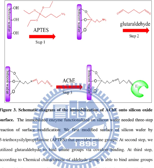
between 2000 and 2008... 42 Figure 3 Schematic diagram of the immobilization of AChE onto
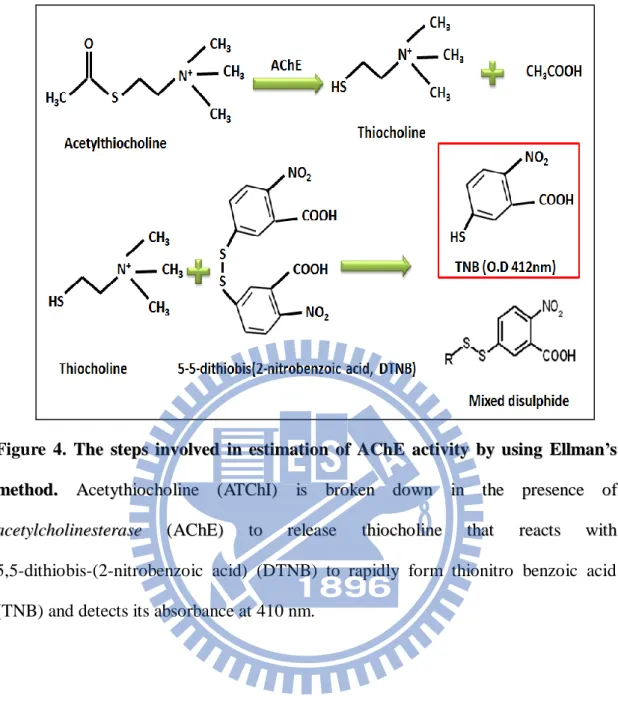
silicon oxide surface... 43 Figure 4 TThheesstteeppssiinnvvoollvveeddiinneessttiimmaattiioonnooffAACChhEEaaccttiivviittyybbyy
u
ussiinnggEEllllmmaann’’ssmmeetthhoodd... . 44 Figure 5 Schematic diagram of the home-made apparatus for
immobilization of acetylcholinesterase onto the silicon
dioxide surface... 45 Figure 6 Overview of home–made reactor system for
measurement immobilized enzymatic kinetics... 46 Figure 7 A systematic and standardized data analysis... 47 Figure 8 The first automatic program for modifying progress
curves of immobilized-enzyme reaction cycles... 48 Figure 9 The second automatic program for computing initial
approximation of 0 * max H V , 0 * Km and decay
curve about the issue of deactivation of immobilized
enzyme... 49 Figure 10 The third automatic program for finding the final value
viii
Figure 11 The XPS spectra of each immobilization step... 51
Figure 12 Progress curves of enzyme assay for AChE complete reaction... 52
Figure 13 The absorbance of 2-nitro-5-thiobenzoic acid (TNB)... 53
Figure 14 The overview of general crystal with vector coordinate... 54
Figure 15 Effective range of AChE assay... 55
Figure 16 Michaelis-Menten plots for hydrolysis reaction by free AChE... 56
Figure 17 The typical progress curves of enzymatic assays... 57
Figure 18 AChE kinetics. According to the second automatic program, the versus /[S]o plot was used to determine initial V*max/H at high substrate concentration... 58
Figure 19 AChE kinetics. The typical iterative plots (a) and (b) were the output values of the second and third automatic programs... 59
ix
Abbreviation
Abbreviation Full Name
AChE acetylcholinesterase
ATChI acetylthiocholine iodide
DTNB 5,5-dithiobis-(2-nitrobenzoic acid)
TNB 2-nitro-5-thiobenzoic acid
K-PB potassium phosphate buffer
APTES 3-triethoxysilylpropylamine
1
I Introduction
1-1 Introduction of acetylcholinesterase
Acetylcholinesterase (AChE, E.C. 3.1.1.7), also known as AChE , is a tetramer
composed of 4 equal subunits of 70 kDa each. It’s a glycoprotein that exists is several
forms and the membrane-bound globular AChE forms have hydrophobic domains that
anchor them in the membrane phospholipid bilayers [1]. This is an evolutionary
consequence of one of its key active hydrolysis of the neurotransmitter acetylcholine
(ACh) to terminate signaling in cholinergic synapses, including the neuromuscular
junction, so the great speed of the enzyme is essential for rapid modulation of
synaptic activity (Figure 1). In the other hand, Acetylcholinesterase also plays an
important role in cholinergic transmission by catalyzing the rapid hydrolysis of the
neurotransmitter acetylcholine (ACh) into acetate and choline [2]. Moreover,
acetylcholinesterase is also found on other tissue like the red blood cell, muscle,
2
1-2 Introduction of Alzheimer’s disease
Alzheimer's disease (AD) is one of slowly progressive disease of the brain that is
characterized by impairment of memory and eventually by disturbances in reasoning,
planning, language, and perception [3, 4]. In many research, they think that
Alzheimer's disease results from an increase in the production or accumulation of a
specific protein (-amyloid protein) which is in the brain that leads to nerve cell death
[5]. Moreover, many of research find out more about the causes of Alzheimer disease
until now there is no one reason why people get Alzheimer disease. At present,
researchers find older people are more likely to get it, and the risk gets greater the
older the person gets. For instance, the risk is higher for someone who is 85 years old
than it is for someone who is 65. And women are more likely to get it than men [6].
Researchers also think genes handed down from family members can make a person
more likely to get Alzheimer disease. But that doesn't mean everyone related to
someone who has Alzheimer disease will get the disease [7]. Other factors, combined
with genes, may make it more likely that someone will get the disease. Although AD
can’t be cured and is degenerative but we can prevent it from risk factor like high
blood pressure, high cholesterol, down syndrome, or having a head injury. Keeping
the positive side that researchers believe exercise, a healthy diet, and taking steps to
3
1-2.1 Symptoms of
Alzheimer’s disease
Alzheimer disease can affect people in many ways, but the most common symptom
pattern is gradually worsening difficulty in remembering new information because
disruption of brain cell function usually begins in regions involved in forming new
memories [9-11]. When damage spreads, individuals experience as the following are
warning signs of AD:
•Memory loss that disrupts daily life.
•Challenges in planning or solving problems.
•Difficulty completing familiar tasks at home, at work, or at leisure. •Confusion with time or place.
•Trouble understanding visual images and spatial relationships. •New problems with words in speaking or writing.
•Misplacing things and losing the ability to retrace steps. •Decreased or poor judgment.
•Withdrawal from work or social activities. •Changes in mood and personality.
4
1-2.2 Deaths from
Alzheimer’s disease
Nowadays, AD is becoming a more common cause of death as the populations of
the United States and other countries age [12-15]. Even though other major causes of
death continue to experience significant declines, those from AD have continued to
rise. Between 2000 and 2008 (preliminary data), deaths attributed to AD increased
by 66%, whereas those attributed to the number one cause of death, heart disease,
decreased by 13% as shown in Figure 2.
The increase in the number and proportion of death certificates listing AD reflects
both changes in patterns of reporting deaths on death certificates over time as well as
an increase in the actual number of deaths attributable to AD [16-19]. Severe
dementia frequently causes complications such as immobility, swallowing disorders,
and malnutrition [20, 21]. These complications can significantly increase the risk of
developing pneumonia, which has been found in several studies to be the most
commonly identified cause of death among elderly people with AD and other
dementias. The situation has been described as a “blurred distinction between death
with dementia and death from dementia”. Regardless of the cause of death, 61% of
people with AD at the age of 70 are expected to die before the age of 80 as compared
5
1-3 Motivation
I
Inn ppaasstt rreesseeaarrcchh,, deaths attributed to AAllzzhheeiimmeerr’’ss ddiisseeaassee increased by 66% that
means AAllzzhheeiimmeerr’’ssddiisseeaasseebbeeccoommeeaasseerriioouusspprroobblleemmiinn the 21st century . According
to curing Alzheimer’s disease related with enzymatic kinetics of AChE in past report
so that we selected this important enzyme for research [22-27]. Soluble AChE was
already used to study its biological properties and pharmaceutical products in the past
but it might not be a suitable approach to investigate AChE in real situation [28-31].
Thus, it become very important to be able to determine the kinetics of enzyme
immobilized on the planar surfaces within the microfluidic system in order to
evaluating the function of the whole system. So far, there is no such method reported.
In past research, our lab developed a novel kinetic model, based on systematized and
standardized approach, for measuring K*m and V*max of enzyme immobilized on
planar silicon oxide surface within a microfluidic bioreactor [32] In this study, we
further developed the novel kinetic model into automatic program, which was
designed by software of LabVIEW. Then, we could easily get parameters of apparent
K*m and V*max as soon as we used automatic program. Finally, we could compare
enzymatic kinetics of immobilized and soluble enzyme with two kind of kinetic
6
II Material and method
2-1 Experimental material
1
1.. Compound::AAcetylcholinesterase (AChE)-type V-S from Electric eel
C Coommppaannyy::SSiiggmmaa--AAllddrriicchh M Moolleeccuullaarrwweeiigghhtt::228800kkDDaa A Assssaayy::6600%% 2
2.. Compound:: Acetylthiocholine iodide (ATChI)
C Coommppaannyy::SSiiggmmaa--AAllddrriicchh M Moolleeccuullaarrwweeiigghhtt::228899..1188 S SoolluubblleeiinnDDIIwwaatteerr 3
3.. Compound:: 5,5-dithiobis-(2-nitrobenzoic acid) (DTNB)
C Coommppaannyy::SSiiggmmaa--AAllddrriicchh M Moolleeccuullaarrwweeiigghhtt::228899..1188 S Soolluubblleeiinneetthhaannooll
7
4
4.. Compound: Potassium phosphate monobasic (KH2PO4)
C
Coommppaannyy::SSiiggmmaa--AAllddrriicchh
M
Moolleeccuullaarrwweeiigghhtt:: 136.09
5
5.. Compound: Potassium phosphate dibasic (K2HPO4)
C
Coommppaannyy::SSiiggmmaa--AAllddrriicchh
M
Moolleeccuullaarrwweeiigghhtt:: 174.18
6
6.. Compound: Ethanol (CH3CH2OH)
C
Coommppaannyy:: Echo Chemical Co.
M
Moolleeccuullaarrwweeiigghhtt:: 46.07
A
8
7
7.. Compound: (3-Aminopropyl)triethoxysilane (APTES), H2N(CH2)3Si(OC2H5)3
C Coommppaannyy::SSiiggmmaa--AAllddrriicchh. M Moolleeccuullaarrwweeiigghhtt:: 221.37 A Assssaayy::9988%% S Soolluubblleeiinneetthhaannooll 8 8.. CCoommppoouunndd::GGlluuttaarraallddeehhyyddeessoolluuttiioonn,,(CH( 2(CH2CHO)2) C Coommppaannyy::FFlluukkaa((UUSSAA)) M Moolleeccuullaarrwweeiigghhtt::110000..1122 A Assssaayy::~~2255%%iinnHH22OO 9
9.. Compound: Sulfuric acid, (H2SO4)
C Coommppaannyy::SSiiggmmaa--AAllddrriicchh. M Moolleeccuullaarrwweeiigghhtt:: 98.08 A Assssaayy::9999..99%%
9
1
100.. Compound: Hydrogen peroxide solution, (H2O2)
C Coommppaannyy::SSiiggmmaa--AAllddrriicchh. M Moolleeccuullaarrwweeiigghhtt:: 34.01 A Assssaayy::3300%% 1
111.. Phosphate buffer (PB) was prepared in deionized (DI) water and its pH was
adjusted to 8 .
12. Deionized and distilled water DI water, ddH2O
The water we used was purified with filters, reverse osmosis, and deionized system until the resistance was more than 18 MΩ·cm. DI water was used to clean, wash, and be a solvent.
1
133.. P-type Si(100) wafers (14-21 -cm, MEMC, MO, USA)
It is 15 cm diameter, on which 100 nm oxide layers were grown using wet oxidation
10
2-2 Instruments
1. NI LabVIEW 2011
It is a comprehensive development environment that provides engineers and scientists
unprecedented hardware integration and wide-ranging compatibility. On the other
hands, it is also a program used to automate testing and data gathering. It is basically a
graphical programming language in which the user can set up the program to
manipulate and store data.
2. UV–Vis spectroscopy (HITACHI, U-3310, Tokyo, Japan )
UV–Vis uses light in the range of near UV, visible and near infrared. The absorption
in the light range is due to the optical properties of the chemicals involved.
3. Programmable syringe pump (KD Scientific, KDS260P, USA)
We utilized programmable syringe pump to translate reaction solution into the channel
and eluted to a spectrophotometer for the determination of the concentration of
reporter molecules.
4. Hot plate (SHIN KWANG)
After the patterned sample of interest was immersed in the APTES solution for 30 min
in room temperature we banked the patterned sample at 120oC for 30 min.
5. X-ray photoelectron spectroscopy (XPS)
11
2-3 Experiment procedures
2-3.1 Silica pattern formation processes
P-type Si(100) wafers (14-21 -cm, MEMC, MO, USA) with 15 cm diameter were
deposited and etched to form the structure with silicon oxide pattern on poly-Si film.
To prepare the silicon oxide pattern on poly-Si film, the poly-Si film was first
deposited with silane gas (SiH4) at 60 cm3/min and 620oC. Prior to photolithography,
the silicon oxide film was grown by wet oxidation with a gas mixture of hydrogen
(8000 cm3/min) and oxygen (5000 cm3/min) at 978oC. The mask with the pattern of
interest was used to define the photoresist (TMER-iP3650, Tokyo Ohka Kogyo,
Tokyo, Japan) pattern. A 365 nm light emitted from high pressure mercury lamp
(SUV-2001CIL, USHIO, Tokyo, Japan) induced the photo-active reaction for the
photoresist film. After the dissolution of exposure area with 2.38%
tetramethylammonium hydroxide, the plasma was used to etch the silicon oxide film
without passivation by photoresist pattern. The reactive-ion etch system (TE5000,
Tokyo Electron Limited, Tokyo, Japan) was operated at 500W RF power under 0.2
Torr high vacuum, and the gas mixture of 20 cm3/min of CF4, 20 cm3/min of CHF3,
and 400 cm3/min Ar. Finally, the residual photoresist was removed and cleaned by the
mixing chemical of H2SO4 and H2O2 (volume ratio = 3:1) at 120oC for 10 min. The
12
2-3.2 Immobilization of acetylcholinesterase on surface silicon
wafer
In order to immobilization, the piece of silicon oxide wafer would be clean
carefully by the SPM solution (sulfuric-peroxide mixture), H2SO4 and H2O2 (volume
ratio is 3:1) twice and incubated the temperature at 120oC for 30 min. It should be
noted that the cleaning solution is very corrosive and dangerous. After rinsing with
pure water and drying, the sample was immersed in the
(3-aminopropyl)triethoxysilane (APTES, Sigma–Aldrich, MO, USA) solution to
proceed the silanization reaction for 30 min at room temperature to create an
amine-functional surface. The APTES solution was prepared by the following
procedures: preparing the 5% APTES solution by diluting with 95% ethanol.
Following the APTES treatment, the silicon wafer was rinsed with 95% ethanol
thoroughly. Then, the silicon wafer was baked at 120oC for 30 min to complete the
Si–O bond formation. The sample was immersed in the linker solution (12.5%
glutaraldehyde, i.e. pentane-1,5-dial) for 60 minutes in room temperature. The 12.5%
glutaraldehyde solution was diluted with DI water (deionized system until the
resistance was more than 18 MΩ·cm) from 25% glutaraldehyde (in water,
13
((AChE)-type V-S from Electric eel (Sigma-Aldrich)) solution that the powder of
a
acetylcholinesterase (AChE) was dissolved in phosphate buffer (PB buffer, pH 8)) for
30 minutes at room temperature. Then, we washed patterned sample by PB buffer and
dried with nitrogen gas. The overall surface modification is shown in Figure 3.
2-3.3 Enzyme standard assay of soluble AChE
I
Inn tthhiiss rreesseeaarrcchh,, AACChhEE aaccttiivviittyy ooff bbootthh ssoolluubbllee aanndd iimmmmoobbiilliizzeedd eennzzyymmee wwaass
d
deetteerrmmiinneedd aaccccoorrddiinngg ttoo tthhee EEllllmmaann mmeetthhoodd ((SSeeee FFiigguurree 44)).. In determination of
soluble AChE activity, tthheerreeaaccttiioonn solution was prepared by mixing PB solution (pH
8.0, 10 mM), various concentration of aacceettyylltthhiioocchhoolliinnee iiooddiiddee,,00..11mmMMDDTTNNBB aanndd
a
addddeedd aann aapppprroopprriiaattee aammoouunntt ooff tthhee eennzzyymmee [[3311,, 3355--3377]].. After mixing the catalytic
substance with the reactants, the initial product release at the onset of the reaction was
measured using a personal computer and a Hitachi UV–Vis-3310 enzyme reaction
measurement system (a UV–Vis spectrophotometer possessing a
temperature-controlled thermostatted cell holder; Hitachi, Tokyo, Japan). For the
soluble AChE kinetics analysis, the initial reaction of the change in absorbance at 410
nm was recorded (in real-time). The initial rate of the absorption change against the
reaction time was converted to enzyme activity using a molar absorption coefficient
14
Km and Vmax were obtained through nonlinear regression analysis using SigmaPlot
2001 (v. 7.0) and Enzyme Kinetics Module (v. 1.1, SPSS, Chicago, IL USA) software.
The assays were obtained in triplicate; average values were reported. All activity
assay experiments were carried out at room temperature.
2-3.4 Enzyme standard assay of immobilized AChE and reactor
system
Preliminary tests for the immobilization of AChE activity were carried out using
home-made apparatus and checked activity with Ellman’s method. The home-made
apparatus (Figure 5) was designed and used to evaluate the enzyme activity on the
sample surface of interest. The Teflon ring tightly contacted with the substrate and
sealed with the silicon resin glue. Prior to conducting the enzyme immobilization, we
needed to test the reliability of the home-made apparatus to avoid leakage problem.
The clean and APTES immobilization methods for enzyme immobilization were
conducted in the home-made apparatus with the same procedures as mentioned above.
The sample was repeatedly immersed by fresh 10 mM potassium phosphate buffer for
five times to wash away the residual enzyme solution. Observation of the activity of
the enzyme was a direct method to know whether the enzyme was successfully
immobilized or not. The reaction solution was prepared by adding the 1 mM
15
into the solvent of 10 mM potassium phosphate buffer (PB) at pH 8. Then, we added
the above solution into the home-made apparatus. At the reaction time of interest, the
liquid was siphoned out from the home-made apparatus to an UV-Vis
spectrophotometer (Hitachi UV-Vis-3300, Tokyo, Japan) for characterization. We
analyzed the absorbance of 2-nitro-5-thiobenzoic acid (the catalytic product of
5,5-dithiobis-(2-nitrobenzoicacid)) at 410 nm wavelength to determine the activity of
acetylcholinesterase. After we know the enzyme successfully was immobilized onto
surface, we could study further enzyme kinetics assay of immobilized AChE with
home-made micro- fluidic reactor system further. According to the model which have
been published by our lab [32] that we have constructed a novel home-made
micro-fluidic system (Figure 6) for assay enzymatic kinetics parameter of
immobilized enzyme.
This novel bioreactor design has a flow channel, which is made of glass and the
reaction liquid filled in cylinder is continuously pushed by syringe pump. To utilize
this micro-fluidic reactor system for measuring enzymatic kinetics parameter of
immobilized enzyme has two parts. For first part, we should construct baseline in
order to confirm the reaction liquid is stable and unchanging with time. In baseline,
the reaction liquid would not go through flow channel, it only goes through cuvette
16
For second part, the reaction liquid also would continuously go through flow channel
and the reaction liquid is simultaneously catalyzed by immobilized enzyme. Finally,
the product of reaction liquid flow through cuvette and detect its absorbance by
UV-Vis spectrophotometer. In experiment, two different concentrations of
acetylthiocholine iodide (ATChI), 1000 M and 50 M, were used to create
saturating and non-saturating substrate condition, respectively, for the immobilized
AChE-catalyzed reaction with home-made micro-fluidic reactor system. The ATChI
concentrations used were determined according to the Km of free Acetylcholinesterase
from Electrophorus electricus and the reaction mixtures for immobilized AChE
contained ATChI (1000 M or 50 M) and 0.1 mM DTNB in 10 mM potassium
phosphate buffer at pH 8. Injection of the reaction mixtures into the reactor was
controlled by automatic pumping system and operated at desired to have space time ()
at 0.5 min, 1 min, 2 min. The output solution was directed into a quartz flow cell
mounted in the UV-Vis spectrophotometer (Hitachi UV-Vis-3300, Tokyo, Japan) for
TNB detection at 410 nm. TThhee rreessuullttss ooff iimmmmoobbiilliizzeedd kkiinneettiiccss eexxppeerriimmeennttss wweerree a annaallyyzzeeddbbyy theoretical considerations ((aasssshhoowwnniinnaappppeennddiixxII..))tthhaattwwaassccoonnssttrruucctteedd b byy oouurr llaabb.. AAllll ooff aannaallyyssiiss ddaattaa uusseedd rreepprreesseenntt mmeeaann vvaalluuee ddeerriivveedd ffrroomm tthhrreeee d deetteerrmmiinnaattiioonnss..
17
2-4 Development of theoretical model to analytic program based on
LabVIEW
Theoretical model previously developed [32] fits very well to the kinetics of
immobilized enzyme on one-side planar surface (detailed analysis and prediction of
kinetics are given in Appendix I). Unfortunately, this theoretical model was too
complicated to utilize and difficult for further development for automation. According
to process of model, we utilized software of LabVIEW to develop the analysis
program divided into three parts. Therefore, it could easily get the kinetic parameters
of immobilized enzyme as soon as possible by operating the analysis program. In the
first part of the analysis program, we need to modify the raw data of progress curves
of immobilized-enzyme reaction cycles. Hence we set some required parameters such
as flow rate (l/min), space time (min), extinction coefficient of product () and
high/low feed concentration of substrate (M) (Figure 8 (a)). Next, we use LabVIEW
to fit the data points of high/low baseline concentration to get the background during
the eexxppeerriimmeennttss (Figure 8 (b)). Then, the reaction data at high/low concentration will .
be modified by the fitting curve through the "modify" button based on the analysis
program. (Figure 8 (c)). In the second part, we modified reaction data at high/low
concentration to calculate of the kinetic parameters. After successively first
computing, we will get the initial approximation of 0 * max H V , 0 * Km and decay
18
curve about the issue of deactivation of immobilized enzyme (Figure 9). Finally, we
key the modified reaction data at high/low concentration into the third part of the
analysis program. By initial Km* 0 and these modified will get the final
approximation of r * max H V , r *
19
III Results and discussion
3-1 Determining the surface modification of silicon wafer for
immobilization enzyme
In this experiment, we dropped the acetylcholinesterase solution onto a silicon
wafer modified with functional linker for three main steps (Figure 3). Every
immobilized step was following standard flowchart and then we checked each
immobilized step for percentage of elemental analysis that was characterized by X-ray
photoelectron spectroscopy (XPS) (See Figure 11).
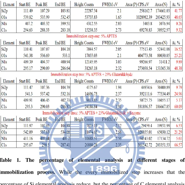
The XPS was used to verify the attachment of the enzymes onto the surfaces of
the functional linker (See Table 1). For the cleaning silicon wafer without surface
modification, the total percentage of elemental analysis indicates that the percentage
of N1s approaches zero. The result implies there is no pollution onto surface. In second
immobilized step, we dropped some 5% APTES solution on surface of silicon wafer.
The result points out the percentage of N1s increases about 10%, it means APTES
reacts with the surface silanol group (Si-OH) to form primary amine group on silicon
dioxide film. In third immobilized step, 25% glutaraldehyde is subsequently used to
react with the surface amine group. Owing to yielding an imine linkage (C=N) with
one end aldehyde group in glutaraldehyde so that we can see the percentage of N1s
20
solution to react with surface aldehyde group. The result shows the percentage of C1s
elevates to 66%. All of result are in agreement with the existing literature for proteins
and enzymes bound to the surface of silicon wafer with surface modification.
3-2 Confirm activity of surface-immobilized AChE
It is obvious that the surface of silicon nitride and poly-Si have no active group for
immobilization. The bio-important enzyme, i.e. AChE, can be assembled onto the
silicon dioxide film. The reaction having three main steps is illustrated in Figure 3.
Hence, the AChE is successfully immobilized onto the surface of silicon oxide film.
The analysis of surface enzyme (AChE) activity is very complex [36, 38]. Figure 12
shows the time course of the changes in Ultraviolet-visible for the assay reaction and
corresponding controls, where specific components that of the reaction mixture were
omitted. Only complete reaction was observed an increase of absorption at 410 nm,
reflecting the enzyme activity of AChE (Figure 12).
Figure 4 illustrates the two simultaneous chemical reactions appeared in the
home-made apparatus (Figure 5). The acetylthiocholines iodide (ATChI) is
transferred into the thiocholine molecule under the catalysis by the AChE enzyme and
translated with 5,5-dithiobis-(2-nitrobenzoic acid) (DTNB) to yielding the products of
2-nitro-5-thiobenzoic acid (TNB). Once the enzyme is still active, the concentration
21
also gradually increases. This reaction design, together with the home-made apparatus,
provides an easy characterization way by using UV-Vis spectrophotometer to evaluate
the enzyme activity after surface immobilization.
In order to confirm the AChE was immobilized tightly onto the patterned SiO2/Si
substrate, we withdrew the reaction mixture from catalytic surface to a cuvette when
reaction had proceeded 2 min and then incubated in a cuvette for more 2 min, in this
period, two times of detection followed at accumulation time 8 min and 20 min
respectively. No increment of TNB concentration was found, that was no TNB
produced if the reaction mixture was removed from catalytic surface. Reload the
previous reaction mixture to catalytic surface at accumulation time 10 min; the
reaction immediately started again. At accumulation time 18 min, we withdrew the
22
3-3 Estimate amount of immobilized AChE on surface silicon wafer
According to coordinate vector of generalized unit cell (Figure 14), a unit cell is
defined by six numbers: the lengths of three unique edges, a, b and c; and five unique
angles, , and Thus, the volume of crystal is obtained by
) 1 ( sin cos sin cos ) cos , cos , cos ( ) 0 , sin , 0 ( ) cos , cos , cos ( sin 0 cos 0 0 ˆ ˆ ˆ
abc abc b b b ac b b b c c a k j i Y Z X V .Setting coordinate vector of "b" point as:
)
2
(
1
cos
cos
cos
cos
cos
cos
2 2 2 2 2 2 2 2 2
b
b
b
b
By projecting thevector of the point c onto the coordinate vector of the point b and
we get
)
3
(
sin
cos
cos
cos
cos
cos
sin
0
cos
cos
cos
c
c
c
.To substitute eq (3) into eq (2); we get
)
4
(
)
sin
cos
cos
cos
2
cos
cos
cos
cos
1
(
cos
2 2 2 2 2
.23
)
5
(
)
cos
cos
cos
2
cos
cos
cos
1
(
)
cos
cos
cos
2
cos
cos
cos
cos
cos
cos
cos
1
(
cos
cos
cos
2
cos
cos
cos
sin
cos
sin
2 / 1 2 2 2 2 / 1 2 2 2 2 2 2 2 2 2 2 2 2 2
abc
abc
abc
V
.According to the Protein Data Base, the unit cell of crystallized acetylcholinesterase,
AChE (ID:1EEA), has the following parameters: a = 140.86 Å , b = 201.46 Å , c =
235.77 Å ; 00o, 90.00o, = 90.00o ; the molecular weight of the unit
AChE: 60 KDa . We can roughly estimate the mean diameter D of the unit AChE
(DAChE) as: 2 / 1 2 2 2 3
)
cos
cos
cos
2
cos
cos
cos
1
(
)
(
6
abc
Volume
D
Thus, we obtained:
1/3 2 / 1 2 2 2 3 / 1 ) cos cos cos 2 cos cos cos 1 ( 6 ) ( 6 Volume abc DAChESubstituting the parameters into the above equation, we get:
DAChE 233.78 Å = 2.3378*10-8 m ; r 1.1689*10-8 m = 1.1689*10-7 dm.
We calculate the area of the microfludic channel and get 0.1408 dm2.
As mentioned above, we find out the mean diameter D of the unit AChE (rAChE) and
get the area of the unit AChE:
24
We assume that the unit AChE was immobilized on one plane on the surface. Thus,
we can roughly estimate the amount of AChE immobilized on the surface of a silicon
wafer. mg X mole Da g X mole unit mole unit AChE unit unit dm dm 4 1 2 3 1 2 2 3 1 2 1 2 2 1 4 2 10 * 27 . 3 10 * 466 . 5 10 * 60 ) ( 10 * 466 . 5 10 * 02 . 6 1 10 * 28 . 3 ) ( 10 * 28 . 3 / 10 * 292 . 4 1408 . 0
We estimate that the amount of immobilized AChE is 3.27*10-4 mg on the surface of
a silicon wafer in the region of the microfludic channel. Consequently, we can convert
the unit (V*max/H (M/min)) kinetics of immobilized enzyme into the unit (Vmax
(mole/min/mg))
3-4 Kinetics assay of soluble AChE
For determining soluble AChE activity, we firtst need to know appopriate enzyme
concentration. So we have measured appropriate enzyme activity by fixed saturating
substrate concentration and added in various eznyem concentration [39, 40]. As the
following steps, we analyzed datas that could find its linear range from 0.5 to 4.5 nM
as shown in Figure 15. So that we selected this linear range for our standard assay in
25
Owing to studying enzyme kinetics of AChE, we detected product of
2-nitro-5-thiobenzoic acid (TNB). The complete assay mixture contained the
following components: 4 mM to 0.015 mM ATChI, 0.1 mM DTNB, 10 mM
potassium phosphate buffer (pH8.0) and effective range of AChE concentration in a
final volume of 1000 . In the enzymatic kinetics experiment, figure 16 shows kinetic
parameters for the soluble AChE, KKmm aanndd VVmmaaxx. It were assayed at substrate
concentration from 0.015 to 4 mM ATChI and 0.1 mM DTNB added in 10 mM PB
buffer (pH 8). According to the Michaelis–Menten equation, the KKmmof soluble AChE
was 0.791 0.008 mM and the VVmmaaxxwwaass115588..88775555..550044mmoollee//mmiinn//mmgg..
3-5 Enzymatic activity of surface-immobilized AChE based on
running controls
The typical time course plots of product yield are presented in Figure 17 were the
TNB absorbance responses to high/low feed concentration of substrate with a series of
space times min), after subtracting blank controls with automatic program. It
should be emphasized that assay were set up by the scheme (Figure 6) and obtain
respective running blank controls: (1) free substrate ATChI for AChE assay, (2)
enzyme free, by use of bypass scheme to skip reaction solution directly to UV-Vis
spectrophotometer, for AChE assay. Because the values of TNB absorbance at high
26
concentration of substrate ATChI, bypass was an essential strategy to continuous-flow
assay of surface-immobilized AChE in order to subtract accurate blank controls.
3
3
-
-
6
6
Immobilized AChE kinetics: Utilizing automatic program for
I
iterating scheme to determine V*
max/H, corresponding
deactivation curve, and K*
mAs mentioned above, we have already developed the theoretical model for
automatic program according to scheme of theoretical model. Thus, we could obtain
the kinetic parameters, easily. The determination of V*max/H for immobilized AChE
can be achieved easily using three parts of automatic program under saturating
substrate condition, which was 1000 M substrate ATChI. Due to solubility limit of
substrate ATChI in immobilized AChE assay, the initial approximations of V*max/H
(Figure 18) and corresponding deactivation curve (r = 0 in Figure 19(a) were obtained
from eq (2) using 1000 M ATChI as inlet condition of high substrate concentration.
This decay curve (r = 0) and 50 M ATChI as inlet condition of low substrate
concentration were then used to obtain the initial approximation of K*m (r = 0 in
Figure 19 (b)) from eq (1.2). Substituting < V*max/H >o and < K*m >o as the initial
estimations into the set of two iterating eqs (1.3) and (1.4), the converged results of
immobilized AChE were got, through five successive approximations as Figures 19 (a)
and 19 (b), as follows: the deactivation curve H Vmax* = t e 0.01292 86 . 24 31 . 10
27
with R2 = 0.958, original V*max/H = 25.95 M/min, and average K*m = 45.66 M
(Figure 20). Because the substrate concentration enough high, but not saturating was
satisfied well, the kinetic parameters could be correctly obtained through this iterating
calculation (Figure 7). In table 2, comparing immobilized AChE K*m (45.66 M) with
homogeneous AChE K*m (79.10 M) implied that there was almost no influence on
the affinity of substrate ATChI with immobilized AChE. But an decrease Vmax
(mole/min/mg) in once an enzyme has been immobilized, indicates that the
immobilized enzymes have an apparent lower catalytic rate than that of the free
enzyme does, which may be caused by the change of the conformation. There are
several reasons why a different kinetic behavior is observed with an enzyme
immobilized into a solid support relative to the free enzyme. Firstly, the
immobilization may cause some conformational changes in the enzyme molecules.
Secondly, the immobilized enzyme is located in an environment different from that
when it is the free solution, and this can have a significant effect on the kinetics.
Finally, being a membrane enzyme, AChE would not be at the natural optimal
conformation both in free-state in solution and immobilized-state on supporting
28
IV Conclusions
Nowadays, enzyme immobilization becomes an necessary for biosensing,
bio-regulation and many other bioengineering applications. Because advantage of
limited enzyme includes: surmount the stability, recovery, recyclability disadvantages
of using enzymes in solution, making them industrially and commercially viable.
There are many method for enzyme immobilization, such as adsorption that involves
physical surface interactions between the support matrix and the enzyme and can be
driven by combined hydrogen bonding, electrostatic forces, and hydrophobic effects;
Covalent attachment involves binding amino acid residues (NH2, CO2, SH) of the
enzyme to the support matrix. This method is popular for high surface area support
matrixes with large pore diameters where substrate and product can freely diffuse
without the worry of enzyme leaching. Owing to AChE is not free in solution but a
membrane anchored protein in the organism, we selecte covalent attachment to
immobilize AChE on planar silicon oxide surface in order to simulating AChE
anchored on cell membrane like in vivo.
Following previously kinetic model for the determination of the kinetics of the
immobilized enzyme, we successfully measured the kinetics of the immobilized
enzyme. Besides, well-known procedures are available for the kinetic analysis of
29
immobilized AChE on the surface of a silicon wafer in the region of the microfludic
channel in order to converting the kinetic unit of immobilized enzyme. Then, we
could compare the enzymatic kinetics of immobilized and free enzyme in the same
kinetic unit. Finally, we compared immobilized AChE K*m (45.66 M) with
homogeneous AChE K*m (79.10 M) implied that there was almost no influence on
the affinity of substrate ATChI with immobilized AChE. But an decrease Vmax
(mole/min/mg) in once an enzyme has been immobilized, indicates that the
immobilized enzymes have an apparent lower catalytic rate than that of the free
enzyme does, which may be caused by the change of the conformation. Besides,
according to process of model, we further developed the automatic program for
analysis kinetic parameters of immobilized enzyme in order to getting the kinetic
parameters of immobilized enzyme as soon as possible by operating the analysis
program.
Based on our previously proposed model, we constructed systematic and standard
system for analysis of kinetics of immobilized enzymes. Using this prototype platform,
it allowed us to observe the kinetic in-situ change of immobilized enzyme, and the
advanced fundamental research about kinetic mechanism under different stress
30
V Appendix
II..
Theoretical model of micro-fluidic reactor system
According to the model which have been published by our lab [32] that we have
constructed a novel home-made micro-fluidic system (Figure 6) for assay enzymatic
kinetics parameter of immobilized enzyme. This novel model combined plug flow
approximation, Michaelis-Menten equation, and surface reaction limited condition, to
fit the kinetics of immobilized enzyme on one-side planar surface as eq (1).
H
V
S
Ln
H
V
K
m o/
]
[
)
1
(
/
max* * max *
(1)where space time () is the time required to process the volume of reaction mixture
in reactor, K*m is the Michaelis constant (mol dm-3) for immobilized enzymes on the
planar surface, V*max is maximum reaction rate per unit surface area of catalyst (mol
dm-2 min-1), H is the height of rectangular channel reactor, is reaction conversion
fraction, and [S]o is the substrate concentration at inlet of the channel. Surface
reaction limited condition means that diffusion is fast compared to surface reaction.
To meet this requirement, the ratio of the reaction volume to the catalytic planar
surface must be reduced. We built a micro-fluidic bioreactor with a much smaller
channel height than the diffusion layer in semi-infinite diffusion process, and the
corresponding dynamic model was discussed in detail in our past reference [32]. By
31
precisely predict the kinetics of immobilized enzyme at different inlet concentration
of substrate; details are as followings:
If reaction conversion fraction, , is smaller than 1%, and [S]o is much higher than
K*m, then eq (1) can be degenerated as follows:
) ] [ ( ) ] [ ( , , 0 * max oH m H o K S for S H V (2)
where the subscript H of [S]o,H refers to this saturating assay condition, the subscript 0
of 0 * max H V
refers to initial approximation without regard to K*m factor. This
condition means that the highest available concentration of substrate is much larger
than Km. If we could choose [S]o,H ≥ 19Km, then the error involved in the
approximation of 0 * max H V
based on eq (2) would be less than 5%. Eq (2) provides
us to determine 0 * max H V
using linear regression, and the discrete determined
values of 0 * max H V
could be used to fit a decay curve concerning the issue of
deactivation of immobilized enzyme; therefore the any simultaneous value of
0 * max H V
can be determined in experiment progress. Eq (1) obviously indicates that
we should choose as low as possible the [S]o concentration, as long as the output
concentration of reporter is not beyond the limit of detection, to increase the accuracy
of K*m evaluation after H Vmax*
determined as above. Eq (1) can be arranged into the
32 ) 1 ( ] [ , * 0 0 * max S K Ln H V m L o (1.2)
where the subscript L of [S]o,L refers to at low concentration of substrate, the subscript
0 of Km* 0 refers to an initial approximation. With linear regression of eq (1.2),
and using a set of space time s and the corresponding measured data of conversion
fraction s, we can derive estimated value forKm* 0, the slope of eq (1.2). If the
[S]o,H can be prepared to guarantee the saturating assay condition, then 0 * max
H V
and Km* 0 determined by eq (2) and eq (1.2) respectively, will be good
approximations.
Nevertheless, the saturating substrate condition can’t always be achieved in some
assays because of high-substrate inhibition or limit of substrate solubility. If the
highest available concentration of substrate is larger than the level of 3Km, then
accurate estimates of kinetics for immobilized enzymes can still be achieved. For this
case, considering an iterative scheme, we can re-arrange eq (1) as eq (1.3), and
combine eq (1.3) with eq (1.2) to set up the following set of equations.
(1.4) ) 1 ( ] [ (1.3) } ] [ ) 1 ( { 1 r * , r * max , 1 -r * r * max Ln K S H V S Ln K H V m L o H o m Where r = 1, 2, 3, …, and 0 *
Km obtained by eq (1.2) as the initial approximation
for eq (18.3). For optimum estimations of H Vmax*
and Km*, the detected values of in
33
concentration of substrate, [S]o,H, and low inlet concentration of substrate, [S]o,L ,
respectively. After successively finite computing, we will get repetitions of decimal
places being used for r * max H V and r *
Km , and these values are then the final
approximate solutions to eq (1.3) and eq (1.4), respectively.
As mentioned above, this method will fail or gain a large deviation from true value
of kinetics when the highest available concentration of substrate is far lower than
saturation (i.e., ≤ 3Km). This is similar to the limitations of Michaelis-Menten plot to
estimate the kinetic value of homogeneous catalytic reaction. The strategy of
34
References
1. Thies, W., Bleiler, L. (2011) Alzheimer's disease facts and figures. Alzheimers Dement. 7: 208-44.
2. Rosenberry, T.L. (1975) Acetylcholinesterase. Adv Enzymol Relat Areas Mol Biol. 43: 103-218.
3. Mather, J.A. (2005) Alzheimer's disease. Choice: Current Reviews for Academic Libraries. 43: 323-324.
4. Smith, C. (2009) Alzheimer's Disease: Facing the facts. booklist. 105: 89. 5. Eberhart, G.M. (2003) Encyclopedia of Alzheimer's Disease. College &
Research Libraries News. 64: 277.
6. Otte, R.L. (2001) Alzheimer's disease/heart disease (Book Review). Book Report. 19: 71.
7. Johnson, C. (2007) Focus: Alzheimer's disease resources. Medicine on the Net. 13: 18-18.
8. Martin, R.E. (2010) Plain Talk about Alzheimer's disease: Alzheimer's related dementia and wandering. Christian Librarian. 53: 83-83.
9. Starkstein, Sergio E., Dragovic, Milan., Jorge, Ricardo., Brockman, Simone ., Robinson., Robert, G., (2011) Diagnostic criteria for depression in Alzheimer disease: a study of symptom patterns using latent class analysis. Am J Geriatr Psychiatry. 19: 551-8.
10. Patricia, A. Wilkosz., Chowdari, Kodavali., Elise, A. Weamer., Sachiko Miyahara., Oscar, L. Lopez., Vishwajit, L. Nimgaonkar, Steven, T. DeKosky., Robert, A. Sweet., (2007) Prediction of psychosis onset in Alzheimer disease: the role of depression symptom severity and the HTR2A T102C polymorphism. Am J Med Genet B Neuropsychiatr Genet. 144B: 1054-62.
35
11. Tschanz, J.T., Corcoran, C.D., Schwartz, S., Treiber, K., Green, R.C., Norton, M.C., Mielke, M.M., Piercy, K., Steinberg, M., Rabins, P.V., Leoutsakos, J.M., Welsh-Bohmer, K.A., Breitner, J.C., Lyketsos, C.G. (2011) Progression of cognitive, functional, and neuropsychiatric symptom domains in a population cohort with Alzheimer dementia: the Cache County Dementia Progression study. Am J Geriatr Psychiatry. 19: 532-42.
12. Terry, R.D. (2000) Cell death or synaptic loss in Alzheimer disease. J Neuropathol Exp Neurol. 59: 1118-9.
13. Egan, T. (1990) As memory and music faded, Alzheimer patient met death. 14. Stern, Y., Tang, M.X., Albert, M.S., Brandt, J., Jacobs, D.M., Bell, K., Marder,
K., Sano M., Devanand D., Albert S.M., Bylsma F., Tsai W.Y. (1997) Predicting time to nursing home care and death in individuals with Alzheimer disease. JAMA. 277: 806-12.
15. Pappolla, M.A., Sos, M., Omar, R.A., Bick, R.J., Hickson-Bick, D.L., Reiter, R.J., Efthimiopoulos, S., Robakis, N.K. (1997) Melatonin prevents death of neuroblastoma cells exposed to the Alzheimer amyloid peptide. J Neurosci. 17: 1683-90.
16. Frecker, M.F., W.E.M. PrysePhillips., H.R. Strong. (1995) Alzheimer's disease death certificates. Neurology. 45: 2298-2298.
17. Beard, C.M., Kokmen, E., Sigler, C., Smith, G.E., Petterson, T., O'Brien, P.C. (1996) Cause of death in Alzheimer's disease. Annals of Epidemiology. 6: 195-200.
18. Hoyert, D.L., Rosenberg, H.M. (1997) Alzheimer's disease as a cause of death in the United States. Public Health Reports. 112: 497-505.
19. Yanagisawa, K. (2000) Neuronal death in Alzheimer's disease. Internal Medicine. 39: 328-330.
36
20. Raina, A.K. (2002) Neuronal survival and death in Alzheimer disease. Mapping the Progress of Alzheimer's and Parkinson's Disease. 51: 49-57. 21. Borroni, B., Grassi, M., Costanzi, C., Bianchi, M., Padovani, A. (2009)
Behavioral dimensions and acetylcholinesterase inhibitor-related effect in Alzheimer disease over time: a latent trajectory modeling. Cogn Behav Neuro.22: 222-8.
22. Bartolucci, C., Siotto, M., Ghidini, E., Amari, G., Bolzoni, P.T., Racchi, M., Villetti, G., Delcanale, M., Lamba, D. (2006) Structural determinants of Torpedo californica acetylcholinesterase inhibition by the novel and orally active carbamate based anti-alzheimer drug ganstigmine . J.M.C. 49: 5051-8. 23. Lane, R.M., M. Kivipelto., N.H. Greig. (2004) Acetylcholinesterase and its
inhibition in Alzheimer disease. Clinical Neuropharmacology. 27: 141-9. 24. Kuhl, D.E., Minoshima, S., Frey, K.A., Foster, N.L., Kilbourn, M.R., Koeppe,
R.A. (2000) Limited donepezil inhibition of acetylcholinesterase measured with positron emission tomography in living Alzheimer cerebral cortex. Annals of Neurology. 48: 391-5.
25. Kamal, M.A., Greig, N.H., Alhomida, A.S., Al-Jafari, A.A. (2000) Kinetics of human acetylcholinesterase inhibition by the novel experimental Alzheimer therapeutic agent, tolserine. Biochemical Pharmacology. 60: 561-70.
26. Siek, G.C., Katz, L.S., Fishman, E.B., Korosi, T.S., Marquis, J.K. (1990) Molecular forms of acetylcholinesterase in subcortical areas of normal and Alzheimer disease brain. Biol Psychiatry. 27: 573-80.
27. Barshan Tashnizi, M., Ahmadian, S., Niknam, K., Torabi, S.F., Ranaei Siadat S.O. (2005) Covalent immobilization of Drosophila acetylcholinesterase for biosensor applications. Biotechnol Appl Biochem. 52: 257-64.
37
28. Du, D., Chen, S., Cai, J., Zhang, A. (2007) Immobilization of acetylcholinesterase on gold nanoparticles embedded in sol-gel film for amperometric detection of organophosphorous insecticide. Biosens Bioelectron. 23: 130-4.
29. Tumturk, H., Sahin, F., Demirel, G. (2007) A new method for immobilization of acetylcholinesterase. Bioprocess Biosyst Eng. 30: 141-5.
30. Sahin, F., Demirel, G., Tumturk, H. (2005) A novel matrix for the immobilization of acetylcholinesterase. Int J Biol Macromol. 37: 148-53 31. Milkani, E., Khaing, A.M., Huang, F., Gibson, D.G., Gridley, S., Garceau, N.,
Lambert, C.R., McGimpsey, W.G. (2010) Immobilization of acetylcholinesterase in lipid membranes deposited on self-assembled monolayers. Langmuir. 26: 18884-92.
32. Lee, C.C., Chiang, H.P., Li, K.L., Ko, F.H., Su, C.Y., Yang, Y.S. (2009) Surface Reaction Limited Model for the Evaluation of Immobilized Enzyme on Planar Surfaces. Anal. Chem. 81: 2737-2744.
33. Marinov, I., Gabrovska, K., Velichkova, J., Godjevargova, T. (2009) Immobilization of acetylcholinesterase on nanostructure polyacrylonitrile membranes. Int J Biol Macromol. 44: 338-45.
34. Cesar, Mateo., Jose, M. Palomo., Gloria, Fernandez-Lorente., Jose, M. Guisan., Roberto, Fernandez-Lafuente. (2007) Improvement of enzyme activity, stability and selectivity via immobilization techniques. Enzyme and Microbial Technology. 40: 1451-1463.
35. Delouise, L.A., Miller, B.L. (2005) Enzyme immobilization in porous silicon: quantitative analysis of the kinetic parameters for glutathione-S-transferases. Anal Chem. 77: 1950-6.
38
acetylcholinesterase on new modified acrylonitrile copolymer membranes. Journal of Molecular Catalysis B: Enzymatic. 55: 169-176.
37. Sanllorente-Mendez., Dominguez-Renedo, S.O., Arcos-Martinez, M.J. (2010) Immobilization of acetylcholinesterase on screen-printed electrodes. Application to the determination of arsenic(III). 10: 2119-28.
38. Fatranská, M., Kiss, A., Oprsalová, Z., Kvetnanský, R. (1989) Acetylcholinesterase and choline acetyltransferase activity in some hypothalamic nuclei under immobilization stress in rats. Endocrinol Exp. 23: 3-10.
39. Barr, R.D., Koekebakker, M., Lawson, A.A. (1998) Acetylcholinesterase in the human erythron. II. Biochemical assay. Am J Hematol. 28: 260-5.
40. Kiely, J.S., Moos, W.H., Pavia, M.R., Schwarz, R.D., Woodard, G.L. (1991) A silica gel plate-based qualitative assay for acetylcholinesterase activity: a mass method to screen for potential inhibitors. Anal Biochem. 196: 439-42.
39
Table 1. The percentage of elemental analysis at different stages of immobilization process. While the every immobilized step increases that the percentage of Si elemental analysis reduce, but the percentage of C elemental analysis raises. Thus, we could confirm that the enzyme immobilized onto surface silicon wafer, successfully.
40
Immobilized AChE Free AChE Assay condition Immobilization support Silicon oxide none ATChI
Vmax (mole/min/mg) 7.2 0.6 159 5 0.1 mM DTNB Km (M) 46 3 79 8 10 mM PB (pH 8) Kcat (min-1) 433 36 44485 1541
Table 2. Experiment-determined catalytic parameters of turnover numbers (kcat)
and Michaelis constants (Km) for the soluble and planar surface–immobilized
41
Figure 1. The mechanism of action of acetylcholinesteras (AChE) cholinergic nerve transmission is terminated by the enzyme acetylcholinesterase (AChE). AChE is found both on the post-synaptic membrane of cholinergic synapses and in other tissues like red blood cells. Acetylcholine (ACh) binds to AChE and is hydrolysed to acetate and choline. This inactivates the ACh and the nerve impulse is halted. AChE inhibitors prevent the hydrolysis of ACh, which increases the concentration of ACh in the synaptic cleft; AChE inhibitors are widely used in the treatment of Alzheimer’s disease.
42
Figure 2. Percentage changes in selected causes of death (all ages) between 2000 and 2008 [1].
43
Figure 3. Schematic diagram of the immobilization of AChE onto silicon oxide surface. TThheeiimmmmoobbiilliizzeeddeennzzyymmee ffuunnccttiioonnaalliizzeeddoonnssiilliiccoonnwwaaffeerr nneeeeddeeddtthhrreeee--sstteepp r
reeaaccttiioonn ooff ssuurrffaaccee mmooddiiffiiccaattiioonn:: WWee ffiirrsstt mmooddiiffiieedd ssuurrffaaccee ooff ssiilliiccoonn wwaaffeerr bbyy
3-triethoxysilylpropylamine (APTES) that provided amine groups. At second step, we utilized glutaraldehyde to bind amine groups via covalent binding. At third step, according to Chemical characteristic of aldehyde group is able to bind amine groups of acetylcholinesterase (AChE) that would form disulfide bind via covalent binding.


![Figure 2. Percentage changes in selected causes of death (all ages) between 2000 and 2008 [1]](https://thumb-ap.123doks.com/thumbv2/9libinfo/8490054.184651/53.892.138.764.126.829/figure-percentage-changes-selected-causes-death-ages.webp)