Evaluation of dermal thermal damage by
multiphoton autofluorescence and
second-harmonic-generation microscopy
Ming-Gu Lin* Tsung-Lin Yang* Cheng-Tien Chiang Hsien-Ching Kao Jin-Ning Lee Wen LoNational Taiwan University Department of Physics Taipei, Taiwan
Shiou-Hwa Jee
National Taiwan University Hospital and National Taiwan University College of Medicine
Department of Dermatology Taipei, Taiwan
Yang-Fang Chen Chen-Yuan Dong National Taiwan University Department of Physics Taipei, Taiwan
Sung-Jan Lin
National Taiwan University Hospital and National Taiwan University College of Medicine
Department of Dermatology Taipei, Taiwan
Abstract. We attempt to characterize the degree of skin thermal dam-age by using multiphoton microscopy to characterize dermal thermal damage. Our results show that dermal collagen and elastic fibers dis-play different susceptibility to thermal injury. Morphologically, dermal collagen starts to denature at60° C while fracture and aggregation of elastic fibers do not occur until65° C. With increasing temperatures, the structures of both elastic and collagen fibers deteriorate. While second-harmonic-generation 共SHG兲 imaging is helpful in identifying the denaturation temperature of collagen, autofluorescence 共AF兲 im-aging can help to identify the structural alternations of tissue at higher temperatures when SHG signals have decayed. We also employ a ratiometric approach based on the AF-to-SHG index of dermis共ASID兲 to characterize the degree of dermal thermal damage. Use of the ASID index can bypass the difficulty in analyzing inhomogeneous dermal fibers and show that dermal collagen starts to denature at60° C. Our results suggest that with additional developments, multiphoton mi-croscopy has potential to be developed into an effective in vivo im-aging technique to monitor and characterize dermal thermal damage.
© 2006 Society of Photo-Optical Instrumentation Engineers. 关DOI: 10.1117/1.2405347兴
Keywords: multiphoton imaging; autofluorescence; second-harmonic generation; elastin; collagen; thermal damage.
Paper 06125R received May 18, 2006; revised manuscript received Aug. 21, 2006; accepted for publication Aug. 24, 2006; published online Dec. 28, 2006. This paper is a revision of a paper presented at the SPIE conference on Photonic Therapeutics and Diagnostics, Jan. 2006, San Jose, CA. The paper presented there appears 共un-refereed兲 in SPIE Proceedings Vol. 6078A.
1 Introduction
The determination of the extent of thermal damage to skin has important clinical relevance in dermatology. In the case of burn victims, accurate assessment of the boundary of dam-aged tissues can help the clinicians in their removal and lead to recovery. In addition, thermal damage can be an unwanted side effect resulting from skin treatment procedures. Specifi-cally, the use of lasers in skin rejuvenation, pigment treat-ment, hair removal, and treatment of vascular lesions can lead to the deposition of thermal energy outside the target tissue, causing unwanted thermal injuries.1–5Other treatment modali-ties including intense pulse light and radiofrequency can also produce thermal damages.6,7 Unless an effective monitoring technique is developed, the identification of appropriate pa-rameters to enhance treatment and minimize the unwanted thermal injury will continue to be based on trial-and-error practices. Therefore, the development of a minimally invasive
technique capable of monitoring the extent of skin thermal damage is invaluable to clinical dermatology.
A number of techniques have proven to be useful in char-acterizing skin thermal damage. While histological analysis is in common use,1other techniques such as reflected confocal microscopy, indocyanine green fluorescence imaging, optical coherence tomography 共OCT兲, and laser Doppler scanning have been utilized to assess skin thermal damage.8–12An al-ternative approach in assessing skin thermal damage is multi-photon microscopy.13 For the assessment of skin conditions, multiphoton microscopy offers several distinct advantages. First, imaging using the point-like excitation volume results in high-contrast images without confocal detection.14,15In addi-tion, the limited excitation volume reduces the overall speci-men damage. Finally, the near-infrared excitation photons are absorbed and scattered less by tissue constituents and deeper sample imaging depths can be achieved.16,17
In the case of skin imaging, multiphoton microscopy offers another distinct advantage. The abundant collagen fibers in the dermis are capable of generating intense
second-1083-3668/2006/11共6兲/064006/6/$22.00 © 2006 SPIE
*These authors contributed equally to this work.
Address all correspondence to Chen-Yuan Dong, Tel.: 8862-2-3366-5155, Fax: 886-2-2363-9984, E-mail: [email protected]; and Sung-Jan Lin, Tel: 886-2-23562141, Fax: 886-2-23934177, E-mail: [email protected]
harmonic-generation 共SHG兲 signals.14,18 While fluorescence emission typically occurs at longer wavelengths than the wavelength of the combined photons used in multiphoton ex-citation, SHG signal occurs at precisely half the wavelength of the excitation wavelength.19–23Therefore, the clear spectral separation between the SHG collagen fibers and autofluores-cent elastic fibers can be used to identify thermal changes associated with each tissue type and can potentially lead to a quantitative parameter characterizing the extent of thermal damage of the dermis.18,22,23In this work, we used the com-bined imaging modality of multiphoton autofluorescence共AF兲 and SHG microscopy to image and characterize skin dermal damage. While the combination of forward and backward SHG has been proven to be useful in elucidating the SHG mechanism in rat tail tendons and the imaging of the cornea and sclera,24,25we favor the use of backscattering geometry in SHG detection. Our choice is motivated by the clear advan-tage the backscattering approach offers in potential clinical applications.
2 Materials and Methods
The multiphoton microscope system used in this study is similar to the one previously described.18 A diode-pumped solid-state 共DPSS兲 laser 共Millennia X, Spectra Physics, Mountain View, Calif.兲 pumped titanium-sapphire laser sys-tem 共Tsunami, Spectra Physics兲 was used as the excitation source. The 780-nm output of the laser system was guided toward a modified commercial upright microscope共E800, Ni-kon, Japan兲. Prior to entering the microscope, the excitation source was angularly deflected by an x-y scanning system 共Model 6220, Cambridge Technology, Cambridge, Mass兲. The input of the upright microscope was modified to accommo-date a beam expander. The excitation source was beam ex-panded and reflected toward the focusing objective共Nikon S Fluor 40x, NA 1.30兲 by a primary dichroic 共700DCSPXRUV-3p, Chroma Technology, Rockingham, Vermont兲. The primary dichroic is a short-pass filter that transmits below the wave-length range of 350 to700 nm. The power at the sample was
35 mW. The nonlinear AF and SHG signal were generated at the sample focal plane and collected in the epi-illuminated or backscattering geometry by the same focusing objective. After passing through the primary dichroic, the AF and SHG signals were separated into two separate channels where they are de-tected by independent photomultiplier tubes 共R7400P, Hamamatsu, Japan兲. The AF and SHG signals were separated by a secondary dichroic 共435DCXR, Chroma Technology兲. The secondary dichroic is a long-pass filter that reflects below and transmits above 435 nm. The SHG signal was further filtered by a SHG filter共HQ390/20, Chroma Technology兲 cen-tered at390 nm with a bandwidth of 20 nm while the AF was detected by a bandpass filter 共E700sp-2P-E435lp, Chroma Technology兲 for broadband fluorescence detection between 435 and700 nm. To acquire a global image of the thermally treated specimens at high resolution, an x-y sample position-ing stage 共H101, Prior Scientific, UK兲 was used to translate the skin specimen after each imaged frame. A10⫻10 array of overlapping and individually beam scanned images, each with an area of110⫻110m,was then assembled.
Human foreskin specimens were used in this study. The human foreskin was cut into individual pieces and placed in a phosphate buffered saline 共PBS兲 for 20 min at the desired temperature. Temperatures separated by5 ° C intervals rang-ing from25° C to 95° C were used. After the thermal treat-ment cycle, the specimens were removed, inverted, and mounted on slides for dermal imaging. Since the back side of the foreskin corresponds to the dermal layer, the foreskin is the ideal specimen for investigating dermal changes without histological preparation. In order to avoid surface artifacts, all images were acquired at approximate 30m below the sample surface.
3 Results
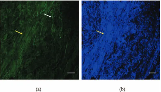
Large-area multiphoton AF and SHG images of the skin are shown in Fig. 1. We observed interesting trends in SHG and AF within the skin dermis as a function of temperature.
sistent with our previous work,18 intensely autofluorescent elastic fibers can be observed in the skin 关Fig. 1共a兲, white arrow兴. On the other hand, we found that while the foreskin dermal collagen fibers are capable of generating intense SHG signals, they are also autofluorescent 关Fig. 1共a兲, yellow ar-row兴, although at lower level than the elastic fibers. In the isolated SHG image, collagen fibers are clearly visible and elastic fibers do not produce SHG signals关Fig. 1共b兲, yellow arrow兴.
For our study, we thermally treated the foreskin dermis at 15 different temperatures from 25° C to 90° C 共5°C inter-vals兲, and representative images of the combined AF and SHG data are shown in Fig. 2. We found that the appearance and organization of dermal fibers change as the temperature in-creases. First, as Fig. 2 shows, the general tendency is for the SHG intensity from dermal collagen to decrease with increas-ing temperatures. At a sufficiently high temperature of60° C, the fibrous structures of dermal collagen in SHG images start to be disrupted. Since SHG signals of collagen are sensitive to structural changes,22 the decrease of SHG indicates that the collagen fibers in the dermis begin to be disrupted at this temperature. As the temperature is further raised, the SHG signals further deteriorate and the presence of collagen fibers can not be easily visualized in the isolated SHG images. On the contrary, the AF of collagen fibers remains to be visible. Although the SHG intensity of collagen is progressively weakened above 60° C, the AF signals can still be used to resolve collagen structures. From AF signals, dermal collagen preserves a fibrous structure at the temperatures lower than 60° C. At 60° C, the fibrous structures of collagen start to be disrupted. At75° C, a great proportion of collagen is molten into an amorphous structure. As the temperature is further raised, the collagen in the dermis appears to have become molten. This is consistent with the histological results show-ing a homogenized structure of collagen 共data not shown兲. Hence, AF images can help to trace the presence of denatured collagen beyond the denaturation temperature. In addition, the AF images from our results also indicate that, after the dis-ruption of structures responsible for SHG signals, collagen continues to undergo structural modifications at higher tem-peratures.
Compared with the collagen fibers, the elastic fibers appear to be less significantly affected by the thermal treatment. Mor-phologically, the elastic fibers still display a fine fibrous struc-ture at60° C, the temperature at which collagen SHG signals start to fade. However, when the temperature is raised to 65° C, an interesting result is revealed by the AF image. Au-tofluorescent elastic fibers start to fracture and become aggre-gated关Fig. 2共d兲, red arrow兴. As temperature is further raised, the fibrous structures are progressively disrupted and the ag-gregations become more evident. We attempted to character-ize the structural changes of elastic fibers by histological pro-cedures. However, the thermally denatured collagen affects the elastic staining and made the analysis of elastic fiber struc-tural alternations impossible. Judging from the serial images obtained at different temperatures, our results suggest that the small autofluorescent aggregates originate from the disrupted elastic fibers.
We also attempted to quantify our observations. Our ap-proach is to calculate the average SHG and AF intensities
across the entire large area image at each temperature. The temperature dependence of SHG and AF signals is shown in Fig. 3. The plots show an interesting trend. It is notable that the AF only fluctuates with increasing temperature关Fig. 3共a兲兴. This result is consistent with the AF images showing that collagen can still be traced by its AF even at high tempera-tures. On the other hand, the SHG signal shows a different trend. At sufficiently high temperatures, the SHG signal tends to decrease关Fig. 3共b兲兴.
However, since the fiber distribution within the dermis is not homogeneous, the intensity variations shown in Fig. 3 may be due to the fiber distributions and morphological fea-tures of the regions being imaged. Therefore, to avoid this artifact, we used the ratiometric definition of AF-to-SHG in-dex of dermis共ASID兲 of
ASID = A/S 共1兲
to characterize the thermal effects on the dermis. In this defi-nition, A is the average AF intensity and S is the average SHG intensity of the images.
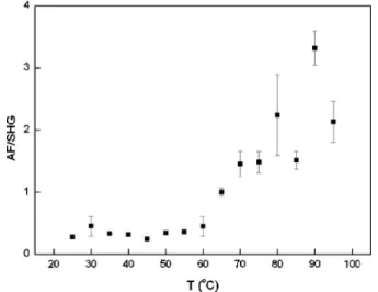
In this nomenclature, a highly autofluorescent sample 共relative to SHG signal兲 would have a higher ASID while a specimen containing intense SHG signal共compared with AF兲 would have a lower ASID. For the thermally treated skin specimens, we computed the ASID from the average AF and SHG intensities obtained across the entire image and the re-sults are plotted in Fig. 4. At temperatures lower than60° C, the ASID is almost constant. This result also solidifies our use of a ratiometric approach in characterizing the thermal dam-age of the dermis in that the ratiometric analysis is not af-fected by the inhomogeneous fiber distribution. As the plot shows, the ASID starts to increase at60° C and a rising trend of ASID is observed when the temperature is further elevated. This index is more sensitive in predicting dermal thermal damage as compared with the SHG intensities alone.
4 Discussion
Our results support the fact that qualitative and quantitative multiphoton AF and SHG microscopy is effective in identify-ing the extent of human dermal thermal damage. Consistent with our previous reports on rat tail tendons,22we observed that at sufficiently high temperatures, the collagen fiber struc-tures in the dermis become disrupted, corresponding to a de-crease in the SHG intensity. Compared with rat tail tendons, the inhomogeneous composition of dermal structures can make quantitative analysis difficult. The isolated SHG inten-sity can fluctuate before the denaturation temperature and complicates the analysis attempts. We observed a parallel change of SHG intensity and AF of the dermis before the denaturation temperature of 60° C. This indicates that the fluctuation of AF and SHG intensity can be accounted for by the inhomogeneous distribution of fibers in the regions im-aged. However, when a ratiometric approach utilizing ASID is used for analysis, this index is shown to be able to better characterize the degree of dermal thermal damage. We found that AF can serve as a reference to compensate for the inho-mogeneous distribution of fibers in the selected regions. Hence, this index is very useful in analysis of the relatively inhomogeneous structures such as dermis.
Fig. 2 Combined AF/SHG images of dermal structures treated thermally at 25, 55, 60, 65, 80, and 95° C共bars 110m兲. SHG signals are blue and
The intensity of AF from collagen shows a different trend of changes associated with thermal treatment. Since SHG is sensitive to the three-dimensional packing of collagen mol-ecules, the disruption of SHG at60° C and higher temperature indicates that collagen structures responsible for SHG are dis-rupted. It is suggested that thermal treatment breaks the hy-drogen bonds that stabilize the native triple helical molecular structure of collagen, leading to an irreversible transformation of collagen molecules into a random coil.26,27 Hence, SHG can be used as a tool to monitor the preservation of original hydrogen bonds in collagen during thermal treatment. How-ever, the AF of collagen is preserved even at temperatures above 60° C. It is suggested that collagen AF is from the cross-linkers of collagen fibers.28Judging from our results, the cross-linkers are less susceptible to the heating process. This is of significance in that after the disruption of SHG signals, AF can still be used to trace the presence of structurally al-tered collagen. Using AF as a guide, we observed that after the hydrogen bonds are disrupted, collagen tissue in the der-mis continues to undergo structural modifications at higher temperature. The fibrous structures are gradually replaced by a molten amorphous structure. In addition to monitoring col-lagen thermal damage, this can be used to monitor the healing
and remodeling process of collagen after thermal injuries. In addition, the morphological structures of autofluorescent elastic fibers also change with increasing temperatures. Spe-cifically, we observed that long fine fibrous elastic fibers started to fracture into short fibers and condense into intensely autofluorescent aggregates at65° C.
5 Conclusion
The thermal damage to dermis can be morphologically and quantitatively analyzed by mulitphoton AF and SHG imaging. Collagen and elastic fibers have different susceptibility to thermal damage and their structural changes respond accord-ingly. The quantitative changes are confirmed by the analysis of the ASID factor we introduce to characterize the relative changes of AF to SHG intensities. The ASID index can bypass artifacts associated with the inhomogeneous distributions of the dermal fibers and shows that the denaturation temperature of dermal collagen is 60° C. This study demonstrates that multiphoton microscopy has the potential to be developed into an in vivo diagnostic technique capable of assessing thermal dermal damage and the remodeling process associated with thermal injuries.
Acknowledgment
Support for this work is provided by the National Research Program for Genomic Medicine of the National Science Council, Taiwan 共NSC 94-3112-B-002-015-Y and NSC 93-3112-B-002-034兲.
References
1. P. H. Koster, C. M. van der Horst, M. J. van Gemert, and A. C. van der Wal, “Histologic evaluation of skin damage after overlapping and nonoverlapping flashlamp pumped pulsed dye laser pulses: a study on normal human skin as a model for port wine stains,” Lasers Surg. Med. 28, 176–181共2001兲.
2. M. Niemz, Laser-Tissue Interactions, Springer-Verlag, Berlin共2002兲. 3. R. E. Fitzpatrick, M. P. Goldman, N. M. Satur, and W. D. Tope, “Pulsed carbon dioxide laser resurfacing of photoaged facial skin,” Arch. Dermatol. 132, 395–402共1996兲.
Fig. 3 Dependence of共a兲 autofluorescence intensity and 共b兲 SHG
in-tensity on temperatures ranging from 25 to 95° C共error bars indicate calculated standard deviations兲.
Fig. 4 Dependence of autofluorescence to
second-harmonic-generation index of dermis共ASID兲 on temperatures 共error bars indi-cate calculated standard deviations兲.
4. K. A. Khatri, E. V. Ross, J. M. Grevelink, C. M. Magro, and R. R. Anderson, “Comparison of erbium: YAG and carbon dioxide lasers in resurfacing of facial rhytides,” Arch. Dermatol. 135, 391–397共1999兲. 5. D. J. Goldberg, “Full-face nonablative dermal remodeling with a
1320 nm Nd: YAG laser,” Dermatol. Surg. 26, 915–918共2000兲. 6. D. J. Goldberg and K. B. Cutler, “Nonablative treatment of rhytides
with intense pulsed light,” Lasers Surg. Med. 26, 196–200共2000兲. 7. R. Fitzpatrick, R. Geronemus, D. Goldberg, M. Kaminer, S. Kilmer,
and J. Ruiz-Esparza, “Multicenter study of noninvasive radiofre-quency for periorbital tissue tightening,” Lasers Surg. Med. 33, 232– 242共2003兲.
8. L. T. Vo, P. Anikijenko, W. J. McLaren, P. M. Delaney, D. H. Barkla, and R. G. King, “Autofluorescence of skin burns detected by fiber-optic confocal imaging: evidence that cool water treatment limits progressive thermal damage in anesthetized hairless mice,” J. Trauma
51, 98–104共2001兲.
9. L. T. Vo, G. D. Papworth, P. M. Delaney, D. H. Barkla, and R. G. King, “A study of vascular response to thermal injury on hairless mice by fibre optic confocal imaging, laser doppler flowmetry and conventional histology,” Burns 24, 319–324共1998兲.
10. R. L. Sheridan, K. T. Schomaker, L. C. Lucchina, J. Hurley, L. M. Yin, R. G. Tompkins, M. Jerath, A. Torri, K. W. Greaves, and D. P. Bua, “Burn depth estimation by use of indocyanine green fluores-cence: initial human trial,” J. Burn Care Rehabil. 16, 602–604 共1995兲.
11. B. H. Park, C. Saxer, S. M. Srinivas, J. S. Nelson, and J. F. de Boer, “In vivo burn depth determination by high-speed fiber-based polar-ization sensitive optical coherence tomography,” J. Biomed. Opt. 6, 474–479共2001兲.
12. Z. B. Ziazi, T. J. Essex, R. Papini, D. Scott, N. R. Mclean, and M. J. Black, “New laser-doppler scanner, a valuable adjunct in burn depth assessment,” Burns 19, 485–489共1993兲.
13. A. T. Yeh, B. Kao, W. G. Jung, Z. Chen, J. S. Nelson, and B. J. Tromberg, “Imaging wound healing using optical coherence tomog-raphy and multiphoton microscopy in an in vitro skin-equivalent tis-sue model,” J. Biomed. Opt. 9, 248–253共2004兲.
14. K. Konig and I. Riemann, “High-resolution multiphoton tomography of human skin with subcellular spatial resolution and picosecond time resolution,” J. Biomed. Opt. 8, 432–439共2003兲.
15. Y. Sun, J. W. Su, W. Lo, S. J. Lin, S. H. Jee, and C. Y. Dong, “Multiphoton polarization imaging of the stratum corneum and the dermis in ex-vivo human skin,” Opt. Express 11, 3377–3384共2003兲. 16. W. Denk, J. H. Strickler, and W. W. Webb, “Two-photon laser
scan-ning fluorescence microscopy,” Science 248, 73–76共1990兲.
17. P. T. C. So, C. Y. Dong, B. R. Masters, and K. M. Berland, “Two-photon excitation fluorescence microscopy,” Annu. Rev. Biomed. Eng. 2, 399–429共2000兲.
18. S. J. Lin, R. J. Wu, H. Y. Tan, W. Lo, W. C. Lin, T. H. Young, C. J Hsu, J. S. Chen, S. H. Jee, and C. Y. Dong, “Evaluating cutaneous photoaging by use of multiphoton fluorescence and second harmonic generation microscopy,” Opt. Lett. 30, 2275–2277共2005兲. 19. A. Zoumi, A. Yeh, and B. J. Tromberg, “Imaging cells and
extracel-lular matrix in vivo by using second-harmonic generation and two-photon excited fluorescence,” Proc. Natl. Acad. Sci. U.S.A. 99, 11014–11019共2002兲.
20. W. R. Zipfel, R. M. Williams, R. Christie, A. Y. Nikitin, B. T. Hy-man, and W. W. Webb, “Live tissue intrinsic emission microscopy using multiphoton-excited native fluorescence and second harmonic generation,” Proc. Natl. Acad. Sci. U.S.A. 100, 7075–7080共2003兲. 21. P. J. Campagnola and L. M. Loew, “Second-harmonic imaging
mi-croscopy for visualizing biomolecular arrays in cells, tissues and or-ganisms,” Nat. Biotechnol. 21, 1356–1360共2003兲.
22. S. J. Lin, C. Y. Hsiao, Y. Sun, W. Lo, W. C. Lin, G. J. Jan, S. H. Jee, and C. Y. Dong, “Monitoring the thermally induced structural transi-tions of collagen using second harmonic generation microscopy,” Opt. Lett. 30, 622–624共2005兲.
23. S. J. Lin, W. Lo, H. Y. Tan, J. Y. Chan, W. L. Chen, S. H. Wang, Y. Sun, W. C. Lin, J. S. Chen, C. J. Hsu, J. W. Tjiu, H. S. Yu, S. H. Jee, and C. Y. Dong, “Prediction of heat-induced collagen shrinkage by use of second harmonic generation microscopy,” J. Biomed. Opt. 11, 034020共2006兲.
24. M. Han, G. Giese, and J. F. Bille, “Second harmonic generation im-aging of collagen fibrils in cornea and sclera,” Opt. Express 13共15兲, 5791–5797共2005兲.
25. R. M. Williams, W. R. Zipfel, and W. W. Webb, “Interpreting second-harmonic generation images of collagen I fibrils,” Biophys. J. 88共2兲, 1377–1386共2005兲.
26. N. T. Wright and J. D. Humphrey, “Denaturation of collagen via heating: an irreversible rate process,” Annu. Rev. Biomed. Eng. 4, 109–128共2002兲.
27. K. Hayashi, G. Thabit III, J. J. Bogdanske, L. N. Mascio, and M. D. Markel, “The effect of nonablative laser energy on the ultrastructure of joint capsular collagen,” Arthroscopy 12, 474–481共1996兲. 28. N. Kollias, R. Gillies, M. Moran, I. E. Kochevar, and R. R. Anderson,
“Endogeneous skin fluorescence includes bands that may serve as quantitative markers of aging and photoaging,” J. Invest. Dermatol.