98 8 19 98 8 23 (medical illustration) Microsoft PowerPoint 2003 Microsoft PowerPoint Microsoft PowerPoint 2009;6:53-61 (medical illustration) [1] (medical illustrator) [2] Adobe Adobe Systems Incorporated
Photoshop Illustrator FreeHand Corel Corel Corporation
PhotoImpact Paint Shop Pro Photo CorelDRAW Graphics Suite Corel DESIGNER Technical Suite
Painter [3] 1 2 3 4 5 6 7
(computed tomography, CT)
[4] (positron
emis-sion tomography, PET) (PET/CT) Microsoft Office PowerPoint 2003 PowerPoint (controls) (toolbar) PowerPoint PowerPoint (menu) (menu item) (status bar) [5] Omar Lababede [4,6] 1.
(Fill Effects) (tool
tip) 2. (Lines) 3. 5. (Add or Remove Buttons)
(Drawing) (Select Multiple
Objects) 2 6. PowerPoint (proof of concept) 49 (carcinoembry-onic antigen) -18 (F-18 fluorodeoxyglucose, F-18 FDG) -18 -18 (Fig. 1) -18
PowerPoint
(Curve) (Freeform)
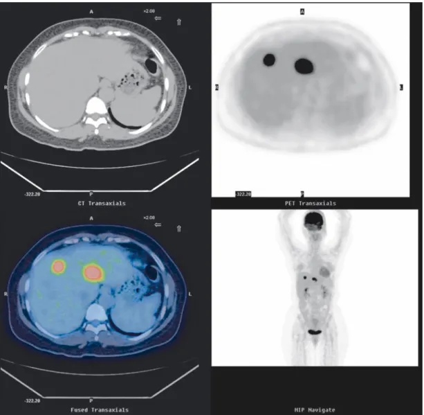
Fig. 1 Rendering F-18 FDG PET/CT display. The transaxial views of CT images (upper left) and corresponding PET images
(upper right) fused to the PET/CT images (lower left) are displayed. In addition, the maximum intensity projection view (lower left) is demonstrated.
(vector object) (Bring to Front) 200% 3 85% (anchor point)
(path) (line
seg-m e n t )
(direction lines)
(direction point)
(Auto
Point) (Smooth Point) (Straight
Point) (Corner Point)
PowerPoint 2007 Alt Ctrl Ctrl PowerPoint 100% -18 RGB (R = 128 G = 0 B = 1 R = 255 G = 0 B = 0) Fig. 2 A Fig. 2 B Fig. 2 C (Fig. 3) 76%
Fig. 3 Delete (*.bmp) PowerPoint PowerPoint PowerPoint PowerPoint (Fig. 4A) (Fig. 4B) (Fig. 4C) Fig. 4D
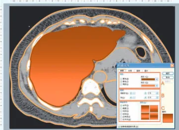
Fig. 2 Applying colors and textures of vector objects on CT
template image. (Microsoft product screen shot reprinted with permission from Microsoft Corporation.)
Fig. 3 Applying colors and textures of vector objects on
PET template image. (Microsoft product screen shot reprint-ed with permission from Microsoft Corporation.)
(atlas) (Fig. 4A) (Fig. 4B) (Fig. 4C) Omar Lababede [4,6] Max Brödel [7]
(a picture is worth more than a thousand words) 1024
32 32
Fig. 4 (A) Exhibiting the final drawing of CT illustration after adding labels; (B) Exhibiting the final drawing of PET
illustra-tion after adding labels; (C) Exhibiting the final drawing of PET/CT fusion illustraillustra-tion; (D) Exhibiting the PET/CT fusion illus-tration from directly adding the PET foci on CT illusillus-tration.
[8]
[9]
(bitmap graphic) (vector graphic)
[10]
(bitmap image) (raster image)
x y
Adobe Photoshop Corel PhotoImpact Microsoft
bmp pcx gif jpg tif (object-oriented images)
eps al cdr
Core CorelDraw Adobe Illustrator
PowerPoint 95%[11] PowerPoint
Richard H. Daffner PowerPoint [12] PowerPoint PowerPoint [13] [14] PowerPoint 2003 90 PowerPoint 2007 PowerPoint PowerPoint 2003 RGB CMYK
Fig. 4D
[15]
Association of Medical Illustrators Institute of Medical Illustrators
Journal of Biological Photography Journal of Visual Communica-tion in Medicine
PowerPoint
1. Tsafrir J, Ohry A. Medical illustration: from caves to cyberspace. Health Info Libr J 2001;18:99-109.
2. Corl FM, Garland MR, Fishman EK. Role of computer
part 1, introduction to the image-manipulation com-mands. AJR Am J Roentgenol 2004;183:847-851. 6. Lababede O, Meziane M. Medical illustration techniques
for PowerPoint: part 1, The basics. AJR Am J Roentgenol 2007;188:W379-383.
7. Schultheiss D, Engel RM, Crosby RW, et al. Max Brödel (1870-1941) and medical illustration in urology. J Urol 2000;164:1137-1142.
8. Hansen CL. Digital image processing for clinicians, part I: Basics of image formation. J Nucl Cardiol 2002;9:343-349.
9. Yam CS, Kruskal J, Larson M. Creating animated GIF files for electronic presentations using Photoshop. AJR Am J Roentgenol 2007;188:W485-490.
10. Tunuguntla R, Rodriguez O, Ruiz JG, et al. Computer-based animations and static graphics as medical student aids in learning home safety assessment: a randomized controlled trial. Med Teach 2008;30:815-817.
11. LaPorte RE, Linkov F, Villasenor T, et al. Papyrus to PowerPoint (P 2 P): metamorphosis of scientific commu-nication. BMJ 2002;325:1478-1481.
12. Daffner RH. On improvement of scientific presentations: using PowerPoint. AJR Am J Roentgenol 2003;181:47-49.
13. Schreibman KL. Getting images into PowerPoint. AJR Am J Roentgenol 2001;177:1271-1272.
14. Yam CS. Using PowerPoint to create high-resolution images for journal publications. AJR Am J Roentgenol 2005;185:273-276.
15. Burger C. Image coregistration and coregistered image rendering. In: von Schulthess GK, ed. Molecular Anatomic Imaging: PET-CT and SPECT-CT Integrated Modality Imaging. 2nd ed. Philadelphia: Lippincott Williams & Wilkins; 2006:74-84.
Received 8/19/2009; accepted 8/23/2009.
For correspondence and reprints contact: Dr. Jainn-Shiun Chiu
Address: Department of Nuclear Medicine, Taipei Medical University - Shuang Ho Hospital. No. 291, Zhongzheng Rd., Zhonghe City, Taipei County 235, Taiwan
Background: Medical illustration is a multi-disciplinary knowledge that integrates the life science and visual com-munication. Through the visual implication of imaging, medical knowledge may be concretized to popularize medical education. On the basis of PET and CT images, the purpose of this study is to apply presentation software for draw-ing the medical illustrations of PET, CT, and PET/CT fusion images.
Methods: We used Microsoft PowerPoint 2003, a kind of presentation software, to be the painting toolkit in our investigation. The images of PET and CT were utilized to be the templates, respectively. The built-in vector graphic tools of PowerPoint were selected to perform the processes of multi-layer copy-painting method based on the serial drafting workflow. Medical illustrations will be drawn depicting the contours and colors of organs on PET and CT images.
Results: With the help of multi-layer copy-painting method based on the serial drafting workflow by using Microsoft PowerPoint, the illustrations of PET, CT, and PET/CT were easily limned. The similarity and difference between anatomical and functional images were successfully presented. At the same time, we also add the illustrations of metabolic foci on the CT illustration.
Conclusion: Through conveying intension of medical imaging, medical illustration not only aids the learning of medical knowledge but also transmits the health education. We disclosed the hidden functions of graphical ability in presentation software and the clinical staff can easily limn the medical illustrations. Therefore, our study validated the feasibility of employing presentation software to portray the medical illustration.
Key words: Medical illustration, presentation software, PET/CT, vector drawing J Nucl Med Tech 2009;6:53-61
Applying Presentation Software to Draw Medical
Illustrations of PET/CT
Kuang-Ching Chiu
1, Yuh-Feng Lin
2,7, Wei-Chih Shen
3, Yu-Chuan Li
4,
Yuh-Feng Wang
5, Jian-Guo Liao
5, Che-Ming Yang
6, Jainn-Shiun Chiu
4,6 1Department of Medical Education and Research, Taipei Medical University - Shuang Ho Hospital, Taipei, Taiwan;
2
Divsion of Nephrology, Department of Internal Medicine, Taipei Medical University – Shuang Ho Hospital, Taipei, Taiwan;
3
Department of Computer Science and Information Engineering, Asia University, Taichung, Taiwan;
4
Graduate Institute of Biomedical Informatics, Taipei Medical University, Taipei, Taiwan;
5
Department of Nuclear Medicine, Dalin Tzu Chi General Hospital, Chiayi, Taiwan;
6Department of Nuclear Medicine, Taipei Medical University – Shuang Ho Hospital, Taipei, Taiwan;
7