Cycling motion soft tissue artifacts quantification on the thigh and
shank using a novel CT-to-Fluoroscopy registration method
以三維動態 X 光創新比對方法量化腳踏車踩踏過程腿部皮膚移動誤差
NSC (101-2627-B-002-007-) 101/08/01 ~ 102/10/31
Yu-Huan Wu1, Cheng-Chung Lin1, Jia-Da Li1, Tsung-Chi Lin1, Mei-Ying Kuo3, Tung-Wu Lu1,2,* 1 Institute of Biomedical Engineering, National Taiwan University, Taipei, Taiwan 2 Department of Orthopaedic Surgery, School of Medicine, National Taiwan University, Taiwan
3 School of Physical Therapy, China Medical University, Taiwan
Abstract- Cycling plays an important role in our daily lives. Skin maker-based motion analysis has been used to assess the functional performance of cycling exercises. However, the associated soft tissue artifacts (STA) of the markers affected the accuracy.
The aims of the study were to quantify the STA on the thigh and shank during cycling exercises using a novel technique. Seven healthy adults wearing skin markers performed cycling movement under simultaneous surveillance of a motion capture system and a fluoroscopy system. Considerable STA’s were found during cycling exercises with greater STA on the thigh. The STA around the knee joint during the cycling movement were quantified for the first time, which will be helpful for developing guidelines for using skin markers and STA-compensation methods. 摘要- 騎乘腳踏車在日常生活扮演重要角色。以 反光標記球為基準的動作分析方法已被用來評 估腳踏車踩踏的功能性表現。然而,這個方法的 準確度會因為皮膚移動誤差降低。此研究的目的 在於使用三維動態 X 光創新比對方法量化腳踏車 踩踏過程腿部之皮膚移動。七位健康成人受試者 在腿部黏貼反光標記球,同時以動作捕捉系統與 三維動態 X 光系統捕捉踩踏腳踏車動作。結果顯 示,在腳踏車踩踏過程中大腿相較小腿伴隨較多 之皮膚移動,移動方向因部位而有差異。本研究 首次成功將腳踏車踩踏過程膝關節鄰近之皮膚 移動量化,這些量化資訊將有助於發展成為使用 反光標記球量測下考量皮膚移動誤差之補償準 則。
Introduction- Cycling plays an important role in transportation, recreation, and sport in our daily lives but cycling-related overuse injuries are not uncommon. Skin maker-based motion analysis has been used to assess the functional performance of cycling exercises and potential injury risks. However, the associated soft tissue artifacts (STA) of the markers affected the accuracy of calculated
mechanical variables [1, 2]. Therefore, knowledge of STA will be helpful for a better interpretation of the results obtained and further development of STA-compensation methods. The aims of the study were to quantify the STA of selected markers on the thigh and shank during cycling exercises using a novel technique.
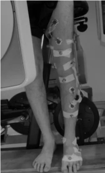
Methods- Seven healthy adults wearing skin markers on the medial and lateral femoral epicondyles (MFC and LFC), thigh (THIL, THIC, THIM), tibial tuberosity (TT), fibular head (FIB), shank (SHA), and medial and lateral malleoli (MA) performed cycling movement under simultaneous surveillance of a motion capture system and a fluoroscopy system (Fig.1). They also received a CT scan so that their
femoral and tibial bone poses could be obtained using a fluoroscopy-to-CT registration method [1]. The STA of the skin markers were then calculated as the marker movement relative to the underlying bone [3].
Fig. 1: Markers attached on left thigh, left shank, and left foot.
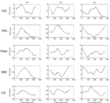
Results & Discussion- Considerable STA’s were found during cycling exercises with greater STA on the thigh. MFC and LFC were displaced posteriorly from their true positions with knee flexion, and moved anteriorly with knee extension. THIC had greater STA among the technical markers on the thigh and moved mainly along the proximal/distal direction (Fig.2, 3). TT moved within a small range throughout the cycling movement, while FIB moved mainly along the proximal/distal direction (Fig.4, 5). The STA around the knee joint during the cycling movement were quantified for the first time in the literature, which will be helpful for developing guidelines for using skin markers and STA-compensation methods.
Fig. 2: Movement of the thigh markers relative to the underlying femur during the cycling movement.
Fig. 3: Movement of the thigh markers relative to the underlying femur during the cycling movement. The STA were expressed in terms of the errors along the anterior/posterior, proximal/distal and medial/lateral directions.
Fig. 4: Movement of the shank markers relative to the underlying tibia during the cycling movement.
Fig. 5: Movement of the shank markers relative to the underlying tibia during the cycling movement. The STA were expressed in terms of the errors along the anterior/posterior, proximal/distal and medial/lateral directions.
References–
[1] Tsai, T.Y., et al. (2011) J Biomech, 44: 1182–1188
[2] Kuo, M.Y., et al. (2011) Gait Posture, 33: 379-384.
[3] Tsai, T.Y., et al. (2010) Med Phys, 37: 1273-1284.