行 政 院 國 家 科 學 委 員 會 補 助 專 題 研 究 計 畫
█ 成 果 報 告
□期中進度報告
提昇私大研發能量專案
-中藥於動脈硬化基因體之研究 (2/2)
Genomic Research of Herbal Medicine on Atherosclerosis
計畫類別:□ 個別型計畫 █ 整合型計畫
計畫編號:
NSC91-2745-B039-001 and NSC92-2745-B039-001
執行期間:2002 年 8 月 1 日至 2004 年 7 月 31 日
計畫主持人:
: 洪瑞松教授
共同主持人:
張文正
教授、
吳介信
副教授、
陳卓昇
副教授、
謝文聰
副
教授、
余雅美
副教授、
謝慶良
教授、
周明加
副教授、
江
素瑛
助理教授等
計畫參與人員: 研究生與研究助理等
成果報告類型(依經費核定清單規定繳交):
□精簡報告 █完整報告
處理方式:除產學合作研究計畫、提升產業技術及人才培育研究計
畫、列管計畫及下列情形者外,得立即公開查詢
□涉及專利或其他智慧財產權,□一年□二年後可公開查詢
執行單位:中國醫藥大學
中 華 民 國 九十三 年 十 月 二十六 日
中文摘要 心血管疾病(譬如:粥狀動脈硬化、高血壓、心肌梗塞、腦中風等)高居國人 前十大死因,因血管病變所造成動脈硬化(arteriosclerosis)會導致血管管徑的狹 宰,甚至完全栓塞,最後導致組織細胞缺血缺氧而死亡。動脈硬化相關疾病之治 療,在中西醫學領域,均有相當長時期的經驗與績效,然而中藥於動脈硬化相關 疾病治療,其療效與作用機轉尚未經全面性的科學量化與探討。本計畫整合了本 校與附設醫院之醫療研究團隊,利用現代化的生物科技及系統性的基礎研究,針 對中醫常用之治療心血管疾病的中藥,如丹參(Labiatae salvia miltiorrhiza)、厚朴 (Magnoliaceae magnolia officinalis)、紅花(Compositae carthamus tinctorius)、霍山 石斛(Dendrobium huoshanense)等與其有效成分,探討其抗粥狀動脈硬化的作用與 其分子機轉,以期望將中藥提升到國際的醫療舞台。粥狀動脈硬化是一種長期性 的發炎反應疾病,主要是因為高脂飲食、血液中膽固醇及低密度脂蛋白的含量過 高,受到自由基氧化,進而形成內皮增生及粥狀動脈斑塊之現象。本團隊利用已 建立標準的動物動脈粥狀硬化模式,並以正常與高膽固醇飼餵組之紐西蘭大白兔 主動脈弓之 RNA 進行基因晶片分析,進而篩選出粥狀動脈硬化病程中具有特異 性表現的相關基因,藉全面且大規模的方式篩選特異性表現的基因群,作為未來 評估抗粥狀脈硬化功效之高效率的工具。經分析顯色結果後挑選了 57 個基因, 以半定量聚合酶鏈鎖反應的技術驗證在正常與高膽固醇飼餵組間基因群的表現 差異。並進一步以即時定量的聚合酶鏈鎖反應技術(Real-time PCR)定量粥狀動脈 硬化相關基因的表達,結果顯示所選擇的中藥萃取物皆能明顯降低原先受高膽固 醇飼料所誘發的特定基因表現,且達統計上顯著差異。再藉由檢測所選擇的中藥 萃取物對粥狀動脈硬化相關基因的表達、血液脂質生化值(總膽固醇、低密度脂蛋 白、三酸甘油酯)、抗氧化酵素活性(SOD、catalase)、組織內氧化自由基與氧化 DNA 傷害(8-hydroxy-2’-deoxyguanosine)累積等的影響,與輔以觀察所選擇的中藥 萃取物對於主動脈粥狀動脈硬化斑塊堆積形成的抑制作用,進而證實丹參、紅 花、厚朴、石斛具有改善粥狀動脈硬化的效能。另實驗結果發現紅花、丹皮在一 定 劑 量 下 具 有 保 護 氧 化 低 密 度 脂 蛋 白(oxLDL) 對 人 類 臍 靜 脈 血 管 內 皮 細 胞 (HUVEC)所誘發細胞凋亡的作用。同時我們利用已建立的缺血-再灌流誘發大鼠 局部缺血性腦梗塞模型,實驗結果發現丹參可降低大鼠因腦缺血-再灌流後所導致 的梗塞面積與氧化自由基。綜合本實驗結果顯示丹參、厚朴、紅花與石斛對於預 防及避免粥狀動脈硬化有良好的效用,可作為治療動脈粥狀硬化之藥物的開發。 關鍵詞:粥狀動脈硬化、丹參、厚朴、紅花、石斛、氧化傷害
英文摘要
Atherosclerosis, a chronic inflammatory disease, is the leading cause of heart disease and stroke-related deaths. Salvia miltiorrhiza
、
Magnolia officinalis、
Carthamus Tinctorius and Dendrobium officinale are Traditional Chinese medicines commonlyused in treating cardiovascular diseases. The increasing evidence of their anti-oxidative effects has prompted us to study their anti-arteriosclerotic effects and the underlying mechanisms. To determine the effects in lowing the formation of the atheroma and molecular mechanisms of Chinese herb treatment, aortas from normal and atherosclerotic New Zealand White (NZW) rabbits were used to extract total RNA. Human cDNA microarray were performed to identify specific genes expression between the normal and the cholesterol group. Real-time quantitative RT-PCR was used to determine five specific gene expressions during atherogenesis. The results of cholesterol group showed over-expression in those five genes. The gene expressions of Chinese herb essence groups were significantly decreased as compared to cholesterol group. These markers may provide mechanistic information for the pathogenesis of arteriosclerosis and highlight potential intervention strategies. Furthermore, the total cholesterol, LDL-cholesterol, triacylglycerol and lucigenin-CL were decreased in the Chinese herb group as compared to the cholesterol group. The elevated amounts of oxidative DNA damage marker (8-hydroxy-2’-deoxyguanosine) in liver tissues and isolated aorta segments of cholesterol-fed rabbits were significantly reduced by Chinese herbs. The intimal surface of the thoratic aorta was covered with atherosclerotic lesions in the control group. Chinese herbs could significantly reduce atherosclerotic lesions on rabbit aorta. Moreover, Carthamus
tinctorius elicited the markedly protective effects on oxLDL-induced cytotoxicity and
apoptosis in human umbilical vein endothelial cells (HUVEC). Further studies on an established rat model of focal cerebral infarct showed that pretreatment with intraperitoneal injection of Salvia miltorrhiza bunge reduced the area of cerebral infarct and also reduced the luminol-CL counts of peripheral blood in ischemia-reperfusion injured rats. Taking together, these data suggest that Salvia
miltiorrhiza
、
Magnolia officinalis、
Carthamus Tinctorius and Dendrobium officinalemay be useful and effective candidates to prevent atherosclerosis.
Keywords:atherosclerosis, Salvia miltiorrhiza
、
Magnoliaceae Magnolia officinalis、
計畫緣由與目的 計畫目標 根據行政院衛生署之89 年統計資料顯示,腦血管疾病、心臟疾病、及高血 壓性疾病分佔國人死因之第 2、3、11 順位。這三大死因或多或少均與動脈硬化 有關連性。因此全國各醫療單位莫不投入大量的時間與金錢,人力及物力,結合 醫學、藥物、生化、及分子生物科技,謀求因應之道。中國醫藥學院附設醫院即 設置心臟血管中心,提供最先進的診斷與治療。學院與醫院組成心血管疾病之研 究團隊與實驗室,亦全力投入臨床及基礎之研究。 動脈硬化相關疾病之治療,在中西醫學領域,均有相當長時期的經驗與績 效。中藥為中華文化幾千年來之重要精髓,然而其療效與作用機轉,尚未經全面 性的科學量化與探討。唯有經過系統性的基礎研究,以現代化的生物科技來確定 其機轉,才能將中藥提升到國際的醫療舞台。 長期以來,丹參,厚朴,紅花,及石斛等中藥,常被用於治療心臟血管相關 的疾病,例如於「補陽還五湯」。本計畫整合了本校與附設醫院之醫療研究團隊, 針對這 4 種中藥(或其有效成分),從數個不同的角度,逐一探討其作用機轉, 並運用基因晶片之生物科技,研發製造可供診斷之利器。本計畫是結合學院及附 設醫院的研究團隊技術、資源及人力,確認中藥治療動脈硬化或血管再阻塞的機 轉原理,做有系統研究的整合性群體研究。本計畫不但可以提供這4 種中藥治療 動脈硬化或血管再阻塞的機轉原理,確認並進一步鎖定引起動脈硬化或血管再阻 塞的相關基因進行深入探討,也讓我們可以進一步鎖定引起這些疾病的相關基 因,在學術上及臨床治療上均具有重要的意義。 計畫研究成果 本團隊利用已建立標準的動物動脈粥狀硬化模式,並以正常與高膽固醇飼餵 組之紐西蘭大白兔主動脈弓之 RNA 進行基因晶片分析,進而篩選出粥狀動脈硬 化病程中具有特異性表現的相關基因,藉全面且大規模的方式篩選特異性表現的 基因群,作為未來評估抗粥狀脈硬化功效之高效率的工具。經分析顯色結果後挑 選了 57 個基因,以半定量聚合酶鏈鎖反應的技術驗證在正常與高膽固醇飼餵組 間基因群的表現差異,高膽固醇飼餵組較正常飼餵組能顯著誘發粥狀動脈硬化病 程中特定基因群的表現,如IL-lra,SAA,TNF-bata,與 iNOS 等。並進一步以即 時定量的聚合酶鏈鎖反應技術(Real-time PCR)定量粥狀動脈硬化相關基因的表 達,結果顯示所選擇的中藥萃取物皆能明顯降低原先受高膽固醇飼料所誘發的特 定基因表現,且達統計上顯著差異。中草藥粗萃取物處理能降低受高膽固醇飼餵
再藉由檢測所選擇的中藥萃取物對粥狀動脈硬化相關基因的表達、血液脂 質生化值(總膽固醇、低密度脂蛋白、三酸甘油酯)、抗氧化酵素活性(SOD、 catalase)、組織內氧化自由基與氧化 DNA 傷害(8-hydroxy-2’-deoxyguanosine)累積 等的影響。實驗結果發現高餵食高膽固醇4、8 週之兔子動物模式,中藥可於血 液中降低膽固醇、三酸甘油脂、低密度脂蛋白等生化參數值。血液自由基結果顯 示於8 週時厚朴、丹參、紅花、石斛組與膽固醇組相比有明顯差異,表示中藥對 降低血液自由基在8 週時有一定的效果。肝臟之超氧自由基含量結果顯示各組與 膽固醇組相比有明顯差異,表示中藥對於降低組織自由基有一定的效果。中藥對 於高膽固醇誘導之兔子動物模式下,其可顯現一抗氧化之結果,進而改善動物體 中心血管系統氧化傷害之程度。輔以觀察所選擇的中藥萃取物對於主動脈粥狀動 脈硬化斑塊堆積形成的抑制作用,主動脈血管染色結果顯示各組與膽固醇組相比 有明顯差異,中藥對於降低血管脂質沉積有一定效果,進而證實丹參、紅花、厚 朴、石斛具有改善粥狀動脈硬化的效能。 另實驗結果發現紅花、丹皮在一定劑量下具有保護氧化低密度脂蛋白 (oxLDL)對人類臍靜脈血管內皮細胞(HUVEC)所誘發細胞凋亡的作用。同時我們 利用已建立的缺血-再灌流誘發大鼠局部缺血性腦梗塞模型,實驗結果發現丹參 可降低大鼠因腦缺血-再灌流後所導致的梗塞面積與氧化自由基。綜合本實驗結果 顯示丹參、厚朴、紅花與石斛對於預防及避免粥狀動脈硬化有良好的效用,可作 為治療動脈粥狀硬化之藥物的開發。 已發表與準備發表研究成果
1. Weng-Cheng Chang, Yuan-Man Hsu, Chieh-Hsi Wu, Ya-Mei Yu, Pao-Lung Yin, Su-Yin Chiangand Jui-Sung Hung, Inhibitory Effect of Magnolia Officinalis on Aortic Oxidative Stress and Apoptosis in Hyperlipidemic Rabbits (Submitted). 2. Yuan-Man Hsu, Chieh-Hsi Wu, Ya-Mei Yu, Chien-Hwain Chen, Weng-Cheng Chang and Jui-Sung Hung, Inhibitory Effect of Salvia Miltiorrhiza on Aortic Oxidative Stress and Apoptosis in Hyperlipidemic Rabbits (Submitted).
3. Weng-Cheng Chang, Ya-Mei Yu, Chieh-Hsi Wu, Yueh-He Tseng, and Kuen-Yuh Wu, Reduction of oxidative stress and atherosclerosis in hyperlipidemic rabbits by Dioscorea rhizome (Submitted).
4. Chieh-Hsi Wu, Ho-Chih Chen, Weng-Cheng Chang, and Jui-Sung Hung, Characterizing the Inhibitory Mechanisms of Magnolol on Rat Smooth Muscle Cell Proliferation (Submitted).
5. Chieh-Hsi Wu, Ho-Chih Chen, and Weng-Cheng Chang, Characterizing the Inhibitory Mechanisms of Magnolol on Rat Smooth Muscle Cell Proliferation (Submitted).
Chinese herb on vascular responsiveness in cholesterol-fed rabbit-isolated arteries (In preparation)
7. J-S Chen, P-U Chou, W-C Chang and J-S Hung, Attenuation of
8. baroreflex sensitivity in rabbits with hyperchlesterolemia: role of nitric oxide (In preparation).
9. SY Chiang, MY Lin, JJ Chu, YM Yu, WC Chang and JS Hung, Inhibitory Effects of Paeonia Suffruticosa and Carthamus Tinctorius on oxLDL-induced Cytotoxicity and Apoptosis in Human Umbilical Vein Endothelial Cells (In preparation).
10. Chieh-His Wu, Ho-Chih Chen, Weng-Chen Cheng and Jui-Sung Hung, The antiatherogenic effects of Honodiol, Magnolol and Alismatis Rhizoma. (In preparation)
11. WC Chang, CH Wu, YM Yu, YS Chang, and JS Hung, Effects of three Chinese essences on gene expression during atherogenesis (In preparation)
12. Lo CJ. Lin JG. Kuo JS. Chiang SY. Chen SC. Liao ET. Hsieh CL. Effect of salvia miltiorrhiza bunge on cerebral infarct in ischemia-reperfusion injured rats.
研究成果發表與準備發表的摘要與部分的圖表
1. Weng-Cheng Chang, Yuan-Man Hsu, Chieh-Hsi Wu, Ya-Mei Yu, Pao-Lung Yin, Su-Yin Chiang and Jui-Sung Hung, Inhibitory Effect of Magnolia Officinalis on Aortic Oxidative Stress and Apoptosis in Hyperlipidemic Rabbits (Submitted).
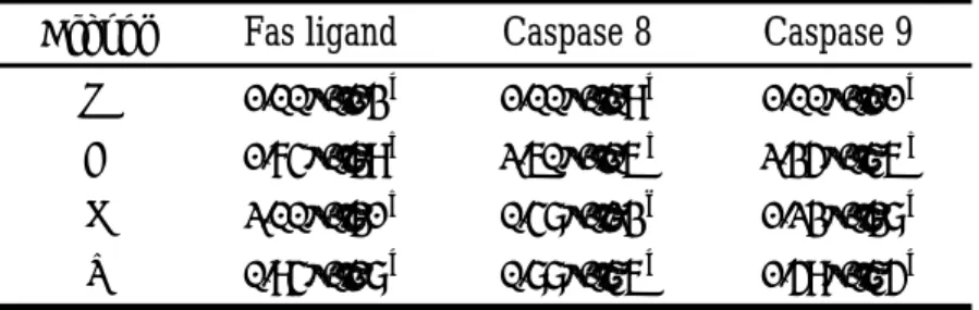
Atherosclerosis is the leading cause of mortality and morbidity in industrialized countries. Twenty hyperlipidemic rabbits were served one of the following: a high-fat and cholesterol diet (cholesterol group, 10% corn oil and 0.5% cholesterol), a high-cholesterol diet supplemented with probucol (200 mg/kg), lovastatin (200 mg/kg), or Magnolia officinalis extract (300 mg/kg). The plasma lipids, oxidative stress (measured by free radical, MDA and oxidative DNA damage) and lipid lesions significantly decreased in the Magnolia officinalis, probucol and lovastatin groups when compared with the cholesterol group (Table 1 and 2, Figure 1). Moreover, the expressions of caspase 8 and caspase 9 in the aortic arches were also markedly lowered by those diets while Fas ligand levels were not changed (Table 3). Therefore, the results indicate that anti-atherogenic effect of Magnolia officinalis is involved with a decreases oxidative stress and with the down-regulation of apoptosis related gene expression in hyperlipidemic rabbits.
Table 1. Effect of probucol, lovastatin and Magnolia officinalis on serum TG, TC, LDL-C, and HDL-C levels Groups TG TC LDL-C HDL-C N 0.77±0.27 a 1.30±0.25 a 0.67±0.20 a 0.76±0.04 a C 3.34±0.82 b 29.49±2.22 b 22.26±3.18 b 2.85±0.28 b P 2.20±0.15 b 8.43±0.80 c 6.67±1.18 c 1.61±0.18 c L 0.77±0.09 a 13.50±3.12 d 10.67±2.53 d 2.96±0.20 b M 0.88±0.15 a 7.09±0.38 c 5.6±0.56 c 2.00±1.08 b All values are the means±SD. Unit is mmole/L.
Abbreviation: N: normal; C: cholesterol; P: probucol; L: lovastatin; M: Magnolia officinalis; TG: triglycerol; TC: total cholesterol; LDL: low-density lipoprotein; HDL: high-density lipoprotein.
a-d : Data with different superscripts in the same column are significantly different at
Table 2. Effect of probucol, lovastatin and Magnolia officinalis on the presence of MDA, Luminol-CL and Lucigenin-CL
Groups MDA ( nmol/ml ) Luminol-CL (Count/WBC) Lucigenin-CL (Count/WBC) N 13.61±3.46 a 0.19±0.09 a 1.27±0.35 a C 57.06±4.89 b 1.57±0.87 b 6.95±0.14 b P 6.80±0.89 c 0.15±0.12 a 1.52±0.25 a L 12.62±5.36 a 0.40±0.04 c 0.99±0.25 a M 18.64±3.30 a 0.16±0.08 a 1.28±0.22 a Results are expressed as the means±SD.
Abbreviation: N: normal; C: cholesterol; P: probucol; L: lovastatin; M: Magnolia officinalis.
a-c: Data with different superscripts in the same column are significantly different at
p<0.05.
Table 3. Effect of probucol, lovastatin and Magnolia officinalis on the expression of Fas ligand, caspase 8 and caspase9
Groups
Fas ligand Caspase 8 Caspase 9N 1.00±0.03 a 1.00±0.02 a 1.00±0.01 a C 1.74±0.09 b 2.60±0.10 b 2.35±0.90 b P 1.50±0.20 b 1.25±0.08 a 1.57±0.20 a L 2.00±0.01 b 0.48±0.10 c 1.23±0.30 a M 1.36±0.22 b 1.58±0.03 a 0.66±0.20 c
All values are the means±SD. Results are expressed as fold increase over normal group as measured by quantitative immunoblotting.
Abbreviation: N: normal; C: cholesterol; P: probucol; L: lovastatin; M: Magnolia officinalis.
a-c: Data with different superscripts in the same column are significantly different at
0 10 20 30 40 50 60 70 80 90 100 N C P L M groups 8-OHdG/10 6 dG * * *
Fig 1. Effect of probucol, lovastatin and Magnolia officinalis on the level of 8-OHdG in aortas. Results are expressed as the means±SD. Abbreviation: N: normal; C: cholesterol: P: probucol; L: lovastatin; M: Magnolia officinalis. *: Compare with the control group; p<0.05.
0 5 10 15 20 25 30 35 40 45 50 N C groupsP L M % sur
face lipid lesion
* * * *
Fig 2. Effects of probucol, lovastatin and Magnolia officinalis on the development of lipid lesions in aorta. Results are expressed as the means±SD. Abbreviation: N: normal; C: cholesterol: P: probucol; L: lovastatin; M: Magnolia officinalis.
2. Yuan-Man Hsu, Chieh-Hsi Wu, Ya-Mei Yu, Chien-Hwain Chen, Weng-Cheng Chang and Jui-Sung Hung, Inhibitory Effect of Salvia Miltiorrhiza on Aortic Oxidative Stress and Apoptosis in Hyperlipidemic Rabbits (Submitted).
The critical features in atherosclerosis development are caused by free radicals (ROS) formation, twenty hyperlipidemic rabbits were served one of the following: a high-fat and cholesterol diet (control group, 10% corn oil and 0.5% cholesterol), a high-cholesterol diet supplemented with lovastatin (20 mg/kg) or Salvia miltiorrhiza extract (300 mg/kg). The plasma lipid, oxidative stress (measured by free radical, MDA and oxidative DNA damage) and lipid lesions significantly decreased in the Salvia miltiorrhiza and lovastatin groups when compared with the control group (Table1 and Table 2). Moreover, the expressions of Fas ligand, caspase 8 and caspase 9 in the aortic arches were also markedly lowered (Table 3). Therefore, the results indicate that the decrease of atherosclerosis lesions by Salvia miltiorrhiza is mainly due to the clearance of free radicals (Figure 1) and the reduction of abnormal expression and/or dysfunction of the apoptosis-regulating genes caused by oxidative stress (Figure 2).
Table 1. Effect of lovastatin and Salvia miltiorrhiza on serum TG, TC, LDL-C, and HDL-C levels Groups TC TG LDL-C HDL-L N 1.30±0.25 a 0.77±0.27 a 0.67±0.20 a 0.76±0.04 a C 29.49±2.22 b 3.34±0.82 b 22.26±3.18 b 2.85±0.28 b L 13.50±3.12 c 0.77±0.09 d 10.67±3.53 c 2.96±0.2 b S 22.95±1.36 d 1.20±0.15 c 19.51±1.26 b 2.40±1.38 b All values are the means±SD. Unit is mmole/L.
Abbreviation : N : normal; C : cholesterol; L : lovastatin; S : Salvia miltiorrhiza; TG : triglycerol; TC : total cholesterol; LDL : low density lipoprotein; HDL : high density lipoprotein.
a-d : Data with different superscripts in the same column are significantly different at
Table 2 Effect of lovastatin and Salvia miltiorrhiza on the presence of MDA, Luminol-CL and Lucigenin-CL
Groups MDA Luminol-CL Lucigenin-CL
N 13.61±3.46 a 0.19±0.09 a 1.27±0.35 a
C 57.06±4.89 b 1.57±0.87 b 6.95±0.14 b
L 12.62±5.36 a 0.40±0.04 c 0.99±0.25 a
S 14.51±6.03 a 0.40±0.11c 1.13±0.29 a
Results are expressed as the means±SD.
Abbreviation : N : normal; C : cholesterol; L : lovastatin; S : Salvia miltiorrhiza. a-c : Data with different superscripts in the same column are significantly different at
p<0.05.
Table 3 Effect of lovastatin and Salvia miltiorrhiza on the expression of Fas ligand, caspase 8 and caspase9
Groups Fas ligand Caspase 8 Caspase 9
N 1.00±0.13 a 1.00±0.09 a 1.00±0.11 a
C 1.74±0.39 b 2.60±1.06 b 2.35±0.46 b
L 2.00±0.31 b 0.48±0.23 c 1.23±0.38 a
S 0.94±0.18 a 0.88±0.46 a 1.59±0.45 a
All values are the means±SD. Results are expressed as fold increase over normal group as measured by quantitative immunoblotting.
Abbreviation : N : normal; C : cholesterol; L : lovastatin; S : Salvia miltiorrhiza. a-c : Data with different superscripts in the same column are significantly different at
0 20 40 60 80 100 N C L S groups 8-O H dG /10^ 6dG
Fig 1. Effect of lovastatin and Salvia miltiorrhiza on the level of 8-OHdG in aortas. Results are expressed as the means±SD. Abbreviation : N : normal; C : cholesterol : L : lovastatin; S : Salvia miltiorrhiza.
*: compare with the control (cholesterol) group; p<0.05. #: compare with the normal group; p<0.05.
0 10 20 30 40 50 N C L S groups % s ur
face lipid les
ion
s
Fig 2. Effects of lovastatin and Salvia miltiorrhiza on the development of lipid lesions in aorta. Results are expressed as the means±SD. Abbreviation : N : normal; C : cholesterol; L : lovastatin; S : Salvia miltiorrhiza.
*: compare with the control (cholesterol) group; p<0.05. #: compare with the normal group; p<0.05.
*
*
#
*
*
*
#
*
3. Weng-Cheng Chang, Ya-Mei Yu, Chieh-Hsi Wu, Yueh-He Tseng, and Kuen-Yuh Wu, Reduction of oxidative stress and atherosclerosis in hyperlipidemic rabbits by Dioscorea rhizome (Submitted).
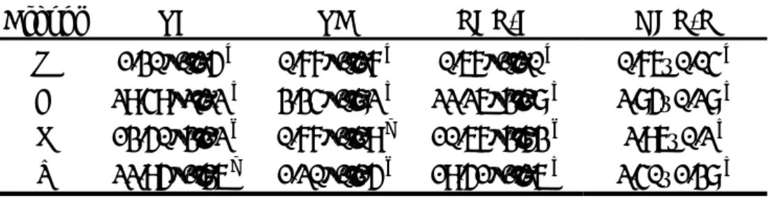
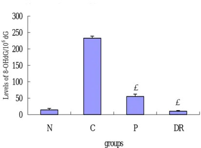
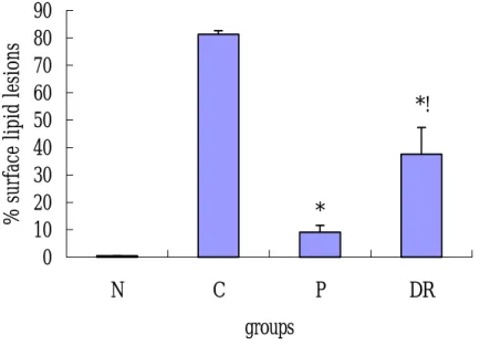
Hyperlipidemia may induce oxidative stress, which is important in the pathogenesis of atherosclerosis. We therefore investigated the antioxidative and antiatherogenic effects of Dioscorea Rhizome (DR) on hyperlipidemic rabbits. The control group was fed chow containing 0.5 % cholesterol and 10 % corn oil. The probucol group and the DR group were fed the same diet as the control group but with the addition of 100 mg/kg probucol and 200 mg/kg DR, respectively. The plasma levels of total cholesterol (TC) and triacylglycerol (TG), T50 of RBC hemolysis, lucigenin-CL (chemiluminescence) and luminol-CL increased in the control group compared to the normal group, and decreased in the probucol group and the DR group compared to the control group (Table 1 and 2). The activity of antioxidant enzymes superoxide dismutase (SOD) and catalase (CAT) were significantly higher in the probucol and DR group than in the control group (Figure 1). The levels of 8-hydroxy-2’-deoxyguanosine (8-OHdG) in liver DNA was lower in the probucol and DR group than in the control group (Figure 2). Eighty percent of the intimal surface of the thoracic aorta was covered with atherosclerotic lesions in the control group but only 40 % of the surface was covered in the DR group (Figure 3). These results suggest that supplementation with DR reduces oxidative stress and attenuate atherosclerosis in hyperlipidemic rabbits
Table 1. Serum GOT、GPT、TG 、TC、LDL-C、and HDL-C levels from rabbits fed with experimental diet for 12 weeks.
Groups Normal Control Probucol DR GOT (U/mL) GPT (U/mL) TG (mmol/L) 39.1±7.6a 50.0±3.2 a 0.4±0.1 a 112.2±5.6b 140.5±7.8 b 2.8±0.1 b 69.8±7.3d 65.3±2.1 d 1.7±0.4 c 94.4±7.3c 99.5±5.2 c 1.4±0.1c TC (mmol/L) 2.0±0.1 a 38.5±0.8 b 16.5±1.1 c 18.1±0.3 c LDL-C (mmol/L) HDL-C (mmol/L) 1.0±0.1a 0.7±0.1a 30.0±0.8b 3.6±0.3b 10.1±1.1c 3.0±0.1b 10.5±0.3c 3.8±0.4b
Note: All values are mean±S.D. Abbreviation: DR (Dioscorea Rhizome) GOT (glutamate oxalacetate transaminase); GPT (glutamate pyruvate transaminase); TG (triacylglycerol); TC (total cholesterol) ; LDL-C (low density lipoprotein cholesterol);HDL-C (high density lipoprotein cholesterol). a-d:Data with different superscripts in the same row are significantly different at P < 0.05.
Table 2. The time required achieving 50 % red blood cell hemolysis (T50) and whole blood free radical content of rabbits fed with experimental diet for 12 weeks.
Groups T50 Lucigenin-CL Luminal-CL (Min) (Counts/WBC) (Counts/WBC) Normal 148.5±20.4 a 4.4±0.2 a 15.8±0.5 a
Control 125.0±12.1 b 15.4±1.7 b 47.2±2.2 b Probucol 149.3±12.0 a 5.8±0.2 a 20.5±7.5a DR 151.0±10.1 a 6.5±0.6 a 28.4±5.0 a
Note: All values are mean±S.D. a-b Data with different superscripts in the same column are significantly different at P < 0.05.
(A) N C P D R 2 2 .9 3 3 .5 3 1 4 5 .6 5 4 2 .2 3 * ! * # 0 2 0 4 0 6 0 8 0 1 0 0 1 2 0 1 4 0 1 6 0 1 8 0 2 0 0 N C P D R g ro u p s U/mg pr otein (B) C P D R 0 .02 3 0 .01 0 2 0 .02 9 65 0.0 1 32 5 * ! * # 0 0 .0 0 5 0 .0 1 0 .0 1 5 0 .0 2 0 .0 2 5 0 .0 3 0 .0 3 5 N C P D R g rou p s k/s ec/mg pr otein
(C) N C P DR 0.694 2.219 1.526 0.525 *! * # 0 0.5 1 1.5 2 2.5 3 N C P DR groups um oles / m in/ m g pr otein
Fig. 1. Liver SOD (A)、catalase (B)、GSH-Px (C) activities of rabbits fed various diets for 12 weeks. All value is means±S.D. Abbreviation: N (normal); C (cholesterol); DR (Dioscorea Rhizome); P (probucol); SOD (superoxide dismutase); GSH-Px (glutathione peroxidase).
# p < 0.05 group control vs. group normal
* p < 0.05 group probucol & DR vs. group control ! p < 0.05 group DR vs. group probucol
N C P DR 14.3 232.8 55.42 10.3 *! * # 0 50 100 150 200 250 300 N C P DR groups Level s of 8-OHdG/ 10 6 dG
Fig. 2 Levels of 8-OHdG in liver from rabbits fed various diets for 12 weeks. All
value is means±S.D. Abbreviation: N (normal); C (control); DR (Dioscorea Rhizome);
P (probucol); CL (chemiluminescence). # p < 0.05 group control vs. group normal
* p < 0.05 group probucol & DR vs. group control ! p < 0.05 group DR vs. group probucol
N C P DR 0.5 81.3 9.1 37.6
*!
*
#
0
10
20
30
40
50
60
70
80
90
N
C
P
DR
groups
% surface l
ip
id
l
esi
ons
Fig. 3. Lipid lesions in the aorta from rabbits fed with experimental diet. Results
are expressed as mean±S.D. Abbreviation: N (normal); C (control); DR (Dioscorea
Rhizome); P (probucol).
# p < 0.05 group control vs. group normal
* p < 0.05 group probucol & DR vs. group control ! p < 0.05 group DR vs. group probucol & normal
4. Chieh-Hsi Wu, Ho-Chih Chen, Weng-Cheng Chang, and Jui-Sung Hung, Characterizing the Inhibitory Mechanisms of Magnolol on Rat Smooth Muscle Cell Proliferation (Submitted).
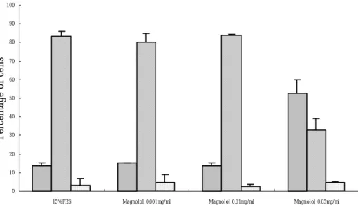
There are about 30 to 50% of people receiving percutaneous transluminal coronary angioplasty (PTCA) suffered from balloon injury-induced restenosis within six months of surgery. The pathological mechanism of restenosis has been attributed to abnormal proliferation of vascular smooth muscle cells. Pretreatment of antioxidants has been expected to reduce restenosis. Since magnolol was found to possess antioxidant effect about 1000 times higher than that of α-tocopherol, this active herbal component can be a potential therapeutic candidate to prevent balloon injury-induced abnormal smooth muscle cell proliferation. In this study, we demonstrated an approximate 61% reduction of smooth muscle cells progressing to S phase by 0.05mg/ml of magnolol via flow cytomertric analysis (Figure 1). BrdU incorporation assay also showed a significant reduction (73%) of DNA synthesis by 0.05mg/ml of magnolol. The protein level of proliferating cell nuclear antigen (PCNA) indicating cell proliferation was suppressed by 0.05mg/ml of magnolol for about 48% (Figure 2). This was in agreement with our current finding that the promoter activity of NF-kB closely related to cell proliferation was also attenuated by 0.05mg/ml of magnolol. The inhibition of cell growth may be mediated by the apoptotic pathway as RIP and caspase 3 protein expression levels were both increased by magnolol in a dose-dependent manner (Figure 3). The antioxidant effect of magnolol was further supported by our finding that MDA formation was significantly inhibited by 0.05mg/ml of magnolol (Figure 4). These studies suggested that magnolol may be a potential pharmacological reagent in preventing abnormal smooth muscle cell proliferation which in turn may be effective for balloon injury-induced restenosis.
Fig 1. Flow cytometric analysis of the inhibitory effects of magnolol on cell cycle
0 10 20 30 40 50 60 70 80 90 100
15%FBS Magnolol 0.001mg/ml Magnolol 0.01mg/ml Magnolol 0.05mg/ml
Percentage of cells
G0/G1
S G2/M
0.001mg/ml, 0.01mg/ml and 0.05mg/ml of magnolol. Each value represents the mean + SD. ** P < 0.01 as compared to 15% FBS, n=6.
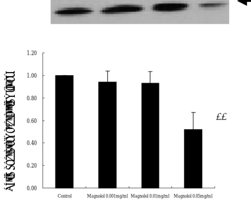
Fig 2. Effects of magnolol on PCNA protein expression level of rat smooth muscle cells. Panel A is the western blot results of PCNA affected by 0.001mg/ml, 0.01mg/ml, 0.05mg/ml of Magnolol. Panel B is the histogram demonstrating the protein expression ratio of PCNA normalized by 15% serum control group. ** P < 0.01, n=6.
0.00 0.20 0.40 0.60 0.80 1.00 1.20
Control Magnolol 0.001mg/ml Magnolol 0.01mg/ml Magnolol 0.05mg/ml
Ratio of protein expressi on level
**
PCNA (36 Kd)A
B
Å Caspase (32kd) 20000 40000 60000 80000 100000*
**
**
Fig 3. Stimulatory effects of Magnolol on caspase-3 protein expression level on rat smooth muscle cells. Panel A is the western blot analysis of caspase-3 affected by 0.001mg/ml, 0.01mg/ml, 0.05mg/ml of magnolol. Panel B is the quantitative analysis of the stimulatory effect of magnolol on caspase-3 expression level. 15% serum serves as the control group. * P < 0.05; ** P < 0.01, n=6.
Fig 4. Antioxidization effects of magnolol on MDA formation in rat serum. The
oxidization effects of 2mM CuSO4 on rat serum was significantly suppressed by
0.05mg/ml of magnolol. DMSO added to serum serves as the control. Each value represents the mean + SD. ** P < 0.01 as compared to the control, n=6.
0 0.05 0.1 0.15 0.2 0.25 0.3 0.35 0.4 Control Magnolol 0.001mg/ml Magnolol 0.01mg/ml Magnolol 0.05mg/ml
**
Optic al De nsity5. Chieh-Hsi Wu, Ho-Chih Chen, and Weng-Cheng Chang, Characterizing the Inhibitory Mechanisms of Magnolol on Rat Smooth Muscle Cell Proliferation (Submitted).
There are about 30 to 50% of people receiving percutaneous transluminal coronary angioplasty (PTCA) suffered from balloon injury-induced restenosis within six months of surgery. The pathological mechanism of restenosis has been attributed to abnormal proliferation of vascular smooth muscle cells. Pretreatment of antioxidants has been expected to reduce restenosis. Since magnolol was found to possess antioxidant effect about 1000 times higher than that of α-tocopherol, this active herbal component can be a potential therapeutic candidate to prevent balloon injury-induced abnormal smooth muscle cell proliferation. In this study, we demonstrated an approximate 61% reduction of smooth muscle cells progressing to S phase by 0.05mg/ml of magnolol via flowcytomertric analysis (Figure 2). BrdU incorporation assay also showed a significant reduction (73%) of DNA synthesis by 0.05mg/ml of magnolol (Figure 1). The protein level of proliferating cell nuclear antigen (PCNA) indicating cell proliferation was suppressed by 0.05mg/ml of magnolol for about 48% (Figure 3). This was in agreement with our current finding that the promoter activity of NF-kB closely related to cell proliferation was also attenuated by 0.05mg/ml of magnolol (Figure 6). The inhibition of cell growth may be mediated by the apoptotic pathway as RIP and caspase 3 protein expression levels were both increased by magnolol in a dose-dependent manner (Figure 4 and 5). The antioxidant effect of magnolol was further supported by our finding that MDA formation was significantly inhibited by 0.05mg/ml of magnolo (Figure 7). These studies suggested that magnolol may be a potential pharmacological reagent in preventing abnormal smooth muscle cell proliferation which in turn may be effective for balloon injury-induced restenosis.
ate 0.100 0.200 0.300 0.400 0.500 0.600 0.700 0.800 0.900 1.000
*
**
**
Fig 1. Effects of Magnolol on BrdU incorporation of rat smooth muscle cells.
Only 0.05mg/ml of magnolol significantly reduces the BrdU incorporation by 76.8 %. ** P < 0.01, n=6.
Fig 2. Flowcytometric analysis of the inhibitory effects of magnolol on cell cycle of rat smooth muscle cells. All the cells were cultured in 15% FBS with addition of 0.001mg/ml, 0.01mg/ml and 0.05mg/ml of magnolol. Each value represents the mean + SD. ** P < 0.01 as compared to 15% FBS, n=6. 0.20 0.40 0.60 0.80 1.00 1.20 expressi on level
**
PCNA (36 Kd)A
B
0 10 20 30 40 50 60 70 80 90 10015%FBS Magnolol 0.001mg/ml Magnolol 0.01mg/ml Magnolol 0.05mg/ml
Percentage of cells
G0/G1
S G2/M
Fig 3. Effects of magnolol on PCNA protein expression level of rat smooth muscle cells. Panel A is the western blot results of PCNA affected by 0.001mg/ml, 0.01mg/ml, 0.05mg/ml of Magnolol. Panel B is the histogram demonstrating the protein expression ratio of PCNA normalized by 15% serum control group. ** P < 0.01, n=6.
Fig 4. The RIP protein expression level affected by magnolol on rat smooth muscle cells. Panel A is the western blot analysis of RIP affected by 0.001mg/ml, 0.01mg/ml, 0.05mg/ml of magnolol. Panel B is the quantitative analysis of the stimulatory effect of magnolol on RIP expression level. 15% serum serves as the control group. * P < 0.05; ** P < 0.01, n=6. 0 10000 20000 30000 40000 50000 60000 Control Magnolol 0.001mg/ml Magnolol 0.01mg/ml Magnolol 0.05mg/ml A rbitra ry U nits Å RIP (74 kd)
*
**
*
Fig 5. Stimulatory effects of Magnolol on caspase-3 protein expression level on rat smooth muscle cells. Panel A is the western blot analysis of caspase-3 affected by 0.001mg/ml, 0.01mg/ml, 0.05mg/ml of magnolol. Panel B is the quantitative analysis of the stimulatory effect of magnolol on caspase-3 expression level. 15% serum serves as the control group. * P < 0.05; ** P < 0.01, n=6.
Å Caspase (32kd) 0 20000 40000 60000 80000 100000 Control Magnolol 0.001mg/ml Magnolol 0.01mg/ml Magnolol 0.05mg/ml
*
**
**
500000 1000000 1500000 2000000 2500000 3000000 T ota l R L U**
Fig 6. Inhibitory effects of magnolol on NF-κB activity in rat smooth muscle cells.
The activation of NF-κB activity was significantly suppressed by 0.05mg/ml of
magnolol. Cells receiving 15% FBS serves as the control. Each value represents the mean + SD. ** P < 0.01 as compared to the control, n=6.
Fig 7. Antioxidization effects of magnolol on MDA formation in rat serum. The
oxidization effects of 2mM CuSO4 on rat serum was significantly suppressed by
0.05mg/ml of magnolol. DMSO added to serum serves as the control. Each value represents the mean + SD. ** P < 0.01 as compared to the control, n=6.
0 0.05 0.1 0.15 0.2 0.25 0.3 0.35 0.4 Control Magnolol 0.001mg/ml Magnolol 0.01mg/ml Magnolol 0.05mg/ml
**
Optic al De nsity6. Wen-Tsong Hsieh, Jui-Sung, Hung, and Wen-Chen Chang, Effect of Some Chinese herb on vascular responsiveness in cholesterol-fed rabbit-isolated arteries (In preparation)
According to the World Health Organisation, cardiovascular disorders are one of the main causes of morbi/mortality in the western world. The effect of chinese herb
(0.3 mg kg−1 day−1), a non-sulphydryl angiotensin-converting enzyme (ACE) inhibitor,
on the vascular responsiveness in aorta isolated from hypercholesterolemic rabbits was examined. Three groups of rabbits (n=30) were used: Group 0 (control group); Group 1 (hypercholesterolemic group, 0.5% (wt/wt) cholesterol-enriched diet) and
Group 2 (hypercholesterolemic+chinese herb 0.3 mg kg−1 day−1). After 18 weeks of
treatment, the rabbits were killed and the thoracic aorta, proximal coronary and mesenteric (5th branch) arteries were isolated, cleaned off and mounted in an organ bath. Chinese herb had no significant effect on plasma cholesterol, high density lipoprotein (HDL) or low density lipoprotein (LDL). Despite the lack of effect of the drug on the above-mentioned parameters, treatment with chinese herb improved endothelium-dependent relaxation induced by acetylcholine in aortic and mesenteric rings from hypercholesterolemic rabbits treated with chinese herb (Figure 1). The
relaxation induced by 10−5 M acetylcholine were 65.0±4.0%; 24.0±9.4% (P<0.01,
n=10) and 51.3±7.0% (P<0.01, n=10) in aortic rings from Groups 0, 1 and 2,
respectively, and 50.0±12.0%; 10.1±10.0% (P<0.01, n=10); 61.0±9.7% (P<0.01,
n=10) in small mesenteric rings from Groups 0, 1 and 2, respectively. In addition, chinese herb treatment improved the increase in serotonin-induced contraction in proximal coronary arteries with respect to the hypercholesterolemic group (Figure 2). On the other hand, we did not find any differences among the group in endothelium-independent relaxation induced by sodium nitroprusside (Figure 3). These results provide evidence that chinese herb restores endothelium-dependent relaxation in hypercholesterolemic rabbit-isolated arteries. These data suggest that chinese herb might have beneficial action in the prevention of vascular alteration involved in atherosclerosis.
nitroprusside-LC tone X Data -2 -1 0 1 2 3 4 5 6 Y Da ta -0.5 0.0 0.5 1.0 1.5 2.0 2.5 control NO-LC baseline LC-nitroprusside Tone X Data -2 0 2 4 6 8 10 Y Da ta 0.0 0.5 1.0 1.5 2.0 2.5 control LC-NO baseline
Fig. 1. Endothelium-independent relaxation induced by sodium nitroprusside (10−8–10−4 M) in noradrenaline-precontracted aorta arteries. Each data shows the mean of each of the 10 experiments and vertical lines indicate the S.E.M. Group 1 (hypercholesterolemic group, 0.5% cholesterol enriched-diet) and Group 2
(hypercholesterolemic+ Ligusticum chuanxiong Hort 0.3 mg kg−1 day−1). ++P<0.01 vs.
Ryanodine-LC Tone X Data -2 -1 0 1 2 3 4 5 6 7 8 To ne (g ra m) 0 1 2 3 4 5 Control Ryanodine LC-Ryanodine Tone X Data -2 -1 0 1 2 3 4 5 6 7 8 Ton e( gr am) 0 1 2 3 4 Control Ryanodine (11K)
Fig. 2. Contractions induced by a single concentration of KCl (120 mM) (panel A) or by a cumulative concentration–response curve induced by ryanodine (10−9–10−4 M) (panel B) in proximal coronary arteries. Each data shows the mean of each of the 10 experiments and vertical lines indicate the S.E.M. Group 1 (hypercholesterolemic group, 0.5% cholesterol enriched-diet) and Group 2
(hypercholesterolemic+ Ligusticum chuanxiong Hort 0.3 mg kg−1 day−1). *P<0.05 vs.
Thas - LC X Data -2 0 2 4 6 8 Y D at a 0 1 2 3 4 5 6 Plot 1 Plot 2 2D Graph 2 X Data -2 0 2 4 6 8 Y Dat a 0.0 0.5 1.0 1.5 2.0 2.5 3.0 3.5 Plot 1 Plot 2
Fig. 3. Contractions induced by 80 mM KCl (panel A) or 10−5
M thasgagine (panel B) in small mesenteric arteries. Each data shows the mean of each of the 10 experiments and vertical lines indicate the S.E.M. Group 0 (control group), Group 1 (hypercholesterolemic group, 0.5% cholesterol enriched-diet) and Group 2 (hypercholesterolemic+chinese herb
0.3 mg kg−1
day−1
). +
7. J-S Chen, P-U Chou, W-C Chang and J-S Hung, Attenuation of baroreflex sensitivity in rabbits with hyperchlesterolemia: role of nitric oxide (In preparation).
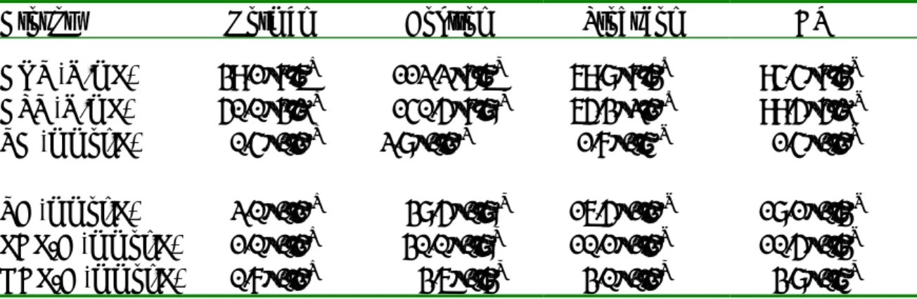
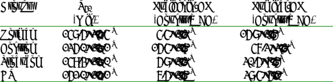
Abnormally high serum cholesterol levels are thought to be an important pathogenic factor in atherogenesis. Baroreflex sensitivity (BRS) was shown to be depressed in hypercholesterolemia. Hypercholesterolemia was also shown to enhance nitric oxide (NO) production in the circulatory system, and it was demonstrated that NO could lead to the attenuation of BRS. Thus, the BRS depression in hypercholesterolemia might be related to NO. The Chinese herb, Magnolia officinalis, was shown to improve hypercholesterolemia, but it is unclear whether it could also improve BRS. In hemorrhage, BRS is depressed and it could be attributed to NO. But it is not clear whether in hypercholesterolemia the BRS depression in hemorrhage is related to the NO or Magnolia officinalis. The present study was to determine whether the BRS depression in hypercholesterolemia, especially in hemorrhage, is mediated by NO. Male New Zealand white rabbits were divided into 3 groups: 1. normal diet (Control group), 2. 0.5% w/w cholesterol (Cholesterol group) diet, or 3. same as Cholesterol group but plus 1% w/w magnolia officiralis crude methanol extract (Magnolia group). The animals were continuously treated under the designated diet for 4 and 8weeks. BRS in the control of heart rate was determined by linear regression method. The results demonstrated that plasma cholesterol was significantly elevated after 8 weeks of treatments in the Cholesterol and Magnolia groups. BRS was not different among these three groups at 4 weeks. However, at 8 weeks, the BRS of the Cholesterol and Magnolia groups was significantly lower than that of the Control, and the BRS of the Magnolia group was greater than that of the Cholesterol group. The arterial blood pressure, heart rate and BRS were not different among these 3 groups at 4 weeks, but arterial blood pressure and BRS of the Cholesterol and Magnolia groups were enhanced at 8 weeks when compared with the Control. It was found that after L-NAME (Nω-nitro-Larginine methyl ester, 20mg/kg, iv.)treatment, only in the Cholesterol group the BRS was significantly enhanced. After hemorrhage, the BRS was depressed in all 3 groups. L-NAME improved the BRS in all 3 groups. The BRS of the Magnolia group after hemorrhage was greater than that of the Cholesterol. The data suggested that BRS was depressed in hypercholesterolemia, and it can be related to NO. Hemorrhage also caused decrease in BRS, and it may also be attributed to the NO. Magnolia apparently improved the BRS of the hypercholesterolemia group, but it is not likely to be medicated by NO.
Table 1. Plasma levels of cholesterol (CHO) and triglycerides (TG) of rabbits fed with various diets at the 8th week.
Group Control(7) Cholesterol(3) Magnolia(9)
CHO (mg/dL) 70±5 781±60* 514±139* TG (mg/dL) 138±5 164±3 191±27
All values are expressed as means ± SE (standard error of means). The
sample sizes are indicated in parentheses. The Control group were fed with normal rabbit chow. The Cholesterol group were fed with normal rabbit chow plus 10﹪(w/w) corn oil and 0.5﹪(w/w) cholesterol. The Magnolia group were fed with the same diet as the Cholesterol group,
but plus l﹪(w/w) Magnolia officinalis methanol extract (see text for
detail).* : P<0.05, compared with the Control group.
Table 2. Baseline systemic blood pressure (BP), heart rate (HR) and BRS of rabbits fed with various diets at the 4th and 8th week.
Group Control Cholesterol Magnolia
Week 4(14) 8(21) 4(9) 8(18) 4(8) 8(12)
BP (mmHg) 95±1 96±2 94±2 109±2* 101±4 109±2*
HR (bpm) 248±8 255±5 233±10 257±5 246±5 274±11
BRS(bpm/mmHg) -2.22±0.29 -2.26±0.21 -1.71±0.35 -1.28±0.05* -1.94±0.22 -1.65±0.11*#
All values are expressed as means ± SE (standard error of means). The
sample sizes are indicated in parentheses. The Control group were fed with normal rabbit chow. The Cholesterol group were fed with normal rabbit chow plus 10﹪(w/w) corn oil and 0.5﹪(w/w) cholesterol. The Magnolia group were fed with the same diet as the Cholesterol group,
but plus l﹪(w/w) Magnolia officinalis methanol extract (see text for
detail).* : P<0.05, compared with the Control group of the same week. #
P<0.05, compared with the Cholesterol group of the same week.
BP(mmHg) HR(bpm)
Group Control(9) Cholesterol(7) Magnolia(4) Control Cholesterol Magnolia
Before 88±4 98±3* 108±4* 247±9 252±9 277±32
After 99±5# 114±4**# 121±5**# 222±15 201±12# 197±30 %Change 12±3 16±2 12±2 -10±6 21±3 -28±6
All values are expressed as means ± SE (standard error of means). The sample
sizes are indicated in parentheses. The Control group were fed with normal rabbit chow. The Cholesterol group were fed with normal rabbit chow plus 10﹪ (w/w) corn oil and 0.5﹪(w/w) cholesterol. The Magnolia group were fed with
the same diet as the Cholesterol group, but plus l﹪(w/w) Magnolia officinalis
methanol extract (see text for detail). * : P<0.05, compared with the Control
group before L-NAME was added . ** : P<0.05, compared with the Control
group after L-NAME was added . # : P<0.05, compared with the same group before L-NAME was added.
Table 4. Systemic blood pressure (BP) and heart rate (HR) profile during induction of hemorrhage in rabbits fed with various diets at the 8th week.
BP (mmH) HR (bpm)
Group Control(8) Cholesterol(9) Magnolia(6) Control Cholesterol Magnolia
Baseline 94±4 112±3 108±3 288±13 271±12 260±20
Peak HR 71±3 78±3 76±6 310±17 296±12 335±20
Lowest HR 58±3 56±3 58±3 274±15 247±10 236±15
Lower plateau 76±8 74±4 66±3 296±10 278±13 293±23
All values are expressed as means ± SE (standard error of means). The sample
sizes are indicated in parentheses. The diet treatment of each group was the same as described in Table 1. Baseline: baseline BP and HR before induction of hemorrhage. Peak HR: phase of hemorrhage when HR reached the highest level. Lowest HR: phase of hemorrhage after HR was decreased from peak HR to a lowest point before developed to a steady plateau. Lower plateau: phase of hemorrhage when criteria of hemorrhage were reached, and both BP and HR reached a plateau (also see text and Fig. 4 for more detail).
B
R
S
(
b
pm
/mmH
g)
-3 -2 -1 4 week 8 weekCntrol Cholesterol Magnolia
*
*
#
Fig 1. BRS of rabbits fed with different diets at the 4th and 8th week.
All values are expressed as means ± SE (standard error of means). The sample
sizes are indicated in parentheses. The Control group were fed with normal rabbit chow. The Cholesterol group were fed with normal rabbit chow plus 10﹪ (w/w) corn oil and 0.5﹪(w/w) cholesterol. The Magnolia group were fed with
the same diet as the Cholesterol group, but plus l﹪(w/w) Magnolia officinalis
methanol extract (see text for detail).* : P<0.05, compared with the Control
group of the same week. #: P<0.05, compared with the Cholesterol group of
Fig. 2. Effects of NO synthase blockade with L-NAME on the systemic blood pressure and heart rate in rabbits fed with different diets.
The Control group were fed with normal rabbit chow. The Cholesterol group were fed with normal rabbit chow plus 10﹪(w/w) corn oil and 0.5﹪(w/w) cholesterol. The Magnolia group were fed with the same diet as the Cholesterol group, but plus l﹪(w/w) Magnolia officinalis
B
R
S
(b
pm/mmH
g)
-3 -2 -1 baseline +L-NAMEControl
Cholesterol
Magnolia
*
*
# #
Fig 3. Effects of NO synthase blockade with L-NAME on BRS in rabbits fed with different diets.
The Control group were fed with normal rabbit chow. The Cholesterol group were fed with normal rabbit chow plus 10﹪(w/w) corn oil and 0.5﹪(w/w) cholesterol. The Magnolia group were fed with the same diet as the
Cholesterol group, but plus l﹪(w/w) Magnolia officinalis methanol extract
(see text for detail).* : P<0.05, compared with the baseline values (before
L-NAME) of the Control group. # : P<0.05, compared with the baseline
Fig. 4. Changes of systemic blood pressure and heart rate during the induction of hemorrhage.
After the Baseline was recorded, procedure of hemorrhage was initiated (arrow). Note that the hemorrhage started with a non hypotensive phase with a gradual increase in heart rate. Then the hemorrhage developed into a hypotensive phase with a further increase of heart rate (Peak HR). However, as hemorrhage progressed into a more severe phase, the heart rate was paradoxically decreased. The hemorrhage was continued until the mean arterial blood pressure dropped below 70 mmHg (Lowest HR), and then the procedure of hemorrhage was terminated. The blood pressure and
heart rate would then level off (Lower plateau). In the tracings, there were three
places showing abrupt decreases in HR. Those were the artifacts caused by flushings of arterial catheter, which were intended to improve the ABP signal.
B
R
S
(b
pm/
mmH
g)
-3 -2 -1 baseline Hemo H+L-NAMEControl Cholesterol Magnolia
*
*
#**
##***
*
**
###Fig.5. Effects of NO synthase blockade with L-NAME on BRS during hemorrhage in rabbits fed with different diets.
P<0.05 was considered statistically significant. Statistical notations are
defined as follows. *: compared with the baseline of the Control group. #: compared with the baseline of the Cholesterol group.**: compared with the hemorrhagic state (Hemo) of the Control group. ##: compared with the
hemorrhagic state (Hemo) of the Cholesterol group.***: compared with the
hemorrhagic state with NO synthase blockade (H+L-NAME) of the Control group. ###: compared with the hemorrhagic state with NO synthase blockade (H+L-NAME) of the Cholesterol group. The Control group were fed with normal rabbit chow. The Cholesterol group were fed with normal rabbit chow plus 10﹪(w/w) corn oil and 0.5﹪(w/w) cholesterol. The Magnolia group were fed with the same diet as the Cholesterol group, but plus l﹪(w/w) Magnolia officinalis methanol extract (see text for detail).
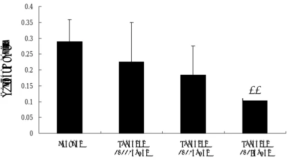
8.SY Chiang, MY Lin, JJ Chu, YM Yu, WC Chang and JS Hung, Inhibitory Effects of Paeonia Suffruticosa and Carthamus Tinctorius on oxLDL-induced Cytotoxicity and Apoptosis in Human Umbilical Vein Endothelial Cells (In preparation).Atherosclerosis, a chronic inflammatory disease, is the leading cause of heart disease and stroke-related deaths. Oxidized low density lipoprotein (oxLDL) is believed to play a key role in causing endothelial cell injury, thereby contributing to atherosclerotic lesion development. Atherosclerosis is characterized by blood stagnation given according to the principles of TCM. Paeonia suffruticosa and Carthamus tinctorius, two commonly used Chinese herbs in traditional Chinese medicine, have the effects of reducing inflammation, promoting blood circulation, and dissipating blood stasis. In this study, we examined the effects of Paeonia suffruticosa and Carthamus tinctorius on oxidized LDL (oxLDL)-induced cytotoxicity and apoptosis in human umbilical vein endothelial cells (HUVEC). Exposures of HUVEC to 0.11~2.0 mg/ml of Paeonia suffruticosa for 24 hours resulted in a dose-dependent increase in cytotoxicity, with survival rates of 81% and 16% at 1.0 and 2.0 mg/ml, respectively. Whereas, Carthamus tinctorius was not cytotoxic to HUVEC at doses lower than 2mg/ml (Figure 1). Paeonia suffruticosa but not Carthamus tinctorius induced apoptosis in HUVEC, as measured by Giemsa staining and flow cytometric analysis (Figure 2,3). Further studies showed that oxLDL enhanced cytotoxicity and apoptosis in a dose-dependent manner in HUVEC (Figure 4). Coadministration with Paeonia suffruticosa (0.11 mg/ml) or Carthamus tinctorius (0.33 mg/ml) at the nontoxic dose levels was able to almost completely block oxLDL-induced cytotoxicity and apoptosis (Figure 5,6). These data showed that Paeonia suffruticosa
and Carthamus tinctorius could significantly protect against oxLDL-induced cell
damage in HUCEC, suggesting their potential use in the treatment of atherosclerosis.
Fig 1. Cytotoxicity induced by Paeonia suffruticosa and Carthamus tinctorius in
HUVECs. After incubation of HUVECs with various concentrations of Paeonia suffruticosa and Carthamus tinctorius for 24 h, cell viability was determined by a MTS assay. Data are the means ± SD of three independent experiments. *p<0.05
0 25 50 75 100 125 0.11 0.33 1.00 2.00 Concentration of dose (mg/ml) ce ll via bility ( % of c ontr ol c ell) Paeonia suffruticosa Carthamus tinctorius
Fig 2. Induction of apoptosis by Paeonia suffruticosa in HUVECs. After 24h incubation of Paeonia suffruticosa to HUVECs, subG1 cells with hypodiploid DNA corresponding to apoptotic cells were quantified by flow cytometric analysis. Data are the means ± SD of three dependent analyses. *p<0.05
FACS analysis of Carthamus tinctorius L .
0 10 20 30
0 0.33 1 2
Concentration of Carthamus tinctorius L. (mg/ml) % s ubG 1 of to ta l c el l Carthamus tinctorius L .
Fig 3. Induction of apoptosis by Carthamus tinctorius in HUVECs. After24h incubation of Carthamus tinctorius to HUVECs, apoptotic cells werequantified by flow cytometric analysis. The subG1 cells with hypodiploid DNA corresponding to apoptotic cells were quantified. Data are the means ± SD of three dependent analyses. 0 10 20 30 40 50 0 0.11 0.33 1
Concentration of Paeonia suffruticosa (mg/ml)
% s ubG 1of t ot al c el l
MTS assay 0 20 40 60 80 100 100 200 300 400 oxLDL (ug/mll) ce ll v ia bil ity ( % o f to ta l c ell )
Fig 4. Cytotoxicity induced by oxLDL in HUVECs. HUVECs were synchonizined in0.1%FBS medium for 24h before incubation with various concentrations ofoxLDL for 24 h. Cell viability was determined by a MTS assay. Data are themeans ± SD of three independent analyses. *p<0.05
IA IB
IC ID
IIA IIB
IIC IID
Fig 5 The protective effects of Paeonia suffruticosa and Carthamus tinctorius on oxLDL-induced cytotoxicity observed under lightmicroscope in HUVECs. I: Phase-contract microscope photographs; II: Giemsa staining microscope photographs (200X). (A) medium control. (B) oxLDL300μg/ml. (C) oxLDL 300μg/ml + Carthamus tinctorius L 0.33mg/ml. (D) oxLDL300μg/ml + Paeonia suffruticosa Andr. 0.11mg/ml.
Fig 6. The protective effects of Paeonia suffruticosa and Carthamus tinctorius on oxLDL-induced apoptosis in HUVECs. After 24h incubation of oxLDL 300μ g/ml with Paeonia suffruticosa 0.11mg/ml or Carthamus tinctorius 0.33mg/ml, subG1 cells with hypodiploid DNA corresponding to apoptotic cells were quantified by flow cytometric analysis. Data are the means ± SD of three dependent analyses. *p<0.05 0 5 10 15 20 25
control oxLDL300μg/ml oxLDL 300μ g/ml+Paeonia suffruticosa 0.11mg/ml oxLDL 300μg/ml +Carthamus tinctorius 0.33mg/ml % subG 1 of tota l c el l
9. Chieh-His Wu, Ho-Chih Chen, Weng-Chen Cheng and Jui-Sung Hung, The antiatherogenic effects of Honodiol, Magnolol and Alismatis Rhizoma. (In preparation)
Chinese medicine has been widely used in protecting cardiovascular function, however, most of the molecular mechanisms remained to be elucidated. It was believed that the mechanisms of these drugs could be related to the antioxidization and lipid lowering effects. Two Chinese medicines, Magnolia Officinalis and Alismatis Rhizoma, have been regarded as an antioxidant and a lipid lowering agent, respectively. We therefore aimed in this study to elucidate the roles of these drugs in protecting cardiovascular functions. Atherosclerosis has been known to be a key injury to elicit cardiovascular diseases. Formation of atherosclerosis is mainly resulted from accumulation of foam cells which were formed by macrophage stuffed with excessive oxidized LDL. Since SOD is a very potent enzyme to scavenge superoxide anions which may aggravate LDL oxidization, it would be rational to look into the relationship between Magnolia Officinalis and SOD gene expression. LDL receptors are believed to clean excessive plasma cholesterol. We therefore assumed that the gene expression of LDL receptor could be upregulated by Alismatis Rhizoma. Honokiol and Magnolol, two pure compounds of Magnolia Officinalis, were tested for their effects on SOD gene expression. We found that the RNA levels of SOD was increased by Honokiol and Magnolol in a dose-dependent manner (figure 1). The
SOD RNA level was increased by100 M of Honokiol for 7 folds as compared to 10
nM while 10 folds by 100 M of Magnolol in comparison to 10 nM. Regarding the
protein levels of SOD, three tested concentrations of Honokiol and Magnolol were
shown to potentiate the protein expression as compared to the controls. While 10 M
of Honokiol was more potent than 0.1 M of Honokiol, no differential effects on
SOD protein expression by 10 M, 1 M or 0.1 M of Magnolol were noticed,
suggesting the different effects of these two compounds on translational efficiency of SOD (Figure 2). The lipid lowering effects of Alismatis Rhizoma was shown by increasing LDL receptor RNA expression levels in a dose-dependent manner (Figure 3). We finally applied these compounds to feed hypercholesterlemic rabbits and evaluated the antiatherosclerotic effects. With Sudan IV staining, we found that these compounds can significantly reduce atherosclerotic lesions on rabbit aorta (Figure 4). Based on these preliminary results, we believe that these Chinese medicines can be effective in preventing atherosclerosis formation. Further evaluation for the clinical application of these medicines on human subjects may benefit those who are suffered from cardiovascular diseases.
圖一: RT-PCR 的結果顯示五種不同濃度的 Honokiol 和 Magnolol 對於 SOD 之 RNA 表達能力的影響。藉由 Cu,Zn-SOD 的引子作用,35 個循環的 PCR 複製出一段 252bp 的 SOD cDNA,亮度越高表示越多的 SOD RNA 被藥物給誘發出來。
圖二,Magnolol 和 Honokiol 對於肝臟細胞超氧歧化酵素之蛋白質表達的影響。 在不同濃度的處理下, 10µM 的 Honokiol 增加超氧歧化酵素之蛋白質表達最為 明顯,而 Magnolol 對於肝臟細胞內的超氧歧化酵素蛋白質表達則沒有呈現一個 濃度依附性的關係。圖中 15%的 serum 和 0.5%的 serum 分別代表正向和負向之 對照組。 圖三: RT-PCR 的結果顯示四種不同濃度的澤瀉對於 LDL receptor 之 RNA 表達能 力的影響。藉由 LDL receptor 的引子作用,35 個循環的 PCR 複製出一段 298bp Magnolol (uM) Honokiol (uM) SOD (21kd) 10 1 0.1 10 1 0.1 15% 0.5% Serum 252bp 10nM 100 nM l 1uM 10 uM 10nM 100nMl 1uM 10uM 100 uM Honokiol 252bp
298bp
1ug/ml 10ug/ml 澤瀉 100ug/ml 1mg/ml
15% Serum
283bp
GAPDH
組,代表各組所加入反應的 RNA 是等量。 圖四,厚朴及澤瀉的膽固醇混合飼料對於粥狀動脈的改善作用。A: 沒有餵食高 膽固醇的兔子主動脈; B: 餵食高膽固醇的兔子主動脈,深黑色代表產生粥狀動 脈硬塊斑的部位(箭頭所指處); C: 餵食 0.1mg/kg 厚朴及澤瀉的膽固醇混合飼料 的兔子主動脈; D: 餵食 1mg/kg 的膽固醇混合飼料的主動脈; E: 餵食 10mg/kg 混合飼料的主動脈; F: 餵食 100mg/kg 混合飼料的主動脈。 表一,餵食 10 周膽固醇的兔子,在不同藥物處理之後其血中總膽固醇含量的比 較,*表示和 1%膽固醇組的比較, p value <0.05; **表示 p value <0.01,n=10。 血中總膽固醇含量 (mg/dl) 正常飼料組 86 + 18 1%膽固醇組 1750 + 365 0.10mg/kg 厚朴及澤瀉 1689 + 253 1mg/kg 厚朴及澤瀉 1320 + 316* 10mg/kg 厚朴及澤瀉 832 + 192** 100mg/kg 厚朴及澤瀉 702 + 201** C. D. E. F. A. B.
11. Lo CJ. Lin JG. Kuo JS. Chiang SY. Chen SC. Liao ET. Hsieh CL. Effect of salvia miltiorrhiza bunge on cerebral infarct in ischemia-reperfusion injured rats. American Journal of Chinese Medicine. 31(2):191-200, 2003.
According to the theory of traditional Chinese medicine, cerebral infarction results from blood stasis, and the method of quickening the blood and dispelling stasis is used to treat cerebral infarct. salvia miltorrhiza bunge (SM) is a Chinese herb, which is considered to have an action of quickening the blood and dispelling stasis, and is frequently used to treat related disorders of blood stasis such as cerebrovascular accident and ischemic heart disease. The aim of the present study was to investigate the effect of SM on cerebral infarct in ischemia-reperfusion injured rats. A total of 30 Sprague-Dawley (SD) rats were studied. A model of focal cerebral infarct was developed by occluding both common carotid arteries and the right middle cerebral artery for 90 minutes. After 24 hours reperfusion, the rats were killed and the brain tissue was stained with 2, 3, 5-triphenyl-tetrazolium chloride (TTC). The areas of cerebral infarct were calculated, and lumino-chemiluminesence (CL) counts and lucigenin-CL counts of peripheral blood taken at this time were measured. The changes in the area of cerebral infarct were used as an index to evaluate the effect of SM on cerebral infarct. The results indicated that pretreatment with intraperitoneal injection of 30 mg/kg and 15 mg/kg SM reduced the area of cerebral infarct and also reduced the luminol-CL counts of peripheral blood in ischemia-reperfusion injured rats. This study has demonstrated that SM can reduce the area of cerebral infarct in ischemia-reperfusion injured rats, suggesting it may be useful in the treatment of cerebral infarct in humans. The therapeutic effect of SM may be partly due to its free radical scavenging activities.