Diclofenac has been widely used, systemically and locally, as an antiinflammatory agent. It has been reported that orally administered diclofenac undergoes hepatic first-pass metabo-lism and considerable gastrointestinal disturbances.1,2)
Trans-dermal delivery is suitable for diclofenac to overcome these two major shortcomings of oral therapy. Iontophoresis is de-fined as the migration of ions when an external electric field is passed through a vehicle containing charged compounds. Based on the literature, the permeation of ionic drugs such as diclofenac can be facilitated by the application of ion-tophoresis.3)
Several variables may influence the transdermal ion-tophoretic permeation of drug molecules, including physico-chemical properties of the drug, the vehicle composition, the electrical factors and skin barrier properties.4,5) The aim of the present study was to investigate the influence of electrical and chemical factors of iontophoresis on in vitro transdermal permeation of diclofenac. Three diclofenac salts with various physicochemical and pharmacokinetic properties including diclofenac sodium (DFS), diclofenac potassium (DFP), and diclofenac diethylammonium (DFD) were utilized as model drugs to compare the differences of these salts in ion-tophoretic behaviors. The electrical and chemical variables examined in this study such as current density, drug concen-tration, ionic strength and iontophoretic application mode can control and optimize the delivery rate of diclofenac salts. Moreover, the magnitude of drug iontophoretic permeability can be influenced by the type and quantity of ions present in vehicle.6)The experiment and mechanism of this effect was
also demonstrated in the present study. MATERIALS AND METHODS
Materials Diclofenac sodium (DFS), diclofenac potas-sium (DFP), and diclofenac diethylammonium (DFD) were gifts kindly provided by Novartis Pharmaceutical Co., Switzerland. Sodium chloride (NaCl), potassium chloride (KCl), and diethylammonium chloride (C4H12ClN) were
sup-plied by Merck Co., Germany. All other chemicals and
sol-vents were of analytical grade.
In Vitro Permeation Experiments The in vitro perme-ation study was performed by using side-by-side glass diffu-sion cells. The skin of female nude mouse (11—12 weeks old) was used as the model membrane in this study. A 8 ml citrate–phosphate buffer (pH 7.4; 0.06M) was used as the medium for receptor compartment. The drug concentration in the donor compartment was 12.5 mM. The drug was more than 99.9% ionized in the donor compartment of pH 7.4 cit-rate–phosphate buffer because of its pKavalue of 4.16.
7)
The available diffusion area between cells was 0.785 cm2. The stirring rate and temperature were kept at 600 rpm and 37 °C. At appropriate intervals, 200ml aliquots of the receptor medium were withdrawn and immediately replaced by an equal volume of fresh buffer.
Application of Iontophoresis A pair of Ag/AgCl wires with an effective working length of 15 mm was immersed in the buffer solution as electrodes, with the cathode in the donor compartment and anode in the receptor compartment. The cathode and anode were each positioned 3 cm from the side of skin. The electrodes were connected to a constant cur-rent power supplier (Model 7651, Yokogawa Co., Japan).
Chromatographic Analysis The amount of DFS, DFP,
and DFD was analyzed by the HPLC method modified from Huang et al.8)The HPLC system consisting of a Hitachi
L-7100 pump, a Hitachi L-7200 sample processor and a Hi-tachi L-4000H UV detector. A 12.5 cm long, 4.0 mm inner diameter stainless steel column with Lichrospher®C-18
col-umn (Merck Co., Germany) was used. An automated integra-tor system (Hitachi D-7500) was used to determine the area under the curve. The mobile phase for diclofenac salts con-sisted of a methanol/0.05% acetic acid solution (65 : 35, v/v) mixture. The flow rate was 1.0 ml/min with UV absorbency monitoring at 260 nm.
Data Analysis The total amount of drug permeating
through the unit diffusion surface and into the receptor was calculated and plotted as a function of time. The flux was calculated by the slope of the linear portion of cumulative amount-time plots for zero-order model and expressed as the
∗To whom correspondence should be addressed. e-mail: [email protected] © 2001 Pharmaceutical Society of Japan
Influence of Electrical and Chemical Factors on Transdermal
Iontophoretic Delivery of Three Diclofenac Salts
Jia-You FANG,a,bRen-Jiunn WANG,aYaw-Bin HUANG,aPao-Chu WU,aand Yi-Hung TSAI*,a
School of Pharmacy, Kaohsiung Medical University,a 100 Shih-Chuan 1st Road, Kaohsiung, Taiwan and Graduate Institute of Pharmaceutical Sciences, Taipei Medical University,b250 Wu-Hsing Street, Taipei, Taiwan.
Received August 10, 2000; accepted December 4, 2000
The aim of this present study was to investigate the in vitro transdermal iontophoretic delivery of three di-clofenac salts—didi-clofenac sodium (DFS), didi-clofenac potassium (DFP), and didi-clofenac diethylammonium (DFD). A series of physicochemical and electrical variables which might affect iontophoretic permeation of diclofenac salts was studied. Application of 0.3 mA/cm2current density significantly increased the transdermal flux of di-clofenac salts as compared to passive transport. The iontophoretic enhancement increased in the order of DFS.DFP.DFD. The permeability coefficient of diclofenac salts all decreased with increasing donor concentra-tion during iontophoresis. The addiconcentra-tion of buffer ions and salt ions such as NaCl, KCl, and C4H12ClN reduced the permeation of diclofenac salts due to competition. However, this effect was lesser for DFD than for DFS and DFP. Comparing the various application modes of iontophoresis, the discontinuous on/off mode showed lower but more constant flux than the continuous mode.
mass of drug passing across 1 cm2of skin over time. The
per-meability coefficient was calculated by dividing the flux by initial drug donor concentration. The area under the curve (AUC) of flux-time plots was calculated by the trapezoidal method. The ratio of the flux of drugs by iontophoresis to the value by passive diffusion (control group) was determined as the enhancement ratio (ER). The retardation ratio (RR) was determined as (drug flux with competitive ion2drug flux without competitive ion)/drug flux without competitive ion.
Statistical Analysis The statistical analysis of the differ-ence between different treatments was detected by using the unpaired Student’s t-test. The 0.05 level of probability was taken as the level of significance. The ANOVA test was also utilized in this present study.
RESULTS AND DISCUSSION
Effect of Current Density The amount of DFS, DFP
and DFD appearing in the receptor compartment of the diffu-sion cells was plotted as a function of time. The three di-clofenac salts permeation with or without iontophoresis in pH 7.4 buffer was performed as shown in Fig. 1. The drug was almost completely ionized (99.9%) in the donor com-partment. There was no significant difference (ANOVA test, p.0.05) among the flux of DFS, DFP and DFD, indicating the counterions did not affect passive transport of diclofenac salts. Application of 0.1 mA/cm2current density during ion-tophoresis was not enough to enhance the permeation of all compounds relative to their passive transport. The number of permeant molecules which passed through skin increased with the increase of strength of the current density according to Faraday’s law.9) Hence a higher current density of 0.3
mA/cm2was conducted to improve the iontophoretic
perme-ation of diclofenac salts. As shown in Fig. 1, the permeperme-ation of diclofenac salts was greatly enhanced by application of 0.3 mA/cm2 current. Unlike the passive permeation result,
the iontophoretic flux and enhancement ratio (ER, flux with iontophoresis/flux without iontophoresis) increased in the order of DFS^DFP.DFD. Yoshida and Roberts have sug-gested that the iontophoretic behavior of anionic solutes in-cluding diclofenac can be best described by the free volume model.10) According to this model, the ion sphere mobility
has been assumed to be proportional to the fractional volume of the space that is accessible to the ion sphere. Therefore, the molar volume as well as solute radius have been shown to be inversely related to iontophoretic mobility.5,11)In addition
to these ionselective properties, the skin also shows size-se-lective effects in iontophoretic transport.12) Application of
iontophoresis may increase the porosity and create pores with effective radii in the lipid matrix.13) Although the
di-clofenac anion was dissociated in the donor compartment during iontophoresis, the radius and mobility of diclofenac anion can be affected by its counterion.7) Previous studies
have shown that the radius of diclofenac anion increased with the increase of the molecular weight and radius of its counte-rion.7,14)Therefore the radius of diclofenac showed a trend of
DFD.DFP.DFS, which has been shown to be inversely re-lated to the iontophoretic enhancement effect of diclofenac salts.
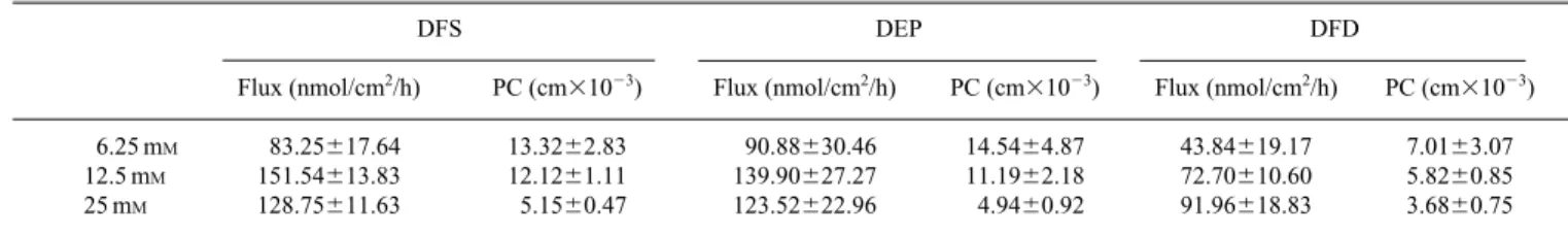
Effect of Donor Drug Concentration Drug
concentra-tion is an important parameter since it provides an easy way
to control the rate of drug delivery in transdermal ion-tophoresis.15)Donor solutions of 6.25, 12.5, and 25 m
Mdrug concentrations were chosen for this study. The flux and meability coefficient values are reported in Table 1. The per-meability coefficient of DFS, DFP and DFD all decreased with increasing donor concentration. This may be due to the lower activity of the drug in more concentrated solution.16)
Moreover, the skin is not an inert tissue and presents some resistance to the movement of ions. Many small ions present in skin or buffer transport part of current density. This could explain the lack of a strictly proportional relationship be-tween the donor concentration and flux.17—19)
Effect of Donor Buffer Ionic Strength As discussed
above, the ions present in buffer can influence the transport of drug during iontophoresis. It is expected that the variation of ionic strength in the donor solution should be important for iontophoretic permeation of diclofenac salts. The ionic
Fig. 1. Cumulative Amount–Time Profiles of Diclofenac Salts across Nude Mouse Skin with or without Iontophoresis
(A) Diclofenac sodium, (B) diclofenac potassium, (C) diclofenac diethylammonium. Each value represents the mean6S.D. (n53).
strength of donor solution was adjusted using citrate–phos-phate buffer with the ionic strength of 0.06 and 0.12M. The result in Fig. 2 shows that the flux of diclofenac salts de-creases as the ionic strength of buffer inde-creases. The low per-meation at high ionic strength could be due to the competi-tion of drug ions and buffer ions for the applied current. Most current density would be carried by buffer ions with relatively high mobilities, the actural fraction of the applied current carried by drug ions would be reduced as the concen-tration of buffer ions increases.20,21)
Previous studies have suggested the possibility that di-clofenac salts form complexes or are weakly dissociated ions in aqueous solution, leading to species such as ion-pairs.22) Diclofenac salts may also permeate across skin as of ion-pairs.7)It is also possible that diclofenac salts form ion-pairs
with buffer species in the solution. The diclofenac flux may decrease as the concentration of cations in buffer increases in the presence of iontophoresis, since ion-pair formation be-tween permeant and these buffer species would lower the percentage of permeant in the free-ionized form.20)
After calculation of the retardation ratio (RR, flux in high ionic strength buffer2flux in low ionic strength buffer/flux in low ionic strength buffer), the effect of ionic strength was weaker in DFD than in DFS and DFP. There are three routes for a drug to permeate the skin: (1) intracellular (transcellu-lar); (2) intercellular (paracellu(transcellu-lar); and (3) transappendageal (shunt). Once into the stratum corneum, drug flux branches to these multiple pathways. Our previous study suggested that transappendageal routes may be a more important path-way for DFD than for DFS and DFP under iontophoresis.6)
Since appendages always show lower resistance than the other pathways,23) the competition between drug ions and buffer ions may be reduced because of the ease of passing
through appendages for these ions.
Effect of Competitive Ion Since the magnitude of di-clofenac iontophoretic delivery could be affected by the ex-traneous ions present in donor, a series of 12.5 mMsalt ions including NaCl, KCl, and C4H12ClN was added to the donor compartment to examine the competitive effect. After the addition of its own counterion (DFS1NaCl, DFP1KCl, DFD1C4H12ClN), the fluxes of DFS, DFP, and DFD were all
significantly reduced (t-test, p,0.05) because of the compet-itive effect (Table 2). The RR value of DFD was lower than that of DFS and DFP which was the same as the result of in-creasing buffer ionic strength.
The effect of the salt ions of NaCl and KCl on the ion-tophoretic permeation of DFS and DFP permeation is shown in Fig. 3. The inhibition of DFS and DFP permeation was greater in NaCl than in KCl. The fraction of the current car-ried by a particular ion is given by its transference number (ti):
14)
Ji5ti· IT/zi· F (1)
where Jiis the flux of ion species I; IT, total current density; zi, valence of ions i; F, Faraday’s constant. When applying
cathodal iontophoresis with NaCl or KCl, the major ion peting with diclofenac anion from donor to receptor com-partment is the negative ion (Cl2). The fraction of total cur-rent carried by the cation or by anion of salt is known as the transference number t1 or t2. The sum of the two transfer-ence number is obviously equal to unity:24)
t11t251 (2)
The transference number is related to the velocities of the ion and the faster-moving ion carrying the greater fraction of current. The velocity of ion also depends on hydration, ion size and ion charge. The sodium ion of NaCl attracts more water of hydration, resulting in a larger hydrated diameter of sodium ion than potassium ion.25)The sodium ion in NaCl
solution hence moves more slowly than the potassium ion in KCl, and hence it has a lower transference number. Accord-ing to Eq. 2, the transference number for the chloride ion of NaCl was larger than that of KCl resulting in a smaller frac-tion of current density carried by diclofenac anion in NaCl-added solution.
Comparison of Continuous Iontophoretic Mode and
Discontinuous (On/Off) Iontophoretic Mode The flux–
time profile for the continuous and discontinuous modes of diclofenac salts during iontophoresis is shown in Fig. 4. This study was conducted at a fixed current of 0.3 mA/cm2. Con-tinuous application of iontophoresis was conducted for 2 h. Discontinuous application of iontophoresis was conducted for a 20 min/10 min on/off cycle. Total current application time was 2 h for both modes. The data in Fig. 4 indicate that
Fig. 2. Iontophoretic Fluxes of Diclofenac Salts in Various Buffer Ion Strengths across Nude Mouse Skin by 0.3 mA/cm2Current Density
Each value represents the mean6S.D. (n53).
DFS DEP DFD
Flux (nmol/cm2/h) PC (cm31023) Flux (nmol/cm2/h) PC (cm31023) Flux (nmol/cm2/h) PC (cm31023)
6.25 mM 83.25617.64 13.3262.83 90.88630.46 14.5464.87 43.84619.17 7.0163.07
12.5 mM 151.54613.83 12.1261.11 139.90627.27 11.1962.18 72.70610.60 5.8260.85
25 mM 128.75611.63 5.1560.47 123.52622.96 4.9460.92 91.96618.83 3.6860.75
the iontophoretic AUC025 h of diclofenac salts is greater for the current applied in a continuous mode than that with a dis-continuous mode. The steady-state permeation could be achieved by the discontinuous mode (Fig. 4). During the cur-rent-off period, the permeant is desorbing from the skin by passive diffusion until the emptying of the drug reservoir in-side the skin.26,27) The desorption time of diclofenac salts from skin after 20 min current-on period may be shorter than 10 min. Hence the maximum diclofenac iontophoretic deliv-ery would never be reached during 20 min/10 min on/off cur-rent application.
The application of an electric field may provide sufficient energy to make conformational changes in skin, which can facilitate the entry of permeant. Such conformational changes could occur in structural proteins or lipis in the skin.26)The continuous current application may cause more severe con-formation of skin than the discontinuous application since the 20 min current-on period of discontinuous mode may not cause meaningful skin alteration and the following 10 min current-off period may reverse the skin to normal status. The flux of diclofenac salts was abruptly reduced after the cut-off of current density in continuous application (Fig. 4). This could be mainly due to lack of driving force of iontophoresis in the later stage of permeation. This result also indicated that the skin structure may be immediately reversible even after iontophoretic application with longer duration.
CONCLUSION
Transdermal iontophoretic delivery offered a strong per-meability and short application time for DFS, DFP, and DFD.
The present study established the basic iontophoretic proper-ties of diclofenac salts throughout the evaluation of electrical and chemical factors. The iontophoretic flux of DFS and DFP was comparable and significantly higher than that of DFD. The donor diclofenac concentration effect showed that the permeability coefficient decreased with increasing donor
Fig. 3. Iontophoretic Fluxes of Diclofenac Sodium (DFS) and Diclofenac Potassium (DFP) after Addition of NaCl or KCl in Donor across Nude Mouse Skin by 0.3 mA/cm2Current Density
Each value represents the mean6S.D. (n53).
Fig. 4. Iontophoretic Flux–Time Profiles of Diclofenac Salts with Contin-uous or DiscontinContin-uous Iontophoretic Application Mode by 0.3 mA/cm2
Cur-rent Density
(A) Diclofenac sodium, (B) diclofenac potassium, (C) diclofenac diethylammonium. Each value represents the mean6S.D. (n53).
Table 2. Effect of 12.5 mMCounterion of Diclofenac Salts Added in Donor Compartment on the 0.3 mA/cm2Iontophoretic Permeation of Its Diclofenac
Anion
DFS DFP DFD
Flux (nmol/cm2/h) RR Flux (nmol/cm2/h) RR Flux (nmol/cm2/h) RR
Control group 151.54613.83 — 139.90627.27 — 72.370610.60 —
1counterion 53.65613.12 20.65 50.52619.16 20.64 32.3161.06 20.56
RR5retardation ratio5(flux with counterion2flux of control group)/flux of control group. Counterion: NaCl for DFS; KCl for DFP; C4H12ClN for DFD. Each data represents the mean6S.D. (n53).
the low diclofenac permeation in high ionic strength could be due to the competition between drug and buffer ions during iontophoresis. A similar result was also observed after addi-tion of diclofenac counterion in the donor compartment. This competition showed a smaller effect for DFD than DFS, and DFP, possibly due to the different transport routes across skin for DFD. The flux of diclofenac salts across the skin re-mained constant although the AUC025 h value of the
discon-tiuous mode was lower than that of the continuous mode.
Acknowlegement The authors are grateful to the
Na-tional Science Council, Taiwan, for the financial support of this study (NSC 88-2314-B-037-017).
REFERENCES
1) Marsh C. C., Schuna A. A., Sundstrom W. R., Pharmacotherapy, 6, 10—25 (1986).
2) Davies N. M., Anderson K. E., Clin. Pharmacokinet., 33, 184—213 (1997).
3) Tyle P., Pharm. Res., 3, 318—326 (1986).
4) Del Terzo S., Behl C. R., Nash R. A., Pharm. Res., 6, 85—90 (1989). 5) Riviere J. E., Heit M. C., Pharm. Res., 14, 687—697 (1997). 6) Fang J. Y., Wang R. J., Huang Y. B., Wu P. C., Tsai Y. H., Biol. Pharm.
Bull., 23, 1357—1362 (2000).
7) Maitani Y., Kugo M., Nagai T., Chem. Pharm. Bull., 42, 1297—1301 (1994).
8) Huang Y. B., Wu P. C., Ko H. M., Tsai Y. H., Int. J. Pharm., 126, 111—117 (1995).
9) Phipps J. B., Padmanabhan R. V., Lattin G. A., J. Pharm. Sci., 78, 365—369 (1989).
(1993).
11) Fang J. Y., Huang Y. B., Wu P. C., Tsai Y. H., Int. J. Pharm., 145, 175—186 (1996).
12) Singh P., Anliker M., Smith G. A., Zavortink D., Maibach H. I., J. Pharm. Sci., 84, 1342—1346 (1995).
13) Inada H., Ghanem A., Higuchi W. I., Pharm. Res., 11, 687—695 (1994).
14) Maitani Y., Kugo M., Nakagaki M., Nagai T., J. Pharm. Sci., 82, 1245—1249. (1993).
15) Behl C. R., Kumar S., Malick A. W., Del Terzo S., Higuchi W. I., Nash R. A., J. Pharm. Sci., 78, 355—360 (1989).
16) Santi P., Catellani P. L., Massimo G., Zanardi G., Colombo P., Int. J. Pharm., 92, 23—28 (1993).
17) Kasting G. B., Merrit E. W., Keister J. C., J. Membr. Sci., 35, 137— 159 (1988).
18) Padmanabhan R. V., Phipps J. B., Lattin G. A., J. Control. Release, 11, 123—135 (1990).
19) Thysman S., Tasset C., Preat V., Int. J. Pharm., 101, 105—113 (1994). 20) Lelawongs P., Liu J. C., Siddiqui O., Chien Y. W., Int. J. Pharm., 56,
13—22 (1989).
21) Fang J. Y., Huang Y. B., Wu P. C., Tsai Y. H., Int. J. Pharm., 143, 47— 58 (1996).
22) Fini A., Fazio G., Gonzalez-Rodriguez M., Cavallari C., Passerini N., Rodriguez L., Int. J. Pharm., 187, 163—173 (1999).
23) Lee R. D., White H. S., Scott E. R., J. Pharm. Sci., 85, 1186—1190 (1996).
24) Martin A., Swarbrick J., Cammarata A., Physical Pharmacy: “Physical Chemical Principles in the Pharmaceutical Sciences”, Lea and Febiger, Philadelphia, 1993, pp.126—127.
25) Gangarosa L. P., Park N. H., Wiggins C. A., Hill J. M., J. Pharmacol. Exp. Ther., 212, 377—381 (1980).
26) Wearley L., Liu J. C., Chien Y. W., J. Control. Release, 9, 231—242 (1989).
27) Fang J. Y., Kuo C. T., Huang Y. B., Wu P. C., Tsai Y. H., Biol. Pharm. Bull., 21, 1117—1120 (1998).