Color doppler ultrasound detection and classification of the
tangential hepatic vein before laparoscopic cholecystectomy
H. M.-P. Yau,1K.-T. Lee,1E.-L. Kao,1H.-Y. Chuang,2S.-H. Chou,1M.-F. Huang1
1
Department of Surgery, Kaohsiung Medical University Hospital, 100 Tzyou 1st Road, Kaohsiung 80708, Taiwan
2Department of Occupational Medicine, Kaohsiung Medical University, Kaohsiung, Taiwan
Received: 6 November 2004/Accepted: 7 April 2005/Online publication: 28July 2005
Abstract
Background: Unexpected fatal bleeding from the gall-bladder bed during laparoscopic cholecystectomy is of-ten associated with injury to the middle hepatic vein. This paper studies whether preoperative color Doppler ultrasound is effective in reducing the risk of injury. Also a venous classification is suggested.
Methods:Between June 1999 and February 2004, 2,146 patients undergoing laparoscopic cholecystectomy by standard method received preoperative color Doppler ultrasound examinations. The closest distance between the hepatic vein and the gallbladder was studied. Also, cases of liver cirrhosis, number of conversions to open cholecystectomy, intraoperative blood loss, operative time, complications, and hospital stay were recorded (group D). At the end of the study, we retrospectively reviewed the same parameter of another 2,146 patients who received laparoscopic cholecystectomy without preoperative color Doppler ultrasound between the period of March 1995 and June 1999 (group ND). Results: In group D, 108patients had cirrhosis. Four hundred and ninety-six patients (27 cases of cirrhosis) had a closest distance of 1 mm or less between the vein and the gallbladder. There were two conversions to open cholecystectomy, but none related to gallbladder bed bleeding. In group ND, there were five conversions, including four cases of gallbladder bed bleeding from the middle hepatic vein and one case of severe adhesion. The conversion rate was significantly higher. In group ND, the mean intraoperative blood loss in the cases of liver cirrhosis was significantly greater. Also, the oper-ative time of patients with the closest vein and gall-bladder distance of 1 mm or less in group D was significantly longer.
Conclusions: Color Doppler ultrasound is an effective method for detecting the presence of potential bleeders. Although the operative time will be a bit longer, the
operation can be done under meticulous care and complete preparation, so that the conversion rate and the risk of fatal hemorrhage can be reduced, especially in patients with liver cirrhosis.
Key words: color Doppler ultrasound — hepatic vein — laparoscopic cholecystectomy — liver cirrhosis
Laparoscopic cholecystectomy (LC) has been widely used as a standard surgical treatment for gallbladder (GB) diseases. One serious intraoperative complication is fatal hemorrhage from the GB bed due to hepatic vein injury during dissection. It is often difficult to control endoscopically and is eventually converted to open cholecystectomy [2, 6, 8, 9, 11]. The situation is often more serious in cases of liver cirrhosis because of the hard texture of the liver and poor hemostatic condition. Brisk bleeding from the GB bed is caused by the injured middle hepatic vein (mHV) [19, 20], which is known to run close to the GB bed [5]. The closer the vessel is to the GB, the higher is the risk of injury.
Since June 1999, we have routinely used color Doppler ultrasound to examine the GB bed of every patient who was going to receive LC. Our purpose for this study is to determine whether Doppler ultrasound is a good method for preoperative detection of the vein so that accidental injury can be prevented. Also, a venous classification based on the anatomical relationship with the GB is suggested for the ease of description. More important, the vein location will be of great help for the surgeon during the dissection.
Materials and methods Study group (Group D)
Between June 1999 and February 2004, we performed color Doppler ultrasound (Toshiba 340A, Tokyo, Japan; with probe 3.75 MHz) on Correspondence to:S.-H. Chou
Surg Endosc (2005) 19: 1377–1380 DOI: 10.1007/s00464-004-2251-y
2,146 consecutive patients who were going to receive LC at our hos-pital (group D). They were all diagnosed with GB disease by the same senior gastroenterologist. The examination focused on the GB bed to determine whether the mHV ran close and tangential to the GB wall. When the closest distance (d) between the mHV and the GB was 1 mm or less, we referred to it as the tangential hepatic vein (THV) (Fig. 1). We further classified the THV into THVR, THVMand THVLfor d at
the right, middle, and left side of the gallbladder, respectively. The measurement of 1 mm was chosen because the possibility of injury for the distance over 1 mm was significantly less under the techniques of our team.
The number of conversions to open cholecystectomy, blood loss, operative time, hospital stay, and complications were recorded.
Retrospective group (Group ND)
We retrospectively reviewed the data of another 2,146 consecutive patients who received LC between March 1995 and June 1999 (group ND). The same parameters as for group D were recorded. No patient in this group received routine preoperative color Doppler ultrasound or had any record focus on the mHV.
The demographic data of the two groups are shown and compared in Table 1.
Statistical analysis
Chi-square test and StudentÕs t-test were used where p < 0.05 was considered statistically significant.
Results
The age, sex ratio, ratio of noncirrhosis to cirrhosis and ratio of Child A and B were not significantly different
between groups D and ND (Table 1). In group D, 108 patients had cirrhosis of Child A or B, 61.2% had GB stone with biliary colic, 20.5% had stone with chronic cholecystitis, and 10.1% had stone with acute cholecys-titis. The rest included GB polyp, polyp with stone, and shrunken GB. Four hundred and ninety-six. patients, including 27 cases of cirrhosis, had THV. Two hundred and eighty-seven cases (58%) were THVR (Fig. 1), 119
cases (24%) were THVMand 90 cases (18%) were THVL.
The rest of the patients had an average GB bed and hepatic vein distance of 6.1 ± 4.6 mm. They all received the standard four-trocar laparoscopic technique, and the GB was resected by retrograde procedure. In the case of positive THV, extra caution was applied during dissec-tion by the surgeon, especially in cirrhotic patients. Se-ven THVR were successfully dissected and clipped
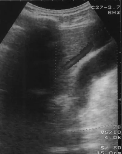
during the procedure (Fig. 2). All were cirrhotic pa-tients. There were two conversions to open cholecys-tectomy, but none related to GB bed bleeding. In group ND, 56.4% had GB stone with biliary colic, 23.95% had stone with chronic cholecystitis, 12.9% had stone with acute cholecystitis. The rest were GB polyp, stone with acute and chronic cholecystitis and shrunken GB. The mean blood loss, operative time, hospital stay and complications of the two groups are shown and com-pared in Table 2. They were not significantly different. The number of conversions to open cholecystectomy in Group ND, mean blood loss in cirrhotic cases in Group Fig. 1. Picture of ultrasound of a huge THVR(blue) that was almost
adherent to the GB. The caliber is over 3 mm.
Table 1. Demographic data for Group D and ND
Group D Group ND P-value
Age (years) 45–67 43–65 NS M:F 1.8:1 1.9:1 NS Noncirrhosis:cirrhosis 2,038:108 2,025:121 NS Child A:B 83:25 91:30 NS By Chi-square test NS, Not significant
Fig. 2. One case of THV (arrow) that was dissected and clipped. 1378
ND and the mean operative time in patients with posi-tive THV were significantly greater.
The complications included bile leakage, hematoma, wound infections, aspiration pneumonia, and ascites, which were not significantly different between the two groups. There were three cases of hospital mortality in group D due to one case of cardiac arrhythmia and two cases of pneumonia, and four deceased in group ND due to three cases of sepsis and one case of cardiac failure.
Both operative results are compared in Table 2.
Discussion
During the past decade, LC has become a popular and even standard treatment for cholelithiasis in the general population, for it allows shorter hospital stay, less operative time, and fewer wound complications [4, 10, 14, 15, 18]. As we know, one of the dangerous compli-cations of LC is unexpected massive bleeding from the THV in the GB bed during dissection. Seventy-five cases of such bleeding in 77,604 LC cases requiring open cholecystectomy due to GB bed bleeding were reported by Deziel et al. [6]. Nenner et al. [11] and Reddick et al. [13] reported that GB bed bleeding was related to injury of the hepatic vein. However, both studies did not focus on the preoperative examination of the hepatic vein and the prevention of fatal hemorrhage. Furthermore, no detailed study about the anatomical relationship be-tween the hepatic vein and GB has been performed.
In 1999, color Doppler ultrasound assessment of the branches of the mHV was reported by Takeyuki et al. [20]. Sixteen percent of patients had THV. However, the study population included only 50 healthy volunteers and a retrospective review of four patients of postlapa-roscopic cholecystectomy. The author did not engage in further prospective study as to whether color Doppler ultrasound was beneficial for the general population or whether the examination actually helped the patients with cirrhosis.
As we know, the incidence of cholelithiasis in cir-rhotic patients is about twice that in noncircir-rhotic pa-tients [3, 12, 17]. The chances of treating a cirrhotic gallbladder stone are great. However, cholecystectomy for these patients presents a great challenge. The high mortality reported is mainly due to the tendency to bleed [1, 7, 16], which causes excessive blood loss. Ara-nha et al. [1] even reported a mortality of 83% in
pa-tients where the PT was elevated >2.5 sec. In order to reduce the risk of GB bed bleeding to a minimum, in addition to improvement of the hemostatic condition, identifying the definite anatomical relationship between the mHV and the GB is very important. Accidental in-jury to the THV is better to be averted, since the hard texture of the liver is difficult to suture and compress in order to stop bleeding.
There were two cases of conversion in group D. One was due to severe adhesion, and the other was due to bleeding from the abdominal wall varices. None was related to GB bed bleeding. However, in group ND, five conversions were noted. All were cirrhotic patients. Four were due to GB bed bleeding from the mHV, and one was due to adhesion. In the general population of both groups, no conversion was noted.
The blood loss in group D was 93.25 ± 24.12 ml, whereas blood loss in Group ND was 98.18 ± 36.22 ml. This was not significantly different. However, when we compared the cirrhotic cases in both groups, which were 150.15 ± 62.016 ml and 240.4 ± 130.008ml, group D was significantly less (StudentÕs t-test). Since the hemo-static condition was not significantly different between the two groups, the difference of blood loss should be mainly due to the injured THV.
The mean operative time was not significantly dif-ferent between D and ND. However, when the operative times of the cases of positive THV were compared with those for the rest, the former were significantly longer (StudentÕs t-test). This can be explained by the fact that more time was spent in meticulous and cautious dis-section.
There was the occasional intraoperative diagnosis of liver cirrhosis. In such a case, a lower than usual wattage was employed (25 watts was suggested to prevent further spark gap thermal injury), in addition to gentle and accurate dissection. We had two such cases, and the procedures were performed uneventfully.
More than that, there is no need to dissect the vein, but if necessary or technically feasible, the THV can be found out and clipped. We have seven such cases, and all were cirrhotic patients.
Finally, we need to emphasize that the authors in the present series have experience in about 7,500 cases of laparoscopic cholecystectomy. There were no technical discrepancies between the two different groups, al-though the patients were not operated on in the same period.
Table 2. Comparison of both groups
Group D Group ND P-value
*Conversion related to GB bed bleeding 0 4 <0.05
Mean blood loss (ml) 93.25 ± 24.13 98. 18 ± 36.22 NS
*Mean blood loss (ml) of cirrhotic patients 150.15 ± 62.016 240.40 ± 130.008<0.05 Mean operative time (minute) 77.36 ± 23.73
*For positive THV: 92.25 ± 22.447
73.00 ± 19.724 NS
<0.05
Complications (number) 11 13 NS
Hospital stay (days) 3.7 ± 1.2 3.8± 1.8 NS
By StudentÕs t-test
NS, Not significant; *significance: P < 0.05
We are not deliberately comparing the results of the two groups because the disease severity or the condi-tions of the populacondi-tions may not be absolutely the same. The authors are simply presenting the applicability and the advantages of this simple noninvasive examination.
Conclusions
The routine prelaparoscopic cholecystectomy color Doppler ultrasound is convenient method for preoper-ative detection of the tangential hepatic vein. It is pop-ular in most hospitals in Taiwan and the cost of examination is low. It has effectively prevented cases of unexpected fatal bleeding and has reduced unnecessary blood loss during dissection, especially in patients with liver cirrhosis. When encountering the potential for bleeding, the surgeon should (i) be well prepared for an emergency conversion to open cholecystectomy before initiating the LC, (ii) use a lower wattage, and (iii) be extremely cautious in dissection, especially in cases of liver cirrhosis.
References
1. Aranha GV, Sontag SJ, Greenlee HB (1982) Cholecystectomy in cirrhotic patients: a formidable operation. Am J Surg 143: 55–60 2. Baev ST, Pozarliev T, Todorov GT (1995) Laparoscopic
chole-cystectomy: 700 consecutive cases. Int Surgl 80: 296–298 3. Bouchier IA (1969) Postmortem study of the frequency of
gall-stones in patients with cirrhosis of the liver. Gut 10: 705–710 4. Colonval P, Navez B, Cambier E, Richir C, de Pierpont B, Scohy
JJ, Guiot P (1997) Is laparoscopic cholecystectomy effective and reliable in acute cholecystitis? Results of a postoperative study of 221 pathologically documented cases. Ann Chir 51: 689–696 5. Cosgrove DO, Arger PH, Coleman BG (1987) Ultrasonic anatomy
of hepatic veins. J Clin Ultrasound 15: 231–235
6. Deziel DJ, Millikan KW, Economous SG, Doolas A, Ko ST, Airan MC (1993) Complications of laparoscopic cholecystectomy: a national survey of 4,292 hospitals and an analysis of 77,604 cases. Am J Surg 165: 9–14
7. Doberneck RC, Sterling WA, Allison DC (1983) Morbidity and mortality after operation in nonbleeding cirrhotic patients. Am J Surg 146: 306–309
8. Kapoor VK, Kumar A, Sikora SS, Kaushik SP (1995) Conver-sions in laparoscopic cholecystectomy—need for a new nomen-clature. Trop Gastroenterol 16: 38–39
9. Lee VS, Chari RS, Cucchiaro G, Meyers WC (1993) Complica-tions of laparoscopic cholecystectomy. Am J Surg 165: 527–532 10. Lujan JA, Parrilla P, Robles R, Main P, Torralba JA,
Garcia-Ayllon J (1998) Laparoscopic cholecystectomy vs open cholecys-tectomy in the treatment of acute cholecystitis: a prospective study. Arch Surg 133: 173–175
11. Nenner RP, Imperato PJ, Alcorn CM (1992) Complications of laparoscopic cholecystectomy in ageriatric population group. NY State J Med 12: 518–520
12. Nicholas P, Rinaudo PA, Conn HO (1972) Increased incidence of cholelithiasis in LaennecÕs cirrhosis: postmortem evaluation of pathogenesis. Gastroenterology 63: 112–121
13. In: Reddick E, Saye WB, Corbitt JD Jr (eds) (1993) Atlas of laparoscopic surgery. Raven Press, New York
14. Schafer M, Krahenbuhl L, Farhadi J, Buchler MW (1998) Cho-lelithiasis—laparoscopy or laparotomy? The Umsch 55: 110–115 15. Schumpelick V, Schippers E (1991) Cholecystectomy: laparoscopic
or conventional? Zeitschr Gastroenterol 29: 659–662
16. Schwartz SI (1981) Biliary tract surgery and cirrhosis: a critical combination. Surgery 90: 577–583
17. Schwesinger WH, Kurtin WE, Levine BA, Page CP (1985) Cir-rhosis and alcoholism as pathogenetic factors in pigment gallstone formation. Ann Surg 201: 319–322
18. Scotte TR, Zucker KA, Bailey RW (1992) Laparoscopic chole-cystectomy: a review of 12,397 patients. Surg Laparosc Endosc 2: 191–198
19. Shen BY, Li HW, Chen M, Zheng MH, Zang L, Jiang SM, Li JW, Jiang Y (2003) Color Doppler ultrasonographic assessment of the risk of injury to major branch of the middle hepatic vein during laparoscopic cholecystectomy. Hepatobili Pancreat Dis Int 2: 126– 130
20. Takeyuki M, Masato K, Katsumaro S, Yasuki U, Ryuzo M, Kazuhiko Y, Susumu K, Yoji Y (1999) Ultrasonographic assess-ment of the risk of injury to branches of the middle hepatic vein during laparoscopic cholecystectomy. Am J Surg 178: 418–421 1380