Congenital or acquired bone defect is a major problem in orthopedic surgery. The use of autoge-nous and banked allogenic bone grafts to induce the formation of bone in bone defects has become common (1). Autografts are preferably used because of their superior efficacy and capability of avoiding transmission of infection. However, it does have drawbacks, including donor site morbidity and limited availability, particularly in children (2). The effectiveness of allografts is limited by problems such as the high cost of bone banking, potential of
graft-related disease transfer, high rates of nonunion and infection, and allograft fractures (3). Despite the efficacy of these conventional methods, problems associated with their use have led to a search for alternatives (4,5). Toward that goal, we evaluated the effect of a collagen-hydroxyapatite/tricalcium phosphate (Col-HA/TCP) microsphere containing rhTGF-b 1 (recombinant human transforming growth factor-b 1) on the healing of metaphyseal bone defects.
Produced by all skeletal tissues, TGF bs potently regulate cell proliferation, differentiation, and matrix synthesis (6,7). It is the capacity of TGFs to promote chondroblastic and osteoblastic differentiation (8), which suggests an autocrine or paracrine role in this process (9), that is of considerable use in bone repair. Using different models, several investigators have analyzed the stimulation of bone healing by TGF b (7,8,10,11). Studies of animals have validated the
Collagen-Hydroxyapatite/Tricalcium Phosphate
Microspheres as a Delivery System for Recombinant
Human Transforming Growth Factor-
b 1
*Jui-Sheng Sun, †Feng-Huei Lin, ‡Yng-Jiin Wang, †Yi-Chau Huang,
§Shan-Chang Chueh, and §Fu-Yin Hsu
*Department of Orthopedic Surgery and Department of Biomedical Engineering, National Taiwan University Hospital; †Graduate Institute of Biomedical Engineering, National Taiwan University; ‡Graduate Institute of Biomedical Engineering, National Yang-Ming University, Taipei; and §Material Research Laboratories, Industrial Technology Research
Institute, Chutung, Hsinchu, Taiwan, ROC
Received August 2002; revised December 2002.
Address correspondence and reprint requests to Dr. Jui-Sheng Sun, Department of Orthopedic Surgery, National Taiwan Uni-versity Hospital, No. 7, Chung-Shan South Road, Taipei, Taiwan, ROC. E-mail: [email protected]; and Yng-Jiin Wang, PhD, Institute of Biomedical Engineering, National Yang-Ming University, 155, Li-Nong St., Sec. 2, Peitou, Taipei, Taiwan, ROC. E-mail: [email protected]
Abstract: The purpose of this study is to evaluate the
carrier capability of collagen-hydroxyapatite/tricalcium phosphate (Col-HA/TCP) microspheres to the rhTGF-b 1 (recombinant human transforming growth factor-b 1). After anesthesia, a bone defect (7.0 mm in diameter and 10.0 mm in depth) was created at the distal femoral condyles of New Zealand white rabbits. These defects were then completely filled with the implant materials. After 5, 7, 9, 11, 13, and 15 weeks, the animals were sacrificed and histological evaluations were performed. The results showed that when the defects were treated with Col-HA/TCP microspheres without rhTGF-b 1, there was only spotty new bone formation during the 15 week experimen-tal period and most of the defect was filled with fibrous
tissue and inflammatory cells, whereas active bone forma-tion with mature marrow tissue formaforma-tion was evident in the defect treated with Col-HA/TCP microspheres con-taining rhTGF-b 1. Collagen-hydroxyapatite/tricalcium phosphate microspheres were expected to be replaced by the regenerated bone structure as the bone reconstruction and bone-remodeling process occurred. It was apparent that bone regeneration was influenced by the addition of rhTGF-b 1. Collagen-hydroxyapatite/tricalcium phosphate microspheres were a good carrier for rhTGF-b 1.
Key Words: Collagen—Hydroxyapatite—Tricalcium
phos-phate—Microsphere—Transforming growth factor—Bone healing.
efficacy and safety of TGFs for bone repair. However, a satisfactory delivery system for rhTGF must be developed before it can be used in humans. Some investigators have described a tricalcium phosphate carrier for rhTGFs (12,13), where colla-gen and hydroxyapatite have been used as bone-filling materials in orthopedic surgery (14). The collagen fibers are known to serve as the scaffold for tissue repair (15) and hydroxyapatite is com-patible and osteoconductive for bone regenera-tion. Although the dense hydroxyapatite disc is not biodegradable, particulate hydroxyapatite can be removed and remodeled in the host (16). Direct implantation of hydroxyapatite or tricalcium phos-phate particles result in the dislocation of material within the tissue. When used as bone grafts, hy-droxyapatite or tricalcium phosphate powders are often mixed with collagen (17), gelatin (18), or fibrin glue (19) to eliminate undesired mobility.
Although most of the collagen matrices are prepared in a slab form, spherical composites of collagen and hydroxyapatite are more versatile in biomedical applications. Other than being used for cell culturing, spherical gel beads have greater flexi-bility in filling different geometric cavities with a closer packing than gels with nonspherical shapes. Microspheres composed of Col-HA/TCP are also injectable for repairing tissue defects. In this study, we used an entirely different strategy of collagen-containing microspheres, which did not induce an immune response from the host (20), as a delivery system for rhTGF. The purpose of this study was to examine the effect of collagen-hydroxyapatite/ tricalcium phosphate microspheres containing rhTGF-b 1 to repair a critical-sized defect in the rabbit distal femur. Bone formation and healing at the site of the defect were evaluated with the use of histological techniques.
MATERIALS AND METHODS Preparation of implanted materials
Microspheres (200–300mm) comprised of biphasic particulate hydroxyapatite/tricalcium phosphate dispersed in fibrous collagen matrices were prepared as described previously (20). Briefly, the procedure involved the droplet formation of a hydroxyap-atite/tricalcium phosphate/collagen mixture (wt/wt; hydroxyapatite 39%; tricalcium phosphate 26%; collagen 35%) emulsified in olive oil, followed by the reconstitution of collagen in the presence of hy-droxyapatite/tricalcium phosphate particles at 37°C. Microspheres sized at 200–300mm could be obtained by controlling the stirring speed of the emulsified
mixture at 400 rpm, when 2% Span 85 was present in the emulsion mixture. The microspheres thus obtained can be used as carriers of growth factors to support the growth of osteoblast cells.
Operation and implantation
Twelve New Zealand white male rabbits with an average weight of 2.5–3.0 kg were used in this study. The animals were fed Purina Laboratory Chow ad libitum and housed in a temperature-, humidity-, and light-controlled environment. Surgical procedures and experimental protocols were approved by and under the supervision of the Medical College’s Animal Research Committee of the National Taiwan University. The rabbits were anesthetized by ketamine (25 mg/kg, s.c., Sintong, Taiwan, ROC) and Combelen (N-[3 ¢-dimethyl-aminopropyl]-3-propionylphenothiazine; 5 mg/kg, s.c.; Bayer AG, Leverkusen, Newbury, England) and local adminis-tration of 0.5% lidocaine. After shaving, disinfecting, and sterile draping of the operation site, the femoral condyles were exposed by lateral longitudinal inci-sion. Initially, a bone defect was created by a 3.2 mm drill and subsequently expanded with a 6 mm drill. All the drill holes were carefully rinsed with Ringer’s solution so that any abraded particles formed during drilling were removed. These defects were then com-pletely filled with the implant materials. The perio-steum, fascia, and skin were sutured layer by layer. After 5, 7, 9, 11, 13, and 15 weeks, the animals were sacrificed with an overdose of intravenous pento-barbital. A total of 12 rabbits divided into six groups for the above experimental time periods were used in the study. In the pilot study, we showed that there was no evidence of bone healing when the defects were left untreated (21). In this study, the defects at both sides were implanted with Col-HA/TCP micro-spheres before the closure of the wound, while in the right side 10mg rhTGF-b 1 (R & D Systems, Inc., Minneapolis, MN, U.S.A.) was injected into the bone defect to mix with the Col-HA/TCP microspheres. The animals were allowed to recover from anes-thesia, and placed in cages until the end of the experiment.
Histological evaluations
The hindlimbs were harvested from the treated animals at the mentioned time periods after opera-tion. Implants and surrounding tissues were removed en bloc, washed in normal saline, and fixed with 4% formaldehyde in phosphate-buffered solution for 18 h, decalcified, dehydrated in alcohol, cleared in xylene, and embedded in paraffin. Sections (5–7mm in thickness) were cut and stained with hematoxylin
and eosin. Representative sections were photo-graphed using light microscopy. Four sections were cut for each implant, parallel to the major axis.
RESULTS Histological evaluation
Only spotty new bone formation within the meta-physeal defect was observed in the group treated with Col-HA/TCP microspheres alone. The large part at the center of the defect remained free of bony tissue during the entire course of the test up to 15
weeks, and was filled with fibrous tissue and inflam-matory cell infiltration. The implanted collagen fibers were still visible at this stage and showed evidence of degeneration. When the results obtained were evaluated as a function of time, it appeared that the inflammatory cell infiltration became more appar-ent as time elapsed. In other words, progressive increased inflammatory reaction within the defect cavity was detected (Figs. 1–6, rhTGF [-]).
When the metaphyseal defect was filled with Col-HA/TCP microspheres containing 0mg of rhTGF for 7 weeks, lymphocyte infiltration and large multinucleated giant cell proliferation were visible.
FIG. 1. Photomicrograph, made at 5 weeks, of a defect filled
with collagen-hydroxyapatite/tricalcium phosphate (Col-HA/TCP) microspheres containing 0 or 10mg of rhTGF-b 1.
Top panel, rhTGF (-): They demonstrate an area of lymphocyte (L.C.) infiltration and areas where the Col-HA/TCP microspheres were solubilized and washed out by solvents during histological preparation (white areas, such as the one with the star). The higher magnification in inset B shows large multinucleated giant cell (G.C.) proliferation (HE stain; bar, 100mm).
Bottom panel, rhTGF (+): They demonstrate more areas of lymphocyte (L.C.) aggregation, and at the higher magnification in inset B show smaller multinucleated giant cells (G.C.) than a defect filled with Col-HA/TCP microspheres containing 10mg of rhTGF-b 1 (HE stain; bar, 100 mm).
FIG. 2. Photomicrograph, made at 7 weeks, of a defect filled with
collagen-hydroxyapatite/tricalcium phosphate (Col-HA/TCP) microspheres containing 0 or 10mg of rhTGF-b 1.
Top panel, rhTGF (-): They demonstrate numerous areas of lymphocyte (L.C.) aggregation and formation of nodules. The Col-HA/TCP microspheres (Col.) are surrounded by lymphoid tissues. The higher magnification in the insets shows lymphoid cell (L.C.) proliferation (HE stain; bar, 100mm).
Bottom panel, rhTGF (+): There are still areas of lymphocyte (L.C.) aggregation and the higher magnification in the insets shows that a defect filled with Col-HA/TCP microspheres (Col.) is well surrounded by regenerated bony trabeculae. In this section, the bone marrow formation (B.M.) is quite evident (HE stain; bar, 100mm).
The Col-HA/TCP microspheres were evident and only scanty bony trabeculae regeneration was found (Figs. 1 and 2, rhTGF [-]). At 9 weeks, marked fibrous tissue proliferation between the areas of Col-HA/TCP microspheres was found and there was numerous large multinucleated giant cell prolif-eration present (Fig. 3, rhTGF [-]). At 11 weeks, lymphoid tissue proliferation and nodule formation became quite evident (Fig. 4, rhTGF [-]). Marked fibrous tissue proliferation between the areas of the
Col-HA/TCP microspheres with only scanty bony trabeculae formation was visible at 13–15 weeks after implantation (Figs. 5 and 6, rhTGF [-]). At 15 weeks after implantation, chronic inflammatory cell infiltra-tion was observed in addiinfiltra-tion to large multinucleated giant cell proliferation (Fig. 6, rhTGF [-]).
A dose of 10mg of rhTGF-b 1 produced numerous isotropically oriented trabeculae. Active new bone formation was evident in the group treated with Col-HA/TCP microspheres with rhTGF-b 1 addition (Figs. 1–6, rhTGF [+]). At 5 weeks postoperative (Fig. 1, rhTGF [+]), lymphocyte aggregation and smaller multinucleated giant cells were quite evident at the
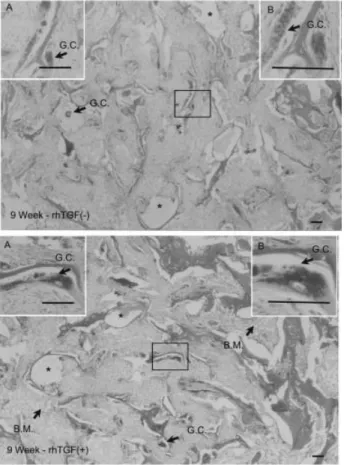
FIG. 3. Photomicrograph, made at 9 weeks, of a defect filled
with collagen-hydroxyapatite/tricalcium phosphate (Col-HA/TCP) microspheres containing 0 or 10mg of rhTGF-b 1.
Top panel, rhTGF (-): They demonstrate marked fibrous tissue proliferation between the areas where the Col-HA/TCP micro-spheres were solubilized and washed out by solvents during histological preparation (white areas, such as the one with the star). The large multinucleated giant cell (G.C.) proliferation is still present and well demonstrated in higher magnification in insets A and B (HE stain; bar, 100mm).
Bottom panel, rhTGF (+): The size of bony trabeculae seemed to decrease in size and there is multinucleated giant cell (G.C.) formation adjacent to the bony trabeculae without collagen-hydroxyapatite/tricalcium phosphate microspheres present. In this section, the bone marrow formation (B.M.) and areas where the Col-HA/TCP microspheres were solubilized and washed out by solvents (white areas, such as the one with the star) are also evident (HE stain; bar, 100mm).
FIG. 4. Photomicrograph, made at 11 weeks, of a defect filled
with collagen-hydroxyapatite/tricalcium phosphate (Col-HA/TCP) microspheres containing 0 or 10mg of rhTGF-b 1.
Top panel, rhTGF (-): They demonstrate marked fibrous tissue proliferation between the areas of the Col-HA/TCP microspheres (Col). Lymphoid tissue proliferation (L.C.) and nodule forma-tion are also evident at this stage. The large multinucleated giant cell (G.C.) proliferation is still present and well demon-strated in higher magnification in insets A and B (HE stain; bar, 100mm).
Bottom panel, rhTGF (+): The size of bony trabeculae is decreased and the size of the Col-HA/TCP microspheres (Col.) is also decreased. The maturation of bone marrow (B.M.) is evident and more pronounced (HE stain; bar, 100mm).
defect that was filled with Col-HA/TCP micro-spheres containing 10mg of rhTGF-b 1. Seven weeks after implantation (Fig. 2, rhTGF [+]), woven bone regeneration was found in the defect cavity in which active bone marrow formation was observed. At this stage, inflammatory cell infiltration with giant cell formation was still quite evident. During 9–11 weeks after implantation (Figs. 3 and 4, rhTGF [+]), the breakdown and dissolution of Col-HA/TCP micro-spheres and bone marrow formation were observed in the histological sections. The regenerated bone decreased in size and there were multinucleated giant cells growing adjacent to the regenerated bone
trabeculae. At 13 weeks after implantation (Fig. 5, rhTGF [+]), the breakdown and dissolution of the Col-HA/TCP microspheres was quite obvious. At the same time, the regenerated bone increased in size and the maturation of bone marrow was even more pronounced. At 15 weeks after implantation (Fig. 6, rhTGF [+]), the implant gradually dissolved and was replaced by the bony structure. The laminar bone appeared, and the Col-HA/TCP microspheres con-tinued to be dissolved, digested, and replaced by the physiological bony marrow tissue, which was filled with active bone marrow cells. It was apparent that bone regeneration was occurring in connection with,
FIG. 5. Photomicrograph, made at 13 weeks, of a defect filled
with collagen-hydroxyapatite/tricalcium phosphate (Col-HA/TCP) microspheres containing 0 or 10mg of rhTGF-b 1.
Top panel, rhTGF (-): They demonstrate scanty bony tra-beculae formation-marked fibrous tissue proliferation between the areas of the Col-HA/TCP microspheres (Col.). Large multi-nucleated giant cell (G.C.) proliferation is present and well demonstrated by higher magnification in insets A and B (HE stain; bar, 100mm).
Bottom panel, rhTGF (+): The size of bony trabeculae increased from that at 11 weeks with active osteoblast formation. The size of Col-HA/TCP microspheres (Col.) decreased. The maturation of bone marrow (B.M.) is evident and more pro-nounced (HE stain; bar, 100mm).
FIG. 6. Photomicrograph, made at 15 weeks, of a defect filled
with collagen-hydroxyapatite/tricalcium phosphate (Col-HA/TCP) microspheres containing 0 or 10mg of rhTGF-b 1.
Top panel, rhTGF (-): They demonstrate scanty bony trabec-ulae formation-marked fibrous tissue proliferation between the areas of the Col-HA/TCP microspheres (Col.). The large multi-nucleated giant cell (G.C.) proliferation is still present and well demonstrated. In the higher magnification in insets A and B, we can also see the chronic inflammatory cell infiltration at this stage (HE stain; bar, 100mm).
Bottom panel, rhTGF (+): The residual Col-HA/TCP micro-spheres (Col.) are well surrounded by bony trabeculae. The bone marrow tissue is well matured (B.M.) and obvious at this stage (HE stain; bar, 100mm).
and influenced by, the rhTGF-b 1, as evidenced by the incorporation of Col-HA/TCP microspheres into the bone trabeculae.
DISCUSSION
TGF-b belongs to a family of related proteins called the TGF-b superfamily. This family of proteins includes the five isoforms of TGF-b (TGF-b 1 through TGF-b 5), BMPs, growth differentiation factors (GDFs), activins, inhibins, and Mulerian sub-stance (9,22–24). TGF-b influences a broad range of cellular activities, including growth, differentiation, and extracellular matrix synthesis. TGF-b is found in many tissues, but is particularly enriched in bone, platelets, and cartilage. It is presumed to be released by platelets after a clot is formed at the time of frac-ture (25). It has been hypothesized that the release of TGF-b 1 is associated with proliferation of periosteal tissue because there is positive immuno-staining for TGF-b 1 in the early fracture-healing period. Several other experimental studies have val-idated the effectiveness of TGFs for the stimulation of bone formation (7,26). Locally delivered TGF-b 1 is the most potent stimulator of bone ingrowth tested to date, exceeding the amount of bone forma-tion obtained following autogenous cancellous bone grafting (27) and many other tested agents (28,29). In other contexts, local delivery of exogenous purified or recombinant TGF-b 1 has been found to enhance fracture healing (30,31), induce skull defect closure (32), accelerate orthotopic osteoin-duction by demineralized bone matrix (33), enhance fixation of ceramic-coated implants (11), and sti-mulate bone marrow osteoprogenitor activity and matrix synthesis (34). It is difficult to draw con-clusions regarding the efficacy of TGF-b on the basis of these studies of experimental fracture healing because different isoforms and doses of growth factor were used and different animal models were employed. Although the results of these studies confirm the hypothesis that TGF-b enhances cellular proliferation, the osteoinductive potential of TGF-b seems limited.
The importance of the delivery system was noted in all of these studies. The ability to deliver a mole-cule so that it will induce a specific biologic effect is critical to the success of growth factor therapy. A number of carrier and delivery systems, including type I collagen, synthetic polymers, and hyaluronic acid gels, have been used to deliver recombinant pro-teins in experimental and clinical models (35). A variety of so-called bone graft substitutes, includ-ing demineralized bone matrix, calcium
phosphate-containing preparations (such as hydroxyapatite, coralline hydroxyapatite, and Bioglass), are also potential carriers for recombinant proteins (35). A highly characterized polymer must be used to en-sure the reproducibility among experiments, tissue biocompatibility, biodegradation, and delivery of growth factors to bone defects. We chose a method of volume expansion-solvent extraction curing to prepare characterized Col-HA/TCP microspheres to deliver selected doses of rhTGF-b 1. The delivery system releases and localizes the TGF, ensuring inter-action with mesenchymal cells that can differentiate into osteoblasts. The delivery system also provides instructional guidance as a template to renew osseous contour.
In our previous experimental studies, rhBMP-4 had been implanted in combination with collagen/ hydroxyapatite microsphere systems (21). However, due to the slow resorption rate and larger size of hydroxyapatite (600mm), there was numerous giant cell formation accompanied by a significant amount of residual hydroxyapatite particles after 2 months post-operation (21). In this study, we added trical-cium phosphate to facilitate the degradation rate of the bioceramics. The main purpose of this study was to determine whether local application of Col-HA/TCP microspheres carried rhTGF-b 1-enhanced bone ingrowth into a metaphyseal defect.
Previous studies have shown that calcium phos-phate treatment of porous-coated implants enhances bone ingrowth (36,37), but in the present study the Col-HA/TCP treatment was not stimulatory. Despite our efforts to optimize the polymer delivery system, multinucleated giant cells were a component of the wound-healing response. There appeared to be more multinucleated giant cells and lymphocytes in the defects filled with a Col-HA/TCP microsphere implant without rhTGF-b than in the defects filled with an implant with rhTGF-b. Multinucleated giant cells were present throughout the 15 weeks of the study (Figs. 1–6). Despite the inflammatory reaction to Col-HA/TCP microspheres, rhTGF-b 1 promoted new bone formation (Figs. 1–6). Moreover, there was no adverse clinical sequelae (such as swelling or the formation of sinus tracts) associated with the im-plants. Remnants of crystalline Col-HA/TCP micro-spheres still can be detected with microscopy at 15 weeks; however, most of them were enclosed within the regenerated bone trabeculae (Fig. 6). Several factors may be responsible for these observations. The rhTGF-b and polymer may have influenced the phenotype, quantity, and activity of cells, thereby affecting the environment of the defect, biodegrada-tion of the polymer, recruitment and stimulabiodegrada-tion of
multinucleated giant cells, and formation of bone. For example, there were fewer multinucleated giant cells in the defects filled with a Col-HA/TCP micros-phere implant containing rhTGF-b 1. The decreased volume of Col-HA/TCP microspheres by 15 weeks may be less stimulatory to granulocytes and multin-ucleated giant cells (Fig. 6). However, complete abro-gation of the multinucleated giant cell response evoked by Col-HA/TCP microspheres may not be possible. If the response is transient, which it appears to be, it may have no clinical relevance. With our model, it is clear that local application of TGF-b 1 has stimulatory effects on intramembranous bone regeneration and bone ingrowth into the bone defect site.
This study showed that local application of rhTGF-b 1 strongly enhanced local rhTGF-bone ingrowth and gap healing and that bone regeneration was stimulated at a remote site (Figs. 2 and 3). Bone ingrowth and gap healing occur via the intramembranous (as opposed to the endochondral) pathway because of previously reported chemotactic, mitogenic, and synthetic effects of TGF-b 1 on cells of the osteoblastic line-age (34,38,39). The morphologic observations of increased trabecular thickness and number (Figs. 5 and 6) are consistent with these proposed mecha-nisms of action. In addition, the effect on trabecular architecture in the gap supported the interpretation that local application of rhTGF-b 1 enhances bone regeneration by stimulating recruitment and prolif-eration of osteoprogenitor cells and by increasing production of the extracellular matrix by committed osteoblasts.
Previous studies have found anabolic remodeling effects at remote sites following local injury to the skeleton (40,41). The mechanisms underlying remote effects on remodeling and regeneration are unknown, but most authors have postulated that local injury leads to the local release and/or synthe-sis of growth factors that then enter circulation (42,43). The present experiment indicates that local application of rhTGF-b 1 can have remote effects on bone regeneration as evidenced by the increase in the volume fraction of the bone in the gaps of the contralateral control limb (Fig. 2). Currently, osteogenic growth peptide and TGF-b 1 seem to be the most likely sources of the systemic remodeling effect (44). It is plausible that the same factors may be involved in the remote stimulation of bone regen-eration. In light of studies showing that systemic administration of TGF-b inhibits immobilization-induced bone loss (45) and stimulates local wound healing (34), the simplest interpretation of our obser-vation is that release of TGF-b from the implant and
transport via circulation stimulated bone regenera-tion at the remote site (46).
Although several recombinant proteins may soon be available as therapeutic growth factors for specific clinical applications, there is concern that a single dose of exogenous protein will not induce an ade-quate biologic response in patients, particularly in situations in which the viability of the host bone and surrounding soft tissues is compromised. To address this potential concern, a better strategy for protein delivery may be gene therapy. An important aspect of gene therapy is the application of appropriate vectors for genes. A major concern related to the use of viral vectors is the subsequent recombination of the defective virus with viruses in the host cell, resulting in the generation of replication-competent viruses with the ability to multiply in the patient. In addition, cells infected with certain viruses (e.g., ade-noviruses) produce not only the transgene product but also other viral proteins (35). The results of the present study show that rhTGF-b 1 delivered in Col-HA/TCP elicits bone formation in metaphyseal bone defects. There was clear morphological evi-dence that 10mg of rhTGF-b 1 promoted the forma-tion of new bone. Furthermore, the rhTGF-b 1 and Col-HA/TCP microspheres were clinically conve-nient to use, biocompatible, and biodegradable, thus increasing the potential therapeutic value of this combination for the stimulation of new bone forma-tion in bone defects in a clinical setting. We conclude that TGF-b 1-containing collagen/hydroxyapatite/ tricalcium phosphate microspheres show promise as an agent to promote bone regeneration of critical size bone defects.
REFERENCES
1. Urist MR, O’Connor BT, Burwell RG, eds. Bone grafts,
deriv-atives and substitutes. Oxford: Butterworth-Heinemann Ltd,
1994:220–34.
2. Dutting A, Thomas W, Lorenz H, Holzt A. Komplikationen nach autologer Knochentransplantation am Entnahmeort.
Zeitschrift fur Ortkopadie und ihre Grenzgebiete 1988;
126:44–7.
3. Kotani S, Yamamuro T, Nakamura T, Kitsugi T, Fujita Y, Kawanabe K, Kokubo T. Enhancement of bone bonding to bioactive ceramic by demineralized bone powder. Clin Orthop 1992;278:226–34.
4. Hollinger JO, Brekke J, Gruskin E, Lee D. Role of bone sub-stitutes. Clin Orthop 1996;324:55–65.
5. Tomford WW. Current concepts review. Transmission of disease through transplantation of musculoskeletal allografts.
J Bone Joint Surg 1995;77A:1742–54.
6. Bolander ME. Regulation of fracture repair by growth factors.
Proc Soc Exp Biol Med 1992;200:165–70.
7. Joyce ME, Jingushi S, Bolander ME. Transforming growth factor-b in the regulation of fracture repair. Orthop Clin North
Am 1990;21:199–209.
8. Joyce ME, Roberts AB, Sporn MB, Bolander ME. Trans-forming growth factor-b and the initiation of chondrogenesis
and osteogenesis in the rat femur. J Cell Biol 1990;110:2195– 207.
9. Roisier RN, O’Keefe RJ, Hicks DG. The potential role of transforming growth factor b in fracture healing. Clin Orthop 1998;355S:S294–300.
10. Einhorn TA, Trippel SB. Growth factor treatment of fractures.
Instr Course Lect 1997;46:483–6.
11. Lind M, Overgaard S, Ongpipattanakul B, Nguyen T, Bunger C, Soballe K. Transforming growth factor-b 1 stimu-lates bone ongrowth to weight-loaded tricalcium phosphate coated implants: an experimental study in dogs. J Bone Joint
Surg 1996;78B:377–82.
12. Heckman JD, Aufdemorte TB, Athanasiou KA, et al. Treat-ment of acute ostectomy defects in the dog radius with rhTGF-b 1. Trans Orthop Res Soc 1995;20:590.
13. Beck LS, Deguzman L, Lee WP, et al. Bone marrow augments the activity of transforming growth factor b 1 in critical sized bone defects. Trans Orthop Res Soc 1996;21:626.
14. Marouf HA, Quayle AA, Sloan P. In vitro and in vivo studies with collagen/hydroxyapatite implants. Int J Oral Maxillofac
Implants 1990;5:148–54.
15. Pachence JM. Collagen-based devices for soft tissue repair.
J Biomed Mater Res 1996;33:35–40.
16. Pohunkova H, Adam M. Reactivity and the fate of some com-posite bioimplants based on collagen in connective tissue.
Bio-materials 1995;16:67–71.
17. Sugaya A, Minabe M, Tamura T, Hori T. Effects on wound healing of hydroxyapatite-collagen complex implants in periodontal osseous defects in the dog. J Periodontal Res 1989;24:284–8.
18. Nagase M, Chen R, Asada Y, Nakijima T. Radiographic and microscopic evaluation of subperiosteally implanted blocks of hydroxyapatite gelatin mixture in rabbits. J Oral Maxillofac
Surg 1989;47:40–5.
19. Wittkampf AR. Augmentation of the maxillary alveolar ridge with hydroxyapatite and fibrin glue. J Oral Maxillofac Surg 1988;46:1019–21.
20. Hsu FY, Chueh SC, Wang YJ. Microspheres of hydroxyap-atite/reconstituted collagen as supports for osteoblast cell growth. Biomaterials 1999;20:1931–6.
21. Wang YJ, Lin FH, Sun JS, Huang YC, Chueh SC, Hsu FY. Collagen-hydroxyapatite microspheres as carriers for bone morphogenic protein-4. Artif Organs 2003;27:162–8. 22. Linkhart TA, Mohan S, Baylink DJ. Growth factors for bone
growth and repair: IGF, TGF b and BMP. Bone 1996;19:1S– 12.
23. Massague J, Wotton D. Transcriptional control by the TGF-b/ Smad signaling system. EMBO J 2000;19:1745–54.
24. Liu F, Hata A, Baker JC, Doody J, Carcamo J, Harland RM, Massague J. A human Mad protein acting as a BMP-regulated transcriptional activator. Nature 1996;381:620–3.
25. Robey PG, Young MF, Flanders KC, Roche NS, Kondaiah P, Reddi AH, Termine JD, Sporn MB, Roberts AB. Osteoblasts synthesize and respond to transforming growth factor-type b (TGF-b) in vitro. J Cell Biol 1987;105:457–63.
26. Centrella M, Canalis E. Isolation of EGF-dependent trans-forming growth factor (TGF b-like) activity from culture medium conditioned by fetal rat calvariae. J Bone Mineral
Res 1987;2:29–36.
27. Kienapfel H, Sumner DR, Turner TM, Urban RM, Galante JO. Efficacy of autograft and freeze-dried allograft to enhance fix-ation of porous coated implants in the presence of interface gaps. J Orthop Res 1992;10:423–33.
28. Kienapfel H, Sprey C, Wilke A, Griss P. Implant fixation by bone ingrowth. J Arthroplasty 1999;14:355–68.
29. Sumner DR, Turner TM, Purchio AF, Gombotz WR, Urban RM, Galante JO. Enhancement of bone ingrowth by trans-forming growth factor-b. J Bone Joint Surg 1995;77A:1135–47. 30. Lind M, Schumacker B, Soballe K, Keller J, Melsen F, Bunger C. Transforming growth factor-b enhances fracture healing in rabbit tibiae. Acta Orthop Scand 1993;64:553–6. 31. Nielsen HM, Anderson TT, Ledet T, Oxlund H. Local
injec-tion of TGF-b increases the strength of tibial fractures in the rat. Acta Orthop Scand 1994;65:37–41.
32. Beck LS, Amento EP, Xu Y, Deguzman L, Lee WP, Nguyen T, Gillett NA. TGF-b 1 induces bone closure of skull defects: temporal dynamics of bone formation in defects exposed to rhTGF-b 1. J Bone Mineral Res 1993;8:753–61.
33. Kibblewhite DJ, Bruce AG, Strong DM, Ott SM, Purchio AF, Larrabee WF Jr. Transforming growth factor-b accelerates osteoinduction in a craniofacial onlay model. Growth Factors 1993;9:185–93.
34. Gazit D, Zilberman Y, Turgeman G, Zhou S, Kahn A. Recom-binant TGF-b 1 stimulates bone marrow osteoprogenitor cell activity and bone matrix synthesis in osteopenic, old male mice. J Cell Biochem 1999;73:379–89.
35. Lieberman JR, Daluiski A, Einhorn TA. The role of growth factors in the repair of bone: biology and clinical applications.
J Bone Joint Surg 2002;84:1032–44.
36. Dalton JE, Cook SD, Thomas KA, Kay JF. The effect of oper-ative fit and hydroxyapatite coating on the mechanical and biological response to porous implants. J Bone Joint Surg 1995;77A:97–110.
37. Burr DB, Mori S, Boyd RD, Sun TC, Blaha JD, Lane L, Parr J. Histomorphometric assessment of the mechanisms for rapid ingrowth of bone to HA/TCP coated implants. J Biomed
Mater Res 1993;27:645–53.
38. Takeuchi Y, Matsumoto T, Ogata E, Shishiba Y. Effects of transforming growth factor b 1 and L-ascorbate on synthesis and distribution of proteoglycans in murine osteoblast-like cells. J Bone Mineral Res 1993;8:823–30.
39. Hock JM, Canalis E, Centrella M. Transforming growth factor-b stimulates factor-bone matrix apposition and factor-bone cell replication in cultured fetal rat calvariae. Endocrinology 1990;126:421–6. 40. Bab I, Gazit D, Massarawa A, Sela J. Removal of tibial marrow induces increased formation of bone and cartilage in rat mandibular condyle. Calcif Tissue Int 1985;37:551–5. 41. Einhorn TA, Simon G, Devlin VJ, Warman J, Sidhu SP, Vigorita
VJ. The osteogenic response to distant skeletal injury. J Bone
Joint Surg 1990;72A:1374–8.
42. Gazit D, Karmish M, Holzman L, Bab I. Regenerating marrow induces systemic increase in osteo- and chondrogenesis.
Endocrinology 1990;126:2607–13.
43. Bab IA, Einhorn TA. Polypeptide factors regulating osteoge-nesis and bone marrow repair. J Cell Biochem 1994;55:358–65. 44. Holbein O, Neidlinger-Wilke C, Suger G, Kinzl L, Claes L. Ilizarov callus distraction produces systemic bone cell mito-gens. J Orthop Res 1995;13:629–38.
45. Machwate M, Zerath E, Holy X, Hott M, Godet D, Lomri A, Marie PJ. Systemic administration of transforming growth factor-b 2 prevents the impaired bone formation and osteope-nia induced by unloading in rats. J Clin Invest 1995;96:1245–53. 46. Summer DR, Turner TM, Urban RM, Leven RM, Hawkins M, Nichols EH, McPherson JM, Galante JO. Locally delivered rhTGF-b 2 enhances bone ingrowth at local and remote sites of skeletal injury. J Orthop Res 2001;19:85–94.