Marine Drugs
ISSN 1660-3397 www.mdpi.com/journal/marinedrugs Article
Steroidal Glycosides from the Soft Coral Sinularia crassa
Chih-Hua Chao 1,2, Kuei-Ju Chou 3, Chiung-Yao Huang 3, Zhi-Hong Wen 3,4, Chi-Hsin Hsu 3,4, Yang-Chang Wu 5, Chang-Feng Dai 6 and Jyh-Horng Sheu 3,4,*
1 Chinese Medicinal Research and Development Center, China Medical University and Hospital, Taichung 404, Taiwan; E-Mail: [email protected] (C.-H.C.)
2 China Medical University, Taichung, 40402, Taiwan
3 Department of Marine Biotechnology and Resources, National Sun Yat-sen University,
Kaohsiung 80424, Taiwan; E-Mails: [email protected] (K.-J.C.); [email protected] (C.-Y.H.); [email protected] (Z.-H.W.); [email protected] (C.-H.H.)
4 Asian Pacific Ocean Research Center, National Sun Yat-sen University, Kaohsiung 80424, Taiwan
5 College of Chinese Medicine, China Medical University, Taichung 40402, Taiwan;
E-Mail: [email protected]
6 Institute of Oceanography, National Taiwan University, Taipei, 10617, Taiwan;
E-Mail: [email protected]
* Author to whom correspondence should be addressed; E-Mail: [email protected];
Tel.: +886-7-5252000 (ext. 5030); Fax: +886-7-5255020.
Received: / Accepted: / Published:
Abstract: One new sterol, crassarosterol A (1), and four new steroidal glycosides, crassarosterols B–E (2–5) were isolated from a Formosan soft coral Sinularia crassa. The absolute configuration of 1 was determined using the Mosher’s method. The absolute configurations for the sugar moieties of 2–5 were determined by HPLC analysis on the o- tolylthiocarbamates derived from the liberated sugar after acid hydrolysis. Compounds 2 and 4 could significantly inhibit the expression of pro-inflammatory iNOS protein at 10 µM.
Steroids 1 and 4 also showed cytotoxicity toward the selected human liver cancer cells.
Keywords: Sinularia crassa; crassarosterols A–E; anti-inflammatory activity; o- tolylthiocarbamate
OPEN ACCESS
1. Introduction
Soft corals were proven to be a rich source of terpenoids[1]. We previously have isolated a series of bioactive cembrane- [2–4] and norcembrane- [5–8] diterpenoids from the Formosan soft corals of the genus Sinularia. Previous chemical investigations on the soft coral Sinularia crassa (Tixier-Durivault, 1951) have led to the isolation of structurally unique steroids and cembranoids, of which some have been shown to exhibit anti-inflammatory [9,10] and 5α-reductase inhibitiory activities, [11]
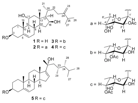
respectively. Our continuing chemical investigation on S. crassa has led to the isolation of one new sterol, crassarosterol A (1), and four new steroidal glycosides, crassarosterols B–E (2–5) (Figure 1).
The structures of 1–5 have been established by extensive spectroscopic analysis, including 2D NMR (1H–1H COSY, HMQC, HMBC, and NOESY) spectroscopic analysis, chemical methods, and HPLC analysis. The anti-inflammatory activity of 1–4 to inhibit up-regulation of the pro-inflammatory iNOS (inducible nitric oxide synthase) and COX-2 (cyclooxygenase-2) proteins in LPS (lipopolysaccharide)- stimulated RAW264.7 macrophage cells and the cytotoxicity of compounds 1–5 against a panel of cancer cell lines including human liver carcinoma (HepG2 and HepG3), human breast carcinoma (MCF-7 and MDA-MB-231), and human lung carcinoma (A-549) were evaluated in order to discover bioactive natural products.
Figure 1. Structures of compounds 1‒5.
2. Results and Discussion
The HRESIMS of crassarosterol A (1) exhibited a pseudomolecular ion peak at m/z 459.2509 [M+Na], consistent with a molecular formula of C28H46O3 and requiring six degrees of unsaturation.
The 13C NMR and DEPT spectroscopic data (Table 1) displayed 28 carbon signals, including five methyls, nine methylenes, ten methines, and four quaternary carbons. The IR spectrum revealed the presence of hydroxy group (3389 cm–1). The carbon resonances at δ 141.2 (C), 121.3 (CH), 156.9 (C), and 106.3 (CH) suggested the presence of two double bonds. The above data coupled with the characteristic 1H NMR signals for methyl groups at δ 0.92 (3H, s), 1.18 (3H, s), 1.04 (3H, d, J = 6.8 Hz), 1.03 (3H, d, J = 7.2 Hz), and 1.03 (3H, d, J = 7.2 Hz) and signals for olefinic protons at δH 5.41
Table 1. 13C NMR sectroscopic data of cmpounds 15.
position 1a 2b 3b 4a 5a
1 39.1, CH2 39.2, CH2 39.2, CH2 39.2, CH2 37.1, CH2
2 31.8, CH2 29.7, CH2 29.5, CH2 29.7, CH2 29.6, CH2
3 71.8, CH 78.2, CH 77.8, CH 78.2, CH 78.3, CH 4 42.7, CH2 39.1, CH2 39.0, CH2 39.1, CH2 38.8, CH2
5 141.2, qC 140.6, qC 140.7, qC 140.5, qC 140.3, qC 6 121.3, CH 121.7, CH 121.6, CH 121.8, CH 122.1, CH 7 31.9, CH2 31.8, CH2 31.8, CH2 31.8, CH2 31.5, CH2
8 31.4, CH 31.3, CH 31.3, CH 31.3, CH 30.4, CH 9 56.9, CH 56.8, CH 56.9, CH 56.8, CH 50.3, CH 10 38.1, qC 38.3, qC 38.3, qC 38.3, qC 36.9, qC 11 68.9, CH 68.9, CH 68.9, CH 68.9, CH 21.0, CH2
12 51.4, CH2 51.4, CH2 51.4, CH2 51.4, CH2 36.2, CH2
13 42.9, qC 42.9, qC 42.9, qC 42.9, qC 47.4, qC 14 53.7, CH 53.6, CH 53.6, CH 53.6, CH 57.9, CH 15 36.3, CH2 36.3, CH2 36.3, CH2 36.3, CH2 31.0, CH2
16 72.5, CH 72.5, CH 72.5, CH 72.5, CH 123.8, CH 17 61.2, CH 61.2, CH 61.2, CH 61.2, CH 160.9, CH 18 14.0, CH3 14.1, CH3 14.1, CH3 14.1, CH3 18.1, CH3
19 19.1, CH3 19.0, CH3 19.0, CH3 19.0, CH3 19.3, CH3
20 29.6, CH 29.6, CH 29.6, CH 29.6, CH 76.0, qC 21 18.2, CH3 18.2, CH3 18.2, CH3 18.2, CH3 29.6, CH3
22 34.8, CH2 34.8, CH2 34.7, CH2 34.7, CH2 49.1, CH2
23 31.2, CH2 31.2, CH2 31.2, CH2 31.2, CH2 29.6, CH 24 156.9, qC 156.9, qC 156.9, qC 156.9, qC 45.5, CH 25 34.1, CH 34.0, CH 34.0, CH 34.0, CH 30.9, CH 26 21.8, CH3 21.8, CH3 21.8, CH3 21.8, CH3 20.9, CH3
27 21.9, CH3 21.9, CH3 21.9, CH3 21.9, CH3 21.5, CH3
28 106.3, CH2 106.3, CH2 106.3, CH2 106.4, CH2 11.6, CH3
29 15.7, CH3
1’ 97.4, CH 94.7, CH 97.2, CH 97.2, CH
2’ 74.0, CH 68.6, CH 70.1, CH 70.1, CH
3’ 66.9, CH 72.1, CH 69.5, CH 69.5, CH
4’ 70.9, CH 72.4, CH 73.0, CH 73.0, CH
5’ 65.8, CH 65.3, CH 65.2, CH 65.2, CH
6’ 16.0, CH3 16.1, CH3 16.2, CH3 16.2, CH3
OAc 170.9, qC 171.5, qC 171.3, qC 171.3, qC
21.2, CH3 21.1, CH3 20.8, CH3 20.8, CH3 a Spectra were measured in CDCl3 (100 MHz); b Spectra were measured in CDCl3 (125 MHz).
(1H, d, J = 5.6 Hz), 4.76 (1H, s), and 4.70 (1H, s) (Table 2) suggested 1 to be a member of 24- methylenecholesterol class [12,13]. The 1H1H COSY correlations allowed the assignment of four separated spin systems (Figure 2). The presence of sp2 methylene substituent at C-24 was further confirmed by the HMBC correlations from H2-28 to C-23, C-24, and C-25 (Figure 2). Likewise, the steroidal nucleus was confirmed by the HMBC correlations from H3-18 to C-12, C-13, C-14, and C-17 and H3-19 to C-1, C-5, C-9, and C-10. The NOE correlations between H3-19/H-1β, H-3/H-1α, and H3- 19/H-11 suggested the α and β orientations for H-3 and H-11, respectively. The absence of an NOE correlation between H3-18/H-17 and the presence of the correlation between H-17/H-16, H-16/H-14, and H-14/H-9 suggested the α orientation for H-9, H-14, H-16, and H-17. Moreover, the β orientation for H-20 was evidenced from the NOE correlations between H3-18/H-20, H3-18/H-12a, and H3-21/H- 12a. The absolute configuration of 1 was determined by the application of Mosher’s method. Analysis of the 1H NMR data of esters 1a and 1b resulted in the determination of a 3S configuration (Figure 3).
Figure 2. Selected 1H1H COSY (▬) and HMBC (→) correlations of 1 and 5 and the fucose residue in 1–5.
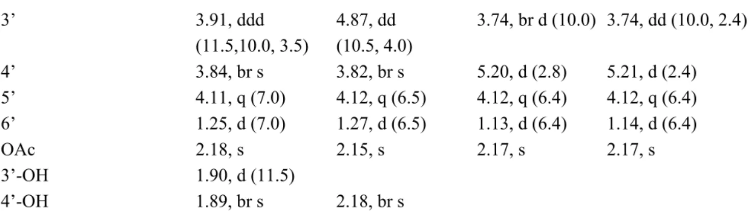
Analysis of the HREIMS and 13C NMR spectroscopic data of crassarosterol B (2) suggested a molecular formula of C36H58O8. The IR spectrum of 2 showed the presence of hydroxy (3461 cm1) and carbonyl (1741 cm1) groups. The latter was identified as an acetoxy group according to the carbon resonances at δ 170.9 (C) and 21.2 (CH3) (Table 1). The 1H NMR spectroscopic data of 2 showed characteristic methyl signals at δ 0.92 (3H, s), 1.18 (3H, s), 1.04 (3H, d, J = 6.8 Hz), 1.03 (3H, d, J = 7.2 Hz), 1.03 (3H, d, J = 7.2 Hz), 5.41 (1H, d, J = 5.6 Hz), 4.76 (1H, s), and 4.70 (1H, s) (Table 2), revealing that 2 has the same 24-methylenecholesterol skeleton as that of 1. By excluding the steroidal moiety and the acetoxy group, the remaining six carbons [δ 97.4 (CH), 74.0 (CH), 66.9 (CH), 70.9 (CH), 65.8 (CH), and 16.0 (CH3)] were ascribed to the presence of a 6’-deoxyhexose residue. The sugar residue was deduced as an α-fucopyranose on the basis of 2D NMR analysis (Figure 2) and the coupling constants of 3JH-1’,H-2’ (4.0 Hz), 3JH-2’,H-3’ (10.0 Hz), 3JH-3’,H-4’ (3.5 Hz), and 3JH-4’,H-5’ (< 1 Hz) (Table 2) [12,14,15]. The acetoxy group attached at C-2’ of the α-fucose residue was evidenced from the downfield chemical shift of H-2’ (δ 5.07). The HMBC correlation from H-1’ to C-3 disclosed that
the α-fucose residue was attached at C-3 of the steroidal aglycone. The absolute configuration of the sugar moiety in 2 was determined by reversed phase HPLC analysis of its o-tolylthiocarbamate [16].
The liberated fucose from acid hydrolysis of 2 was treated with L-cysteine methyl ester followed by reaction with o-tolylisothiocyanate to afford the corresponding o-tolylthiocarbamate derivative. The retention time of the liberated sugar derivative by HPLC analysis was found to be consistent with that of standard L-fucose derivative.
Crassarosterol C (3) gave the same molecular formula as that of 2 based on the analysis of the HRESIMS and 13C NMR spectroscopic data (Table 1). The NMR spectroscopic data of 3 were similar to those of 2, with some exceptions for those of sugar residue. An HMBC correlation from the anomeric proton at δ 5.12 (H-1’) to the carbon signal at δ 77.8 (C-3) connected the fucose residue to C- 3 of the steroidal aglycone. The downfield proton chemical shift at δ 4.87 (1H, dd, J = 10.5, 4.0 Hz) was ascribed to the presence of an acetoxy group at C-3’ (Table 2). The detailed 2D NMR analysis confirmed the above elucidation. Likewise, the L-fucose residue was deduced according to RP HPLC analysis of the corresponding o-tolylthiocarbamate as described above.
Figure 3. 1H NMR chemical shift differences of MTPA esters of 1.
Crassarosterol D (4) was assigned the same molecular formula as those of 2 and 3. A comparison of NMR spectroscopic data of 4 with those of 2 and 3 revealed that an acetoxy group should be located at C-4’ of fucose residue (Tables 1 and 2). This was evidenced by the 1H NMR shift of H-4’ at 5.20 (1H, d, J = 2.8 Hz). In the same manner, RP HPLC anslysis of the corresponding o-tolylthiocarbamate derived from the hydrolyte of 4 allowed the determination of L-fucose moiety.
The HRESIMS and 13C NMR spectroscopic data of crassarosterol E (5) established a molecular formula of C37H60O7. The presence of an acetoxy group was evidenced by the 1H NMR signal at δ 2.17 (3H, s) (Table 1) and 13C NMR signals at δ 171.3 (C) and 20.8 (CH3) (Table 2) as well as the IR absorption band at 1737 cm1. The NMR spectroscopic data appropriate for the sugar moiety of 5 were quite similar to those of 4, suggesting that they shared the same 4’-O-acetylfucose residue. Except for the sugar moiety, the remaining 29 carbon signals as well as the characteristic methyl signals at δ 1.00 (3H, s), 1.05 (3H, s), 1.38 (3H, s), 0.86 (3H, d, J = 6.4 Hz), 0.89 (3H, d, J = 6.4 Hz), 0.76 (3H, d, J = 6.8 Hz), and 0.78 (3H, d, J = 6.4 Hz) (Table 2) revealed that the aglycone of 5 should have a C29
steroidal skeleton [17]. The 23,24-dimethyl-20-hydroxy side chain was deduced by the 1H1H COSY correlations from H2-22 to H3-29 through H-23, from H-24 to both H3-26 and H3-27, and from H-24 to H3-28 as well as the HMBC correlations from H3-21 to C-17, C-20, and C-22 and H3-29 to C-22, C-23, and C-24. This rare steroidal side chain is the same as that of sarcophytosterol isolated previously by
Table 2. 1H NMR Spectroscopic Data of Compounds 15.
# 1, δH (J in Hz)a 2, δH (J in Hz)b 3, δH (J in Hz)b 4, δH (J in Hz)a 5, δH (J in Hz)a 1 a: 2.55, dt
(13.6, 3.6)
a: 2.58, dt (13.5, 3.5)
a: 2.56, dt (13.5, 3.5)
a: 2.58, dt
(14.0, 3.6) a: 1.86, m b: 1.16, m b: 1.16, m b: 1.16, m b: 1.16, m b: 1.10, m 2 a: 1.81, m a: 1.85, m a: 1.79, m a: 1.81, m a: 1.89, m b: 1.58, m b: 1.65, m b: 1.66, m b: 1.64, m b: 1.60, m
3 3.53, m 3.51, m 3.43, m 3.49, m 3.49, m
4 a: 2.30, m a: 2.36, m 2.24, m a: 2.36, m a: 2.36, m
b: 2.26, m b: 2.26, m b: 2.26, m b: 2.24, m
6 5.41, d (5.6) 5.41, d (5.5) 5.40, d (5.5) 5.41, d (5.6) 5.38, br d (3.2) 7 a: 1.99, m a: 1.99, m a: 1.98, m a: 1.99, m a: 2.01, m
b: 1.54, m b: 1.56, m b: 1.54, m b: 1.54, m b: 1.61, m
8 1.50, m 1.50, m 1.49, m 1.50, m 1.66, m
9 0.99, m 0.99, m 0.97, m 0.98, m 1.01, m
11 4.07, td
(10.8, 4.8) 4.07, m 4.06, m 4.07, td
(10.8, 4.4) 1.59, m 12 a: 2.31, m a: 2.31, m a: 2.31, m a: 2.31, m a: 2.10, m
b: 1.18, m b: 1.18, m b: 1.18, m b: 1.18, m b: 1.59, m
14 0.98, m 0.98, m 0.98, m 0.98, m 1.41, m
15 a: 2.24, m a: 2.24, m a: 2.23, m a: 2.23, m a: 2.08, m b: 1.17, m b: 1.16, m b: 1.17, m b: 1.16, m b: 1.87, m
16 4.40, m 4.40, m 4.40, m 4.40, m 5.50, br s
17 1.07, m 1.07, m 1.07, m 1.07, m
18 0.92, s 0.92, s 0.92, s 0.92, s 1.00, s
19 1.18, s 1.18, s 1.17, s 1.18, s 1.05, s
20 1.86, m 1.86, m 1.86, m 1.87, m
21 1.04, d (6.8) 1.04, d (6.8) 1.04, d (6.5) 1.04, d (6.4) 1.38, s 22 a: 1.68, m a: 1.68, m a: 1.68, m a: 1.67, m a: 1.59, m
b: 1.22, m b: 1.22, m b: 1.22, m b: 1.22, m b: 1.48, m 23 a: 2.18, m a: 2.18, m a: 2.18, m a: 2.18, m 1.82, m
b: 1.95, m b: 1.95, m b: 1.95, m b: 1.95, m
24 1.06, m
25 2.24, m 2.25, m 2.25, m 2.25, m 1.42, m
26 1.03, d (7.2) 1.03, d (7.0) 1.03, d (7.0) 1.03, d (6.8) 0.86, d (6.4) 27 1.03, d (7.2) 1.03, d (7.0) 1.03, d (7.0) 1.03, d (6.8) 0.89, d (6.4) 28 a: 4.76, s a: 4.76, s a: 4.76, s a: 4.76, s 0.76, d (6.8)
b: 4.70, s b: 4.70, s b: 4.70, s b: 4.70, s
29 0.78, d (7.2)
1’ 5.02, d (4.0) 5.12, d (4.0) 5.04, d (4.0) 5.04, d (4.0)
2’ 5.07, dd
(10.0, 4.0)
4.02, m 3.93, dd (10.0, 4.0)
3.93, dd (10.0, 4.0)
3’ 3.91, ddd (11.5,10.0, 3.5)
4.87, dd (10.5, 4.0)
3.74, br d (10.0) 3.74, dd (10.0, 2.4) 4’ 3.84, br s 3.82, br s 5.20, d (2.8) 5.21, d (2.4) 5’ 4.11, q (7.0) 4.12, q (6.5) 4.12, q (6.4) 4.12, q (6.4) 6’ 1.25, d (7.0) 1.27, d (6.5) 1.13, d (6.4) 1.14, d (6.4)
OAc 2.18, s 2.15, s 2.17, s 2.17, s
3’-OH 1.90, d (11.5)
4’-OH 1.89, br s 2.18, br s
a Spectra were measured in CDCl3 (400 MHz); b Spectra were measured in CDCl3 (500 MHz).
us from the soft coral Lobophytum sarcophytoides [17].The NMR spectroscopic data for the aglycone moiety of 5 are almost the same as those of sarcophytosterol, except for some minor variations in 1H and 13C chemical shifts at C-2, C-3, and C-4 between both compounds. This is due to the attachment of the sugar residue at C-3 of the steroidal aglycone. Similarly, HPLC analysis of the relevant o- tolylthiocarbamate derived from the hydrolysis of 5 suggested the presence of L-fucose.
The absolute configuration of sterol 1 has been established by Mosher’s method in the present work.
On the basis of biogenesis, the steroidal moieties of the glycosides 2–5 should possess the same absolute configurations as shown in the formulae. Cytotoxicity of steroids 1–5 against HepG2, HepG3, MCF-7, MDA-MB-231, and A-549 cancer cell lines was evaluated. The results showed that 1 exhibited cytotoxicity toward HepG2 cancer cell line with an IC50 value of 14.9 µM, while 4 also showed cytotoxicity toward HepG2 and HepG3 cell lines with IC50 values of 17.6 and 18.9 µM, respectively. The other compounds were found to be inactive (IC50 > 20 μM) toward the above cancer cell lines after 72 h exposure. The anti-inflammatory activity of steroids 1–4 against the accumulation of pro-inflammatory iNOS and COX-2 proteins in RAW264.7 macrophage cells stimulated with LPS was also evaluated using immunoblot analysis. At a concentration of 10 µM (Figure 4), steroid 2 was found to significantly reduce the level of iNOS protein to 12.9 ± 4.3% and 4 could reduce the iNOS espression to 50.1 ± 6.3%.
Figure 4. Effect of compounds 1–4 at 10 μM on the LPS-induced pro-inflammatory iNOS and on COX-2 protein expression of RAW264.7 macrophage cells by immunoblot analysis. (A) Immunoblots for iNOS and β-actin, and relative density of iNOS. (B) Immunoblots for COX-2 and β-actin, and relative density of COX-2. The values are means ± SEM (n = 6). The relative intensity of the LPS alone stimulated group was taken as 100%. Under the same experimental condition, 10 μM CAPE (caffeic acid phenethyl ester; Sigma Chemical. Company, St. Louis, MO) reduced the levels of the iNOS and COX-2 protein to 0.8 ± 4.5 % and 75.6 ± 12.2 %, respectively, relative to the control cells stimulated with LPS. *Significantly different from LPS alone stimulated group (*P < 0.05).
3. Experimental Section
3.1. General Experimental Procedures
The melting point was determined using a Fisher-Johns melting point apparatus. Optical rotations were determined with a JASCO P1020 digital polarimeter. IR spectrum was obtained on a JASCO FT/IR-4100 spectrophotometer. The NMR spectra were recorded on a Varian 400 MR NMR or Varian Unity INOVA 500 FT-NMR instrument at 400 or 500 MHz for 1H (referenced to TMS, δH 0.00 ppm for CDCl3) and 100 or 125 MHz for 13C (referenced to δC 77.0 for CDCl3). ESIMS were recorded by ESI FT-MS on a Bruker APEX II mass spectrometer. Silica gel 60 (Merck, 230400 mesh) and LiChroprep RP-18 (Merck, 40–63 μm) were used for column chromatography. Precoated silica gel plates (Merck, Kieselgel 60 F254, 0.25 mm) and precoated RP-18 F254S plates (Merck, 1.05560) were used for TLC analyses. High-performance liquid chromatography (HPLC) was performed on a Hitachi L-7100 pump equipped with a Hitachi L-7400 UV detector at 210 nm and a semi-preparative reversed- phase column (Merck, Hibar Purospher RP-18e, 5 μm, 250 × 10 mm).
3.2. Animal Material
The soft coral Sinularia crassa was collected by hand using scuba off the coast of Sansiantai, Taitung county, Taiwan, in July 2008, at a depth of 10 m, and was stored in a freezer. This soft coral was identified by one of the authors (C.-F. D.). A voucher specimen (specimen no. SST-03) was deposited in the Department of Marine Biotechnology and Resources, National Sun Yat-sen University.
3.3. Extraction and Isolation
The frozen bodies of S. crassa (1.1 kg fresh wt) were minced and extracted with EtOH (3 × 2 L).
The organic extract was concentrated to an aqueous suspension and was further partitioned between EtOAc and H2O. The EtOAc extract (17.0 g) was fractionated by open column chromatography on silica gel using n-hexane–EtOAc and EtOAc–MeOH mixtures of increasing polarity to yield 32 fractions. Fractions 25, eluting with EtOAc–MeOH (8:1), was further separated by silica gel column chromatography with gradient elution (n-hexane–acetone, 8:1 to 2:1) to yield four subfractions (25A–
25D). Subfraction 25B was subjected to RP-18 column chromatography (MeOH–H2O, gradient, 50–
90%), and subsequently purified by RP-18 HPLC (CH3CN–H2O, 65%) to obtain compounds 1 (6.6 mg) and 5 (1.2 mg). Compounds 4 (1.8 mg) was obtained from subfraction 25C using RP-18 HPLC (CH3CN–H2O, 65%). In the same manner, Subfraction 25D was fractionated over RP-18 gel using gradient elution (MeOH–H2O, gradient, 50–90%) to yield two subfractions (25D-1 and 25D-2).
Subfraction 25D-2 was separated by RP-18 HPLC (CH3CN–H2O, 85%) to yield compounds 2 (1.8 mg) and 3 (1.6 mg).
Crassarosterol A (1): white powder; [α]24D –45 (c 0.66, CHCl3); IR (KBr) vmax 3389, 2962, 2925, 2854, 1462, 1048, 1024 cm‒1; 13C NMR and 1H NMR data, see Tables 1 and 2; ESIMS m/z 453 [M+Na]+; HRESIMS m/z 453.3342 [M+Na]+ (calcd for C28H46O3Na, 453.3344).
Crassarosterol B (2): white powder; [α]24D –34 (c 0.18, CHCl3); IR (KBr) vmax 3461, 2960, 2928, 2868, 1741, 1467, 1377, 1244, 1077, 1030 cm‒1; 13C NMR and 1H NMR data, see Tables 1 and 2;
ESIMS m/z 641 [M+Na]+; HRESIMS m/z 641.4027 [M+Na]+ (calcd for C36H58O8Na, 641.4029).
Crassarosterol C (3): white powder; [α]24D –17 (c 0.16, CHCl3); IR (KBr) vmax 3388, 2963, 2930, 2857, 1732, 1458, 1375, 1258, 1041 cm‒1; 13C NMR and 1H NMR data, see Tables 1 and 2; ESIMS m/z 641 [M+Na]+; HRESIMS m/z 641.4026 [M+Na]+ (calcd for C36H58O8Na, 641.4029).
Crassarosterol D (4): white powder; [α]24D –52 (c 0.18, CHCl3); IR (KBr) vmax 3426, 2960, 2930, 2859, 1735, 1461, 1375, 1247, 1074, 1033 cm‒1; 13C NMR and 1H NMR data, see Tables 1 and 2;
ESIMS m/z 641 [M+Na]+; HRESIMS m/z 641.4026 [M+Na]+ (calcd for C36H58O8Na, 641.4029).
Crassarosterol E (5): white powder; [α]24D –45 (c 0.12, CHCl3); IR (KBr) vmax 3440, 2960, 2925, 2855, 1737, 1461, 1377, 1244, 1074, 1036 cm‒1; 13C NMR and 1H NMR data, see Tables 1 and 2;
ESIMS m/z 639 [M+Na]+; HRESIMS m/z 639.4234 [M+Na]+ (calcd for C37H60O7Na, 639.4237).
3.4. Preparation of (S)-and (R)-MTPA Esters of 1
To a solution of 1 (1.0 mg) in pyridine (0.4 mL) was added (R)-MTPA chloride (25 μL), and the mixture was allowed to stand for 3 h at room temperature. The reaction was quenched by the addition of 1.0 mL of H2O, and the mixture was subsequently extracted with EtOAc (3 × 1.0 mL). The EtOAc layers were combined, dried over anhydrous MgSO4, and evaporated. The residue was subjected to short silica gel column chromatography using n-hexane−EtOAc (3:1) to yield the (S)-MTPA ester, 1a (0.7 mg). The same procedure was used to prepare the (R)-MTPA ester, 1b (1.0 mg from 1.0 mg of 1), with (S)-MTPA chloride. Selected 1H NMR (CDCl3, 400 MHz) of 1a: δ 7.41−7.52 (5H, m, Ph), 5.48 (1H, br d, J = 6.0 Hz, H-6), 4.89 (1H, m, H-3), 4.76 (1H, s, H-28a), 4.70 (1H, s, H-28b), 4.41 (1H, m, H-16), 4.05 (1H, m, H-11), 3.57 (3H, s, OMe), 2.62 (1H, br d, J = 14.0 Hz, H-1a), 2.48 (1H, m, H-4a), 1.85 (1H, m, H-2a), 1.17 (3H, s, H3-19), 1.03 (6H, d, J = 7.2 Hz, H3-26 and 27), 0.92 (3H, s, H3-18);
selected 1H NMR (CDCl3, 400 MHz) of 1b: δ 7.41−7.53 (5H, m, Ph), 5.47 (1H, br d, J = 5.2 Hz, H-6), 4.89 (1H, m, H-3), 4.76 (1H, s, H-28a), 4.70 (1H, s, H-28b), 4.41 (1H, m, H-16), 4.06 (1H, m, H-11), 3.57 (3H, s, OMe), 2.65 (1H, br d, J = 13.6 Hz, H-1a), 2.37 (1H, m, H-4a), 1.77 (1H, m, H-2a), 1.17 (3H, s, H3-19), 1.03 (6H, d, J = 7.2 Hz, H3-26 and 27), 0.92 (3H, s, H3-18).
3.5. Determination of Sugar Configuration
Authentic samples of D-fucose and L-cysteine methyl ester hydrochloride (each 0.5 mg) were dissolved in pyridine (0.1 mL) and heated at 60 °C for 1 h. The mixture was added o- tolylisothiocyanate (0.5 mg in 0.1 mL pyridine) and heated at 60 °C for additional 1 h. The reaction mixture was directly analyzed by reversed-phase HPLC (Mightysil RP-18 GP column; 4.6 × 250 nm;
25% CH3CN in 50 mM H3PO4; 0.8 mL/min; 35°C) and detected at 250 nm to give the retention time of the o-tolylthiocarbamate of sugar. The retention of the o-tolylthiocarbamate derived from L-fucose,
L-cysteine methyl ester, and o-tolylisothiocyanate was obtained by the same manner.
A solution of the glycoside (0.4 mg for each) in 0.6 M HCl/dioxane (1:1 v/v, 0.2 mL) was heated at 80 °C for 24 h. After cooling, the solution was neutralized with Amberlite IRA-400, and the resin was removed by filtration. The filtrate was extracted with EtOAc. The aqueous layer was dried in vacuo and the afforded residue was dissolved in pyridine (0.1 mL) containing L-cysteine methyl ester (0.5 mg), followed by heating at 60 °C for 1 h. A 0.1 mL solution of o-tolylisothiocyanate (0.5 mg) in pyridine was added to the mixture, which was heated at 60 °C for additional 1 h, to yield the corresponding o-tolylthiocarbamate derivative. Reverse phase HPLC analysis of the o- tolylthiocarbamate derivatives derived from the hydrolyte of the glycosides 2–5 showed peaks at 28.2, 28.0, 28.1, and 27.9 min, respectively, while the tR values for standard L-fucose and D-fucose derivatives were observed at 28.0 and 25.4 min, respectively, suggesting the presence of a L-fucose residue in 2–5.
3.6. Cytotoxicity Testing
Cell lines were purchased from the American Type Culture Collection (ATCC). Compounds were assayed for cytotoxicity against human liver carcinoma (HepG2 and HepG3), human breast carcinoma (MCF-7 and MDA-MB-231), and human lung carcinoma (A-549) cells using the MTT [3-(4,5- dimethylthiazol-2-yl)-2,5-diphenyltetrazolium bromide] method [18]. Freshly trypsinized cell suspensions were seeded in 96-well microtiter plates at densities of 5000‒10000 cells per well with tested compounds added from DMSO-diluted stock. After 3 days in culture, attached cells were incubated with MTT (0.5 mg/mL, 1 h) and subsequently dissolved in DMSO. The absorbency at 550 nm was then measured using a microplate reader. The IC50 is the concentration of agent that reduced cell growth by 50% under the experimental conditions.
3.7. In Vitro Anti-Inflammatory Assay
Macrophage (RAW264.7) cell line was purchased from ATCC. In vitro anti-inflammatory activities of tested compounds were measured by examining the inhibition of lipopolysaccharide (LPS) induced upregulation of iNOS and COX-2 proteins in macrophage cells using western blotting analysis [19,20].
4. Conclusions
Prior investigation of the genus sinularia reported some steroidal glycosides; however, all of them were found to possess a 24-methylene substituted side chain [21,22]. 5 is the first example of steroidal glycoside with a 23,24-dimethyl substitited side chain from soft coral of the genus sinularia. Our biological data revealed that 2́-O-acetyl–L-fucose functionality play an important role toward the
inhibition of the pro-inflammatory iNOS. Steroidal glycoside 2 could be useful anti-inflammatory agents, while steroids 1 and 4 have shown their inhibitory activity toward the selected human liver cancer cells.
Acknowledgments
This work was supported by grants from the National Science Council of Taiwan (NSC98-2113-M- 110-002-MY3) and Ministry of Education (98C031702) awarded to J.-H. S.
References
1. Blunt, J.W.; Copp, B.R.; Munro, M.H.G.; Northcote, P.T.; Prinsep, M.R. Marine natural products.
Nat. Prod. Rep. 2011, 28, 196–268.
2. Su, J.-H.; Ahmed, A.F.; Sung, P.-J.; Chao, C.-H.; Kuo, Y.-H.; Sheu, J.-H. Manaarenolides A–I, diterpenoids from the soft coral Sinularia manaarensis. J. Nat. Prod. 2006, 69, 1134–1139.
3. Chao, C.-H.; Wen, Z.-H.; Wu, Y.-C.; Yeh, H.-C.; Sheu, J.-H. Cytotoxic and anti-inflammatory cembranoids from the soft coral Lobophytum crassum. J. Nat. Prod. 2008, 71, 1819–1824.
4. Lu, Y.; Huang, C.-Y.; Lin, Y.-F.; Wen, Z.-H.; Su, J.-H.; Kuo, Y.-H.; Chiang, M.Y.; Sheu, J.-H.
Anti-inflammatory cembranoids from the soft corals Sinularia querciformis and Sinularia granosa. J. Nat. Prod. 2008, 71, 1754–1759.
5. Tseng, Y.-J.; Ahmed, A.F.; Dai, C.-F.; Chiang, M.Y.; Sheu, J.-H. Sinulochmodins A−C, three novel terpenoids from the soft coral Sinularia lochmodes. Org. Lett. 2005, 7, 3813–3816.
6. Ahmed, A.F.; Su, J.-H.; Kuo, Y.-H.; Sheu, J.-H. Scabrolides E−G, three new norditerpenoids from the soft coral Sinularia scabra. J. Nat. Prod. 2004, 67, 2079–2082.
7. Ahmed, A.F.; Shiue, R.-T.; Wang, G.-H.; Dai, C.-F.; Kuo, Y.-H.; Sheu, J.-H. Five novel norcembranoids from Sinularia leptoclados and S. parva. Tetrahedron 2003, 59, 7337–7344.
8. Sheu, J.-H.; Ahmed, A.F.; Shiue, R.-T.; Dai, C.-F.; Kuo, Y.-H. Scabrolides A−D, four new norditerpenoids isolated from the soft coral Sinularia scabra. J. Nat. Prod. 2002, 65, 1904–1908.
9. (a) Radhika, P.; Rao, P.R.; Archana, J.; Rao, N.K. Anti-inflammatory activity of a new sphingosine derivative and cembrenoid diterpene (lobohedleolide) isolated from marine soft corals of Sinularia crassa Tixier-Durivault and Lobophytum species of the Andaman and Nicobar Islands. Biol. Pharm. Bull. 2005, 28, 1311–1313. (b) Anjaneyulu, V.; Radhika, P. Two new sphingosine derivatives from Sinularia crassa Tixier-Durivault of the Andaman and Nicobar Islands. Indian J. Chem. 1999, 38B, 457–460.
10. Chao, C.-H.; Chou, K.-J.; Huang, C.-Y.; Wen, Z.-H.; Hsu, C.-H.; Wu, Y.-C.; Dai, C.-F.; Sheu, J.- H. Bioactive cembranoids from the soft coral Sinularia crassa. Mar. Drugs 2011, 9, 1955–1968.
11. Radhika, P.; Cabeza, M.; Bratoeff, E.; García, G. 5α-Reductase inhibition activity of steroids isolated from marine soft corals. Steroids 2004, 69, 439–444.
12. Kobayashi, M.; Kanda, F.; Damarla, S. R.; Rao, D. V.; Rao, C. B. Marine sterols. XVII.
polyhydroxysterols of the soft corals of the Andaman and Nicobar coasts. (2). Isolation and structures of three 16β-hydroxy steroidal glycosides from an Alcyonium sp. soft coral. Chem.
Pharm. Bull. 1990, 38, 2400–2403.
13. Kobayashi, M.; Krishna, M. M.; Anjaneyulu, V. Marine sterols. XXIV. Isolation of 24- methylenecholestane-1α,3β,3α,6β,16β-pentol from Sinularia sp. of soft coral. Chem. Pharm. Bull.
1992, 40, 2845–2846.
14. Cóbar, O. M.; Rodríguez, A. D.; Padilla, O. L. A new steroidal glycoside from a Caribbean gorgonian, Eunicea sp. J. Nat. Prod. 1997, 60, 1186–1188.
15. Wang, S. -K.; Dai, C. -F.; Duh, C. -Y. Cytotoxic pregnane steroids from the Formosan soft coral Stereonephthya crystalliana. J. Nat. Prod. 2006, 69, 103–106.
16. Tanaka, T.; Nakashima, T.; Ueda, T.; Tomii, K.; Kouno, I. Facile Discrimination of aldose enantiomers by reversed-phase HPLC. Chem. Pharm. Bull. 2007, 55, 899–901.
17. Lu, Y.; Lin, Y. -C.; Wen, Z. -H.; Su, J. -H.; Sung, P. -J.; Hsu, C. -H.; Kuo, Y. -H.; Chiang, M. Y.;
Dai, C. -F.; Sheu, J. -H. Steroid and cembranoids from the Dongsha atoll soft coral Lobophytum sarcophytoides. Tetrahedron, 2010, 66, 7129–7135.
18. Alley, M.C.; Scudiero, D.A.; Monks, A.; Hursey, M.L.; Czerwinski, M.J.; Fine, D.L.; Abbott, B.J.;
Mayo, J.G.; Shoemaker, R.H.; Boyd, M.R. Feasibility of drug screening with panels of human tumor cell lines using a microculture tetrazolium assay. Cancer Res. 1988, 48, 589–601.
19. Jean, Y.-H.; Chen, W.-F.; Sung, C.-S.; Duh, C.-Y.; Huang, S.-Y.; Lin, C.-S.; Tai, M.-H.;
Tzeng, S.-F.; Wen, Z.-H. Capnellene, a natural marine compound derived from soft coral, attenuates chronic constriction injury-induced neuropathic pain in rats. Br. J. Pharmacol. 2009, 158, 713–725.
20. Jean, Y.-H.; Chen, W.-F.; Duh, C.-Y.; Huang, S.-Y.; Hsu, C.-H.; Lin, C.-S.; Sung, C.-S.; Chen, I.- M.; Wen, Z.-H. Inducible nitric oxide synthase and cyclooxygenase-2 participate in anti- inflammatory and analgesic effects of the natural marine compound lemnalol from Formosan soft coral Lemnalia cervicorni. Eur. J. Pharmacol. 2008, 578, 323–331.
21. Anjaneyulu, V.; Rao, K. N.; Radhika, P.; Kobayashi, M. Isolation of 24-methylcholest-5-ene-3 beta,25-diol-3-O-beta-D-arabinopyranoside from Sinularia grandilobata of Andaman and Nicobar Islands. Indian J. Chem., Sect. B 1996, 35, 757–759.
22. Anjaneyulu, V.; Rao, P. V. S.; Radhika, P. A new sphingosine derivative and a new polyhydroxy steroidal glycoside from Sinularia gravis Tixier-Durivault of the Andaman and Nicobar Islands.
Indian J. Chem., Sec. B 1999, 38, 357–360.
Samples Availability: Not available.
© 2011 by the authors; licensee MDPI, Basel, Switzerland. This article is an open access article distributed under the terms and conditions of the Creative Commons Attribution license (http://creativecommons.org/licenses/by/3.0/).