中山醫學院入十問學年度碩士論文
研究所到:醫學研究所
指導教授:李秀雄博士 王朝鐘博士
論文名稱 Milbemycin D 及 VD圍99-11 封感祟廣東住血緣
蟲老鼠治療效泉之生化評估
Biochemical evaluation of ]\生ilbemycin D a盟d VD-99-11o盟 A拘:giostro揖igylus cantonensis infected in rats
研究生:徐玲玉
中華民圈八十五年六月
館僻刷酬金耳問時F團制叭叭4院制制制也請岫對矗闡明抽山明明目中內川川
授權書
(博碩士論文)
本授權書所授權之論文為本人在中山醫學技 醫學研究所
…一…一一一一組 84 學年度第 2 學期所撰碩士學位論文。
論文名稱 Milbemycin D 及 VD-99-11 對感染廢棄廣東住血線蟲老鼠治療敷果之生化評估
串串同意 口不悶意
本人具有著作財產權之論文揖耍,授予盟家厲害館、本人畢業學校及行政院 關家科學委員會科學技衛資料中心,得重製成電子資料檔後 i投錄於該單位之 綱路,並與台灣學衛網路及科技駕路連議,得不限地域時間與次數,以免碟 或紙本重製發行。
---一一一一一叮叮-呵--司---﹒咽_._--._---~
輯同意口不同意
本人具有著作財產權之論文全文資料,授予行政院路家科學委員科學技衛資 料中心,得不限地域時問與次數以徵縮、光碟重製後發行,並得享該中心徵 縮小組製作之研究報告、獎勵代表作、搏碩士論文三擋資料等館新台幣值倍 元之服務 o 本論文因涉及專利等智慧財產權之申請,請將本論文全文延後至
民國一年一月後再公愣。
---一一一←一…一…一一一一句司---
體同意口不同意
本人具有著作財產權之論文全文資斜,授予教育部指定送績之間書館及本人 畢業學校園書館,為學衛研究之宮的以各種方法草製'或為上述目的再授讓 他入以各種方法叢製'不限時賠與地域,推每人以一份為限。
上述授權內容均無須訂立讓與及授權契約書 o 依本授權之發行權為非專屬性發行 權利 o 依本授權所為之收錄、重製、發行及學衛研發利用均為無償。
指導教授姓名:李秀雄
1 、六\J
研究生簽名 :fiL-Li--士… 學號:R帥的5
(親筆正楷)
自期:貝閻 85一年一6一月 B
---且-巴巴一一一一一…一一一---耐._--輛---.---
備註1.上述同意與不同意之攔立若未鉤選,本人摺意視同授權 o 2. 授權第二項者,請再交論文一本予承辦人員 o
3. 本授權書已於民國 85年 4 丹 10 日送語著委會侈正定稿 o
簽署人須知
工,放著作攘麓的巍覽會哇梅里真能說聽聽、覺草案輿實質結警方式繫會鐘內學擴贅料,
均須先得到著作晨才產權人授權,諾分別在三三種利用方式的摺意攝內鈞選並裝置是
各項資料 o
2. 所謂非專屬授權是指被授權人所取得的權利並非獨占性的使辯護,授權入尚可 將相同的權利重覆授權結他人使用;反立即為專屬授權,如果您巴鑫署專屬授 權寄予其他法人或自然入,請勿筆著本授權書。
3. 授權人的權利與義務:
在美國授權搏碩士論文予 UMI 公奇(搏碩士論文全文質科發仔企奇)製作發行 , ~頁交付美金晶亮的出放贅,錯舊年逼七件以上時得事投入 10擎的權利金約美 金 20 元;在闊內本計畫之經費全數由政府支應,收入亦應歸鷗庫,為答謝您的 支持,科資中心特為您援供新台幣 500 元的等值資料服務(1;).研究報告、獎勵 代表作、搏碩士論文三擋為限) ,請逕洽本案聯絡入,地挂電話詳如第 5 項 o 義務方面唯一要注意是,著作人自後不可以主張終止本授權薔雪 {B 您仍可以授 權其他自然人或法入上述的行為。
4. 全閣博碩士論文全文資料散結片整合計葷的宏觀妓益:
在個入方面,您的論文將可永久保存{徵縮技備在理論上可保存八百年,實證 已逾百年)也因為您的授權,使得後進得以透過電腦網路與先碟多管道按索
,您的論文將因而被充分利用。在國家總體利益方面,紙本容易盟影印而造成 裝訂上的傷害,圖書館中孤本的公閱陳列與外倍也有破損之虞'唯有頓政府全 面性的整合,借助科技設備才能一舉完成保存與和用的全方位效益,回憶您過 去尋找資料之不便經驗,學弟與學妹確實 2頁要您的論文與授權番。
5. 本案聯絡電話 (02)7377746 江守盟、王淑貞 她主!l:::台花市和平東路之段 106號 17摟 1702蜜
..._---_...-‘---
研究生姓名:徐玲玉 聯絡電話: (04)3896190-12331 地址: 台中市南區大慶衛工段 113 號中山醫學院寄生蟲學科
本論文為中山醫學院授理學碩士學位之品 備條件之一會經中山醫學院研究所碩士論文
考試委員會審查合格及口試通過。
口試委員
高雄醫學院寄生蟲學科主任顏仝敏博士
榕在~乏
中山醫學院寄生蟲學科主任 李秀雄博士
(論文指導教授) 不乏朮
中山醫學院生化研究所所長 王朝鐘博士
(論文指導教授) 學糾結
中華民國入十五年六月
學生徐玲玉論文題器為說ilbe臨yCl琵 B 及 VD棚剪鵬口
對感接廣東住血線蟲老鼠治療效泉之生化詩估?其論 文已經中山醫學院醫學研究所碩士論文考試委員會 審查合格及口試通過?並由指導教授核時後無誤 O
指導教授:李秀雄博士 簽名:
王朝鐘博士 簽名:
中華民國八十五年六月
這乞謝
本論文首先要感謝兩位恩師 寄生蟲學科主任李秀雄
博士在學業、工作與研究土悉心的指導及生化前所長王朝 鐘博士在生化領域餌,心之指引,得以順利完成型特誌銘謝,
永感謝忱 o 文稿初成復蒙 亮雄學醫學院寄生蟲科主任顏 仝敏博士撥冗詳細指丘,並提供寶貴意見書使之史臻完美雪 白衷感激。
求學期間乎感謝徐成全教授、蔣思?東老師在蛋白質純 化技術之鼎力相助;林玉玲老師、李妙真老師、小械、自
君在生化實驗之全力支持;許振求醫師、林偉隆醫師在為 理切片研究之費心襄助;味美倫老師、蔡崇弘老師在生物 統計學之盡心輔助會廖娟娟小姐在整個實驗過程之全力輔
佐;育芳、婉瑩、俐均、日萱、仲錄和業鑄在資料排版土 之細心協助,使本文得以順利完竣?在此一帶誌謝 o
最後僅將本文獻給摯愛的先生喘文章、可愛的女兒 E 佳 真、最關心我的父母以及所有協助我的良師益友 o
入十五年六月
目錄
中文摘要 1
英文摘要 2
第一章緒論
弟
KRr-BhFF 前、'言、 哇
第二節研究動機 10
第二章材料與方法 11
第二章結果
23第四章討論 27
圈表及說明
32參考文獻 51
中文摘要
以宿主肺臟絃織蛋白變化之現象?評估不同之 r
aminobutyric acid(GABA) 類驅蟲蔡 VD-99-11 及 Milbernycin D' 治療感染廣求住血線蟲第三期幼蟲的大白鼠之治療效 果?作為與傳統治療效泉之比較 9 以 j家討其優劣性。實驗 結泉在感;長廣求住血線蟲而未治療及以 VD-99闕.1 1 治療組之 老鼠肺臟組織微粒體的 SDS且PAGE 電泳分析中發現分子量 22-kDa 有一明顯蛋白質,然未感在及以 Milbemycin 0 治療 組之老鼠肺臟組織的微粒體卻沒有相同蛋白質;使用
superdex 200 column 作連續層析法純化,鑑定比蛋白質其化
學位及免疫性為鐵蛋白 (ferritin) ,再利用等電點電泳法及 離子交換層析法確認其為 L fcrritin 0 觀察應用免疫組織祟 色法之為理切片,在惑:按廣求住血線蟲肺臟組織有發炎現 象,浸潤之吞噬細胞內也有 ferritin 之堆積。因此?推測鐵 蛋白 (ferritin) 為感系廣求住血線蟲致病機轉之重要因 素 o 據此?我們認為以生化方法評估治療效果方法正確同 時也可作為探討致為機轉之研究方法之一。
英文摘要
Protein variation of the lung tissue in the rats
,which infected for
Angi仿trongyluscantonensfs third larvae were treated with
VDθ9-11and lnilbemycin
D歹 c01nparedwith the effects of traditional chemotherapeutics. Sodimn dodecyl sulfate-polyacrylamíde gel elcctrophoresís techniques were used to analyze the biochemistry differences of the lung tissue
叫他 found
the
22次Daprotein was quantitatively increased in the lung micros01nes from the Angiostrongylus
cαntonensisinfected and incured
rats歹 however,it was alInost absent in normal and cured rats. The protein was purified by sequential chromatography on Superdex 200 column
,and was identified chemica
l1y and immunologically as ferritin. Moreover
,using isoelectric focusing and anion exchange
chromatography歹 itwas further known as L ferritin. The distribution of thís
22去Daprotein in the lung tissue of the Angiostrongylus cantonensis infected rats was studies by immunocytochemistry. The positively stained ce
l1s were mainly infíltration macrophages.
2
Fr
OlTIthe
result歹明Tepredicted that there lnay be
SOlne relatíonships between ferritin and pathogenesis of
infect吋Angiostrongylus cantonensis. Therefore
,We think the therapeutic used biochelnical evaluation lnentioned above was correct and could be one of the methods to study pathogenesis
J '、
前言
廣求住血線蟲是一種鼠類寄生蟲?最早於 1933 年由 中函學者躁,心陶先生在中國大陸廣求省玄鼠及褐鼠之肺 臟所發現寄生蟲?命名為 Pulmonen的 cantonensis
1937
年日人拉本 (Matsumoto) 在台灣求岸花蓮之野鼠體內發
現本蟲之感率會為台灣鼠類感奈之第一報告;同年橫)1]
(Yokogawa) 將之命名為斤的mostrongylus
ratti
0 世界第 一個人體報告?是 1945 年在台灣由野村 (N01TIUra) 和林(
Lin) 兩位醫師從一位十五歲疑似腦膜炎的日本男孩腦脊髓液中取得蟲體,經橫川證實為廣泉住血線蟲未成熟蟲 體 1946 年 Dougherty 發現灼的nonema cantonensis 和
Haemostrongylus
ratti 二屬與 Angiostrongylus 屬相同?而 重新命名此寄生蟲為廣求住血線蟲 (Angiostrongylus cαntonensis) 。然而在當時此寄生蟲並未受到廣泛注意;直到 1959 年 Hsieh 發表[台灣之人畜共通寄生蟲病概要]
( Outline of Parasitic Zoonoses ìn Taìwan)
,文章中捉到 1937 年松本和 1945 年野村干口林的發現?此寄生蟲為才受到世界各地的重視。
4
廣東住血線蟲雌性蟲體長 22-32mm 、直徑 O 詔"
0.50mm
,雄性蟲體長 20-25mm 、直徑 0.30-0 .40mm '成 蟲寄生於老鼠肺動脈及心臟 1955 年生活失首先由Mackerras and
Sandars 所描述,其生活史中?需要有軟體動物當為中間宿主?包括陸生螺類、水生螺類及蛤喻等
(Cross歹 1967~ Otsuru歹 1977) ,各種螺類為流行病學的重要 傳祟婊介;此外含有多種動物可當為你蟲宿主?如淡水蝦、
渦蟲、淡水蟹和蛙類(虎皮蛙、金線蛙) ( Alicata歹 1962~
Rosen et
α7.,1967;
Otsuru多 1979~Beck et a
1.,1980; Bowden
,1981)
,老鼠為自然界適當宿主賢人為非適當宿主;當第一期幼蟲由老鼠糞便排出之後?進入中!可宿主體內會由第 一期幼蟲發育為第二期幼蟲再繼續發育形成第三期幼 蟲,適當宿主老鼠由口腔食入感祟第三期幼蟲之中問宿主
後 1-2 天第三期幼蟲會到腦部,進入嗅球(
olfactory
lobes) 和大腦半球 (cerebralhemispheres)'
4-6 天形成第四期幼品,最後至IJ 蜘蛛膜下Jl空 (subarachnoid
space)
, 約7-9 天會形成第五期幼立起, 26-29 天移行五月干勁脈繼續發
育形成成蟲 T 由食入第三期幼蟲至糞便發現第一期幼蟲提 倡生活史需 42-45 天 (Bhaibulaya, 1975) 。3
廣求住血線蟲在非適當宿主只形成大 IJ 、長 7.7-18.3mm 、
直徑 0.03-0.3mm 之第四期幼蟲或第五期幼蟲(未成熟蟲
體) (Alicata
,1970) ,人的感祟因誤食感祟廣求住血線蟲第
三期幼蟲之中間宿主或係蟲宿主,感奈廣東住血線蟲在非 適當宿主體內?幼蟲移行時的機械性傷害及浸潤之嗜酸性
白血球所釋放之毒素及化學物質?是造成中樞神經系統病
害之主要原因( Y osh
Ì111ura
,1994); 人食入感祟性第三期
幼蟲之後,會侵入胃黏膜並進入血管及淋巴管造成胃腸道症狀(Jindrak and A1i cata
,1968) ,也可能侵入肝臟而造成 肝臟股大 (Yii , 1976) ,最後?這些幼蟲會經由血液循環或
用閏神經系統進入腦部(J indralι1968 、 1970~J indrak
and Alicata
,1970)
,造成發燒、嚴重頭痛、噁心或嘔吐、倦怠、頸部優硬、神經錯亂、昏迷甚至造成死亡等中樞神 經系統之為害性 (Alica泊,
1965)
;第四期或第五期幼蟲可在大腦髓質、橋腦、小腦、腦軟朕發現 (Yii ,
1976)
0 人 體感;各除了在腦部發現蟲體之外?此寄生品也會侵犯白衣部(Huang et al 歹 1964) ;另外, 1968 年 Yii et a1., 由一位台 灣五歲女孩之肺臟組織發現成蟲, 1986 年 Hwang et al寸
發現一個九位感祟者的家族中有四位死於肺臟併發症?6
1978 年 Sonakul 在泰國一位 34 歲之婦女肺臟組織也發現 蟲體。
廣泉住血線蟲主要分佈于熱帶及亞熱帶?從南緯 23 度到北緯 23 度?求經 100 度到西經 150 度?其氣候大多溫 暖潮;笠,涵蓋國家有台灣 (Chen , 1979) 、中國大陸 (Yang
et al., 1984) 、求南亞國家包括泰國 (Punyagupta ef al 叮
1970) 、馬來西亞 (Lim
and Ramachandran
, 1979) 、並可達 太平洋群島夏威夷 (Rosen et al 歹 1967) 、大溪地 (Rosen et al., 1961) 及澳洲北部 (Procivand
Brindley歹 1984) 手為造成當此居民嗜伊紅性腦膜炎及腦膜腦炎的主要原因( Alicata歹
1970) 。台灣、泰國及南太平洋地區包括夏威夷、大溪地為 廣求住血線蟲感祟嚴重地區。
台灣由於氣候及文化習慣?廣泉住血線蟲之中間宿 主、係蟲宿主和自然界適當宿主分佈廣泛, 1937 年松本在 台灣花蓮野鼠體內發現此寄生蟲蟲體之後, 1964 年
Kuntz
et al.,1967 年 Cross et 仗, 1972 年 Ch凹, 1975 年 Yii
et a1.,1976 年 Cross and Van Peenen歹和 1977 年
Otsuru歹等會封台灣不同地區之老鼠作流行病學之調查?發 現台灣有七種老鼠為廣求住血線蟲天然終宿主,分別是刺 鼠 (Rαttus coxingα coxzng,蚓、白腹鼠 (R.lose蚓、褐鼠 (R.7
norvegicus) 、家鼠 ( R. rattus subsp.) 、蘭嶼黑鼠仗. 1叫tus
mindanensis) 、澎湖黑鼠 (R. rattus rufescen抄、鬼鼠
( Bandicota indicα nemorivaga) , 其中鬼鼠之感;長率更高達 到%以上; 自然界的中向宿主有二種陸螺為非洲大蝸牛 (Achαtinα fú1ica) (Chiu歹 1964~
Cross
,1967
~Chen
et al.,1971
~ 乳Ten ,1973:
Chen et a1.,1974: Otsuru
et al.,1976:
Otsuru歹 1977) 、薄殼蝸牛 ( Bra砂baena similαris)(Chiu
,1964)
,一 種水螺為田螺 (Cipαngopaludinα chinensis)(Chang and Cross
,1966)
,二種蛤喻為大型企舌瑜 (Laevicαulis alte) (Otsuru,1977) 、小型全舌金會 (~αginulus plebei1的)
(Cross
,1967)
自 然界的保蟲宿主有一種渦蟲類 (planarian) (Cross歹 1967) 、 二稜青蛙由虎皮蛙(如na tigrina) 和金線蛙(如mplancyi) (Otsuru,
1977)
0 台灣從 1945 年發現世界第一個人 體為歷報告之後?間隔將近二十年直到 1964 年 Huang 發 現第二個為歷?但此後即不斷有為歷報告, 1979 年 Chen 統計台灣地區共有 259 個為反報告,死亡率高達 3.1% '
流行地區分佈於南台灣、求台灣,大多款是孩童感祟?並 無性別差異?流行季節於夏季兩季 ι9 月間;在台灣主要
8
感祟途徑大多因吃入未煮熟之非洲大蝸牛仔計,
197
5~Chen
,1979~ Hung and Chen歹 1988) ,而造成民眾相當大之危
害;目前已超過 300 個為歷 (Hwang eta
l.,1986)
;是台灣相當重要的人畜共通寄生蟲病, J主並-NR-適當之治療蕪物予 以治療?因此?以生化技術評估感祟廣求住血線蟲老鼠之
治療效果?以探討其致病機轉及作為發展新,驅蟲祟的參 考。
9
研究動機
自從 1937 年日人拉本 (Matsumoto) 在台灣求岸花蓮 之野鼠體內發現廣東住血線蟲感祟,且 1945 年野村 (Nomura) 及林 (Lin) 二位醫師在台灣發現廣求住血線蟲
世界第一個人,校為例報告之後?這程寄生蟲在台灣是相當 重要的人畜共通寄生蟲為之一 (Yii, 1976~
Chen
,1979)
0雖然?廣求住血線蟲對人類最嚴重的危害,係傷害人 體中樞神經系統會為造成嗜伊紅性腦膜炎及嗜伊紅性腦膜 腦炎之主要原因;但在 1968 年之後,在人體之肺臟也有該
蟲體之發現,因而使患者產生肺炎、肺肉芽腫、肺出血等
症狀 T 造成肺臟組織之為害性 (Yii et al., 1968~Sonakul
,1978~ Hwang
et al.,1986 ) ;而由於廣東住血線蟲如何破球 肺臟組織之玫病機轉方面之研究文獻頗稀,有鑑於此?本 研究利用生化技術分析感祟廣東住血線蟲之老鼠肺臟組織 及以蕪物治療之老鼠肺臟組織之蛋白質變化現萃,以採討 廣泉住血線蟲之致為機轉 T 並作為發展手持,驅蟲蔡之參考。
( ) i
材料與方法
(、動物是染
1 第三五期組蟲是>>交集
從實驗室中所感;祟長廣泉住血線蟲第一期幼蟲之
β
卸10ωn叮1η1可'P功hα01αωri的α gl的正αlb扣r縛叫t
方法:將感;祟吾的 B釘10仰111ψ'ph仰αl仰αωr川j叩j盯α gl的αb加r川甘叫αfωα 壓碎,以
30 之比例加入人工胃蛋白消化液,放入磁性攪拌 子於 37 0C 恆溫箱以磁性攪拌機攪拌 2 小時,取出用紗 布過 i處,加入生理食鹽水稀釋、靜置?每隔 30 分鐘,
再倒掉一半上清液,以生 J史食鹽水清洗至上;青液完全
清 5fït 再用滴管吸取沉澱物,於角平去lJ 顯微鏡下,收集 第三期幼蟲 O
人工 if妥正步改化J逆之庇護f:(使用前 l 小時自己製) 7 gm pepsin' (1 :250。可 Sigma)
8 ml IICI
Dist. water to 1000 ml
11
2. 實驗動鞠分組
使用動物為購白國科會動物中心之 Wistar
strain
四週齡雄性大白鼠?分實驗組、陽性對照組及陰性對 照紐;每組各 5 隻老鼠 O
3. 動鞠聽主義
實驗組及陽性封照組之老鼠,於感吾吾前 12 小時禁
令空腹?每隻老鼠以口餵管餵食 20 隻廣求住血線蟲 第三期幼蟲. I女性對照全且不作任何處理。
二、藥物治療
感染後第六天實驗組老鼠以口飯管分別給予
lnilbemycin
D 及 VD-99啊 1 1 之驅蟲惡毒治療 O1.
Milbemycin D :
5mgl旬之劑量治療,連續投蔡十夭。
2. VD-99-11
0.6mgl句之劑量治療?連續投蕪三天 O
12
三、艾文果評估
1 感染籬蟲數是喜f數
的.第一紛紛孟之紛至
感;長後第八週連緒三天收集老鼠糞便?以褔馬林
"乙 M 濃縮法 (formalin-ether concentration) 計算每去
克糞便中第一期幼蟲數目。
(2). 成品#炭
感;各後第十五週解剖老鼠,計算老鼠肺動脈及心 臟內之成蟲數口
Lì
立盤{拉普{當
的,原獻血是這之亭亭主
未感;長廣東住血線蟲之陰性封照姐、感;長未治療 之惕性對照組及經禁物治療之感;養老鼠解剖後會每隻 老鼠取 1 -h'克之肺臟組織?於;水;谷中儘受切碎,組織 與均質;夜之比例為 1 : 5 '以均質機研磨至組織完全磨 碎均質,於冷凍離心機分別以 600 淘金力離心 10 分 鐘書沉澱物為細胞核,上;青液再以 15歹000 xg 重力離 心 30 分鐘書沉澱物為粒線體,再耳叉上清液以 100歹000
xg
重力離心 60 分鐘 T 沉澱物為微粒體,上清液為細胞質;將細胞核、粒線體及微粒體之 pellete 溶於均質液,
與細胞質一起置於咀20 0C 係存口
均資FJ逆之庇EJ
0.282 g
TrÌs-HCl 歹 pH:7 .4(20mM) ,
67.24 mg EDT A (2mM) ,
10ml glycerol(10%) ,
349 戶!戶 -lnercaptoethanol
'
也H20
to 100 m
l.1.+
( 2). 玉宇 &f{Jt 安分布
取 20μi 之肺臟萃取物?加入 Bio-Rad
protein assay dye 2.5 ml
'室溫反應 ]0 分鐘, 5roIJ 595
nm 吸光 位 D 以 bovineserum al
bumin 為標準液?測定細胞核、粒線體、微粒體及細胞質之蛋白質濃度 O
(3) 宮永分布
j采用 Bio-Rad 'h司 Protean
II xi
cell 之電泳槽作 蛋白質電泳分析。蛋白質電泳的方法:將電泳玻片洗淨擦乾?利用 約 0.75mlηspacer 為間隔裝好電泳片?製備 ]2%
SDS-PAGE 之 separating
gel
'混合均勻後迅速倒入電 泳片中,直到液晶距離 well 約 1.5cm 處為止,加入去 離子 1]<.. ,覆蓋液函,待膠體凝固後 F 將上層水倒掉書再問始製備 4.0% SDS-PAGE 之 stacking
gel
'混合均 勻後倒入電泳片中?將電泳齒梳插入電泳片中?若有氣泡,則上下移動齒梳?使氣;包離間 gel '待 stacking
gel
~疑問後,拔掉齒梳,用二次清水清洗 wel1數次,準備 loading
sample
(Laemmli多 1970) 。15
取 2。他的蛋白質樣本加等量之 sample
buffer ,
於 1000C 煮沸 5 分鐘後?迅速放回冰上等待 loading
,
將所有撿體罵毛細管 tips 小心加至每個 well '加上標
準 proteins
standard lnarker (Bio-Rad)
,將上下電泳糟加 入 SDS 電泳緩街液至電導線被蓋過為止,以電源、供應 器是電?問始以 15mA 電流直到色帶跑至 stackinggel
與 seperating gel 的交界處?調整電流為 30mA '當色 帶跑至底線時,才停止電泳。電泳完畢後?將膠體取
出 T 以 COOlnassie blue 祟色
12
%SDS-PAGE 之 mcji:
12 ml 30 % acrylamide/0.8 % bis ' 9.9 ml d
2H
20,
7.5 ml
1.5 M Tris-HCI
,pH:8.8 '
300μ1
10 % SDS '
300μ10
% Alnmonium persulfàte'
12μ1
TEMED
16
4.0
%
SDS-PAGE 之 IffcJi:
1.3 ml 30
%
acrylamide/0.8%
bis ' 1 ml d2H20 '
2.51nl 0.5 M Tris-HCl, pH:6.8
100μ! 10
%
SDS100μi 10
%
Ammonium persulfate ' 10 μi TEMEDSample buJJel'月之庇護j J
1.0 ml 0.5M Tris-HCl, pH 6.8 1 .6 ml glycerol
1.61111 10
%
SDS0.4 ml ß-lnercaptoethanol
0.4 ml 0.5 %(W/V) bromophenol blue 3.0 ml d2H2
0
5x e!ectrode ln![!er 之府在rj •
15.0 g Tris base (25mM) 72 g glycine (192mM) 3.0 g SDS (0.1
%)
d2H2
0
to 1000 lnl17
0.1
%
Coomαssie blue 之 IffcJf:
1 g coomasie blue
G明250 ~容法令(400 ml methanol ' 100 ml acetic acid ' 500 ml Dis
t.water) 中?;容解後?
過濾、之。
屁在 f逆之 Iffc Jf
:
100 lnllnethanol ' 75 ml acetic acid ' d
2H
20 to 1000 lnl
四、 22-kDa protein 之單雄、純i t.&鑑定
1. 單離、純化
取感祟廣求住血線蟲老鼠肺臟組織均質後的微
粒體 4 ml 事先以 0 .4 5μ111 孔隙的 acetate menbrane j通
濾?再使用 Superdex200 gel filtration column (HR 26/60 phannacia) 層析?使用 501nM pH 7 .4之 Tris
buffer (0.1 M NaCl) ,流速每分鐘 2ml '於 uv 檢測 器下?每次收象 5ml '將每個 peak 所收集的蛋白質 作 SDS-PAGE 電泳分析?結泉第二個 peak 含有分子
18
量 22-kDa 之蛋白質?用 10mM pH7 .4之 Tris
buffer
透析?隔夜後 , l令 J求乾標;再;容於 6M
urea
'使用同 樣的 column 再一次層析?收集含有 22-kDa 蛋白質的第二個 peak '透析、冷凍乾海係存 O
2. 分子靈是灘罷
應用 gel
filtration
HPLC 之方法 T 測定比蛋白之 native 分子量 (Andrews1965
)。丹等純化的 22-kDa 蛋白質使用 TSK-3000
SW ultrapeak COl
UlTIn (7.5 x 300mlTI
Toyo-soda) ,緩街液為 0.1M phosphate buffer (O.lM Na2S04 ' 0.05% NaN
3 'pH
6.5) 流速每分鐘 lml '測量比蛋白質的 retardíng
time '
以已知分子量的蛋白質標準品作 Standardcurve
'計算 其分子量 O3.
22咀kDa 蠶由質 Polydonal antibo卻是聽備及鑑踅 將純化之 22-kDa 蛋白質( 500μgprotein/ml )
,與 等量之 Freund's
complete 佐劑乳化,在日本大白兔之背部皮下注射不同部位?二星期及四星期再分別追加
注射?於最後一次注射十二天之後?收集血液,離心,
取血清?進行各種分析。
19
o.
會inÆJ茫撈潰之芸 (Double immuno{,見伊lsion)將 1
%
agarose 以 100 0C 煮沸卜5 分鐘?冷卻豆 的。C 時再注入 75x15 mm 玻片上?打上四個;呵?在中 央槽中注入製備的抗體會周圍之槽中注入老鼠之liver ferritin (Sigma)
,置於室溫, 24 小時後觀察 Ob.
Æ 按當永 (lmmunoelectrophoresis)如雙相免疫擴散法 (Keyser, 1972) ,玻片先倒入
1 % agarose
'待冷凝後?於 agarose 上?打兩個圓槽?一方自槽注入純化的分子量 22-kDa 之蛋白 質,另一方自槽注入感祟廣東住血線蟲肺臟組織均 質 r皮書然後置於電泳槽,加入 60mM 的巴比妥酸緩 衝液 pH 8.6 之電泳液,電泳完後,在 agarose 中央
挖出一長條形之槽?注入 22- kDa 蛋白質之抗血清放
入湖溼培養且中, 48 小時之後?再脫蛋白?以 Amido
black
朵色觀察。c. 手才紹智泳三芸(
Isoelectric focusing)
使用 pH 梯皮子 10
(Ampholyte '
Bio-Rad) 之 薄層 polyacrylamide gels 進行電泳,首先 15 分鐘 100 伏特?接著 15 分鐘 200 伏特?最後的分鐘 450 伏 特 T 祟色觀察。20
t 旗子三言撈厚非法 (Anion
exchange chromatography)
使用 NeoBQr AQ colUlun (4 ml, Dynachrom AS) 層析 9 緩街液為 pH 7.4, 200mM Tris-HCI ,以鹽梯度
法沖提,記錄其層析題(\九Torwood
et a
l., 1976) 。e- J吾友,ZEZ 堅定成品的nl11unoblotting)
耳叉 5μgi屯f己的 22-kDa 蛋,白質、老鼠肺月改組織{技粒 體均質液及老鼠 1iver ferritin (Sigma) ,力。入 sample buffer 混合均勻, 100 Oc 加熱 5 分鐘使蛋白質變性,
然後 loading 至 120/0 的 SDS司PAGE 電泳?電泳完畢 後;取出膠體會轉移至硝化纖維紙 (Bio-Rad) (Towbin and Gordon, 1984) ,將硝化纖維紙與含 5 0/0 脫脂奶粉 的 PBS 作用 1 小時?加入 rabbit anti-rat 22-kDa protein antiserum (1 :2000 dilution) 於 37 0C 作用 2 小 時習以緩街液清洗,與 primaryantibody 作用後之硝化
纖維紙再進一步與 horseradish peroxidase conjugated goat antiωrabbit IgG antibody (Bio-Rad) 1午用! 小時?以 緩街液清洗,最後以受質 DAB kit (vector) 進行主色反 應?待色帶出現後?以蒸餾水終止呈色反應並清洗之。
21
五、蹄臟組織病理變化觀察
1 竊理協片
將肺臟組織固定於 10% 福馬林?以石蠟包 j史書切
片 5μm 以 hematoxylin闢eOS111 進行染色?封片、觀察 O
2. Immunocytochemistry
製作 5μlTI 之切片 , ì是於 xylene 脫蠟?將切片水化 依序;是於 100% 乙醇一手 95% 乙醇一步 70010 乙醇一手 500/0
乙醇?最後以 PBS 清洗?以合 0.05%(v/v) H
2
02 的甲醇?室混下作用 10 分鐘,去除組織中內生性過氧化酵素,
以 PBS 清洗 3 次,加入合;
10
0/0nonimmune goat
serum 之PBS
'室溫下作用 30 分鐘將之洗掉 I南方口 rabbitanti-rat 22-kDa protein antiserum (1: 1 000) (primary antibody)
,室 溫下作用的分鐘,以 PBS 清洗 9 滴加 biotinylate-goata 抓n社叮札ti卜卜“岫自刊.儡儡-侃.1-
S州t廿re叩p戶子乳加)1沁圳1<.ωi仇1V叫id副lI1 Cω011邪叭jμ戶ug伊at心:l刀cd hor刊S趴叩叫cω)ra吋a叫l吋di芯sh p咒eroxl泊da部se 反應後 ? 以 PBS 清洗,以 diaminobenzid ine 於室溫下祟色 1-4 分 鐘,蒸餾水沖洗?最後以 hematoxylin 作對比祟色。
22
結泉
(、 藥物治療去支持毛
丘克是凝滯幼品豆豆之愛?化
表一顯示使用 milbemycin D 治療感系廣求住血線 蟲之老鼠?糞便未發現第一期幼蟲會解剖後也未在月草 動脈及心臟發琨蟲體?老鼠體重及肺臟重量與陰性對
照組比較?以 Wi 1conson心 um rank 分析,結果 P>
0.05
,二者之間並沒有顯著差異 9 為治癒組;而以VD-99-11 治療感奈之老鼠,其幼蟲數、成蟲數、老鼠 重量及肺臟了重量與陽性對照組比較?以 Wilconson-Sum rank 分析,結果 P>
0.05
'二者之間也沒有顯著差異 為未治癒組。ZJ求是戶手于五字母 'Jt之安化
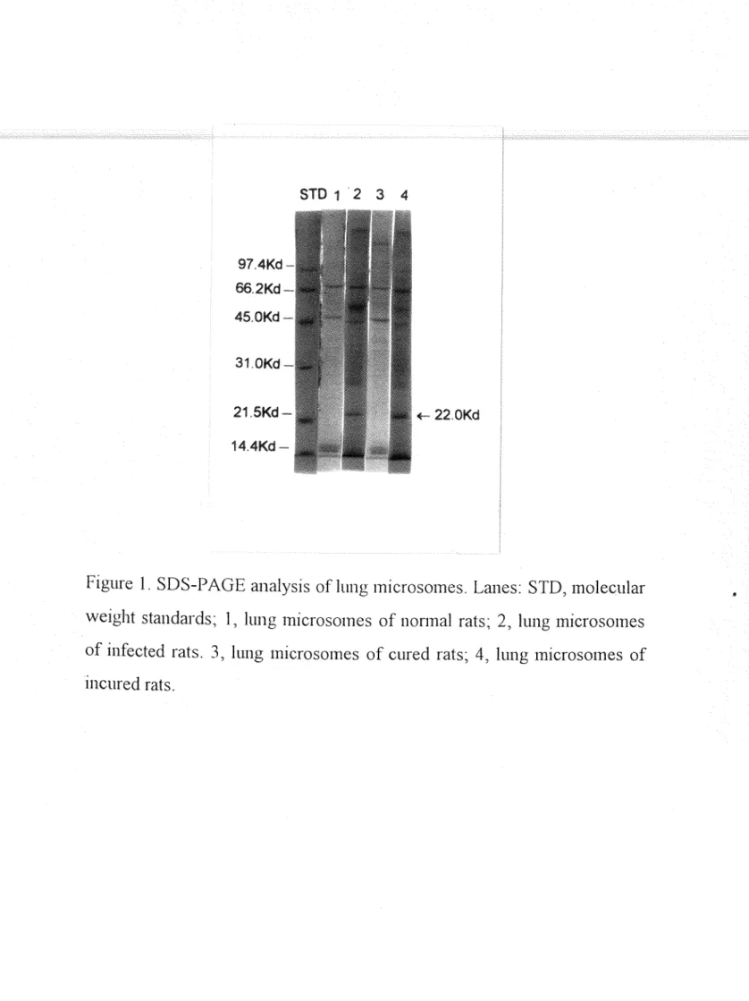
利用 SDS-PAGE 電泳分析老鼠肺臟組織蛋白質之 變化結果如園一,在陽性封照組及未治癒絃之老鼠肺臟 絃織微粒體?發現分子最 22-KDa 有一明顯蛋白質?而 在陰性封照組及治癒組之老鼠肺臟組織微粒體?並未發 現此蛋白質。
23
且. '- 22 …KDa 督白質之主義韓、純it.
將感;長廣泉住血線蟲之老鼠肺膝微粒體?以 Superdex
200 gel filtration
column 作連續層析法,在第一次層析,層折閩中如圈二 a '此蛋白質位於第二個 peak '冉一次 使用 superdex
200 gel filtration
column 層析?收集純度較高之分子量 22-kDa 蛋白質如閻二 b 0
三、 Ferritin 之鑑定
22-kDα 妥&安r native 分子畫、 isoform 之經定
應用 HPLC 以 TSK-3000
SW gel filtration
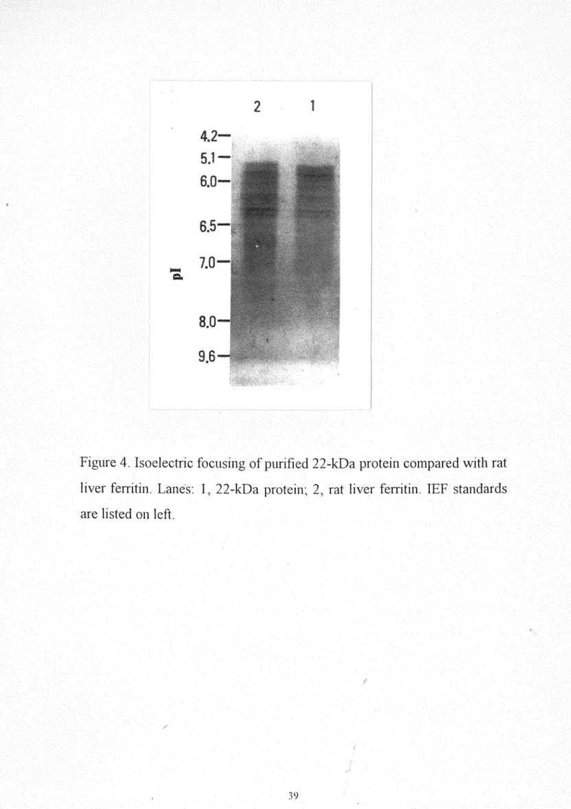
column 層 析,測定 22屯Da 蛋白質 native 分子量約為 440-kDa 0 (圈因為 ferritin 有 isoform '以等電點電泳法測定 22-kDa
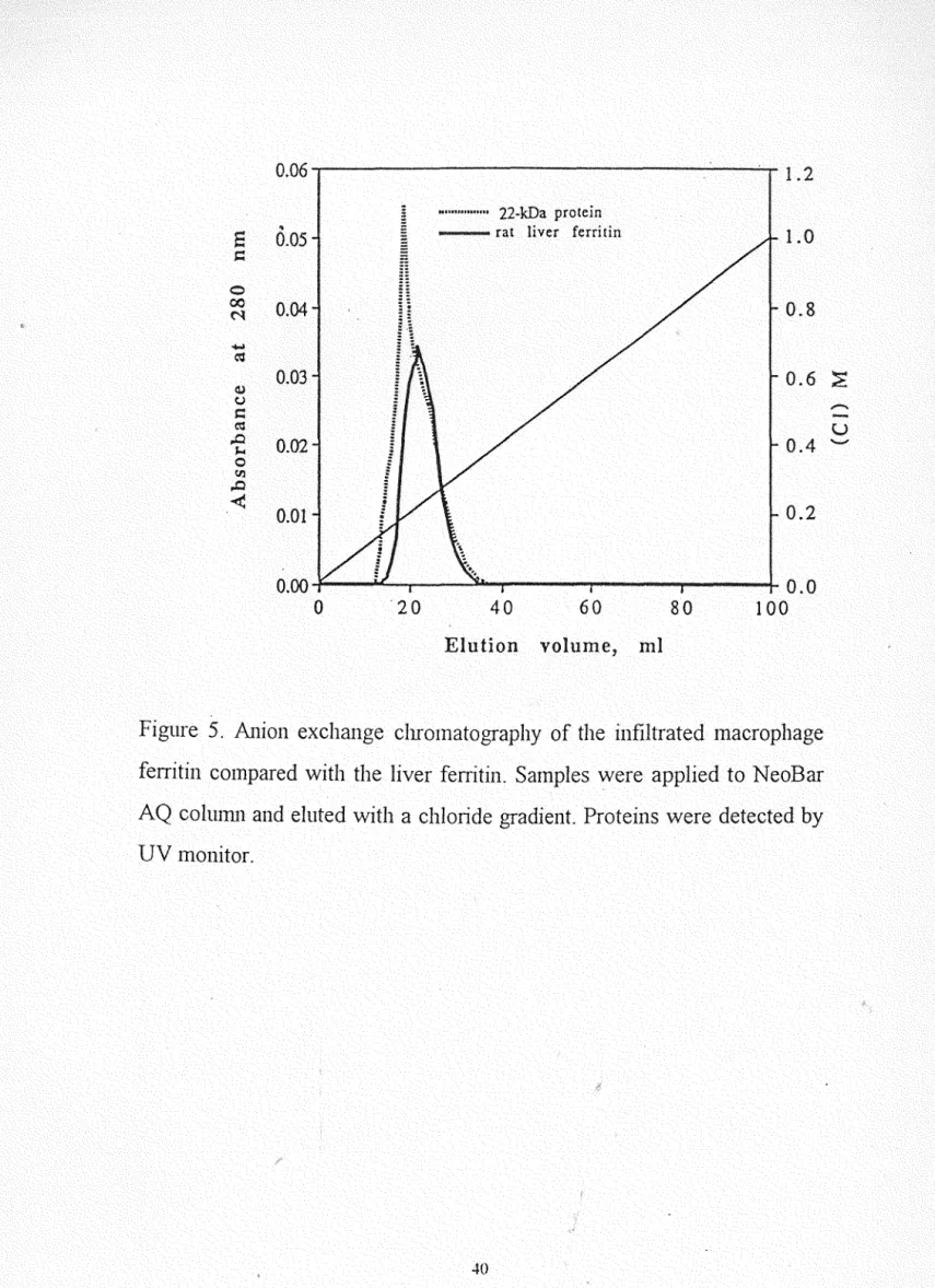
蛋白質之 PI 值為 5.0-6 .4(國四)與 L íèrritin 相似;離子 交換層析法其層析圖與老鼠之 liver ferritin 相類似, (圈五) 均顯著表示 22-kDa 蛋白質為 L
ferritin
024
Polvclonal
antibody 之震級、分布以純化之 22-kDa 蛋白質製備 polyclonal
antibody
,將 製備的 rabbitanti-rat 22-kDa
antiserUlTI 與純化之 22-kDa 蛋白質利用雙柏免疫擴散法觀察?是否有抗原-抗體反應之沉 澱線;由國六可看出中央槽中的兔血清?可分別與過園槽
中純化之 22-kDa 蛋白質產生免疫沉澱線?顯示兔血清確實 有對抗 22-kDa 蛋白質之抗體存在。
為了確認此抗體是否為單一特異性事由免疫電泳之分
析結果,如圈七在純化的 22次Da 蛋白質之一邊,有一條免 疫沉澱線會而在感祟廣求住血線蟲老鼠肺臟組織均質;夜這
邊乎也只有單一條沉澱線,因此,確認此抗血清為具有單 一特異性之抗體?以作為免疫分析使用。
J吾友,ZEZdd手法分布
為了鑑定 22-kDa 蛋白質, ;1存感;長廣求住血線蟲老 鼠肺臟組織均質液、正常老鼠肺臟組織均質液、純化的 22去Da 蛋白質以及老鼠 liver ferritin 作 immunoblotting 分 析?如閏八除了正常老鼠肺臟組織均質液在 22-kDa 沒有蛋 白質存在?其它均可在 22-kDa 發現明顯的蛋白質存在 O
25
由以上之實驗結泉?在感;長廣求住血線蟲及未治癒組 之老鼠肺臟組織微粒體所發現分子受 22-kDa 之蛋白質為
L
ferritin
0四、前職組織之病理變化現象
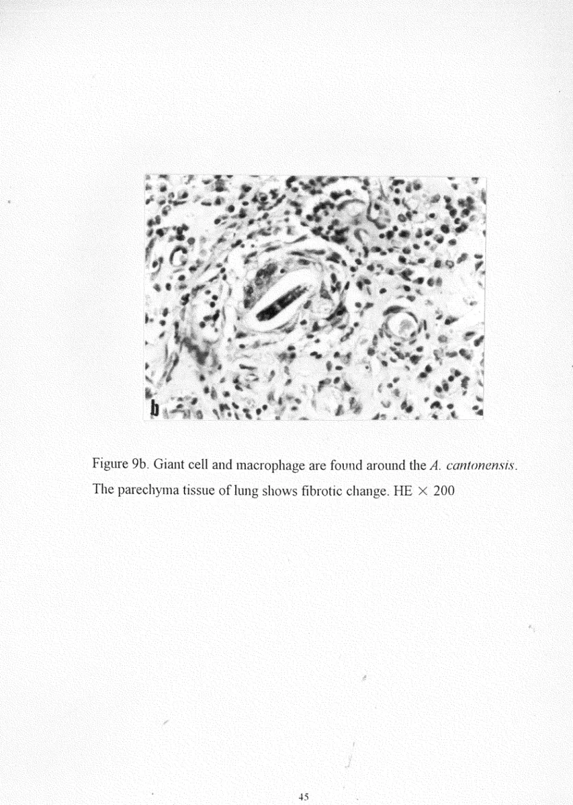
觀察感祟廣東住血線蟲肺臟組織之切片,在肺動脈內 有肉芽月童及血管內膜增生(國九 a) ,巨大細胞及吞噬細胞 浸潤肺臟實質組織,內含蟲卵及幼蟲,且伴有纖維化之現
~(崗九 b) ,浸潤之吞噬細胞會大多留繞於增生之血管用 問(國九 c) ,且發現其細胞質含有細小顆拉(圈九 d) 。
將感:M生廣求住血線蟲之老鼠肺臟組織作免疫組織;各 色法發現增殖的血管附近有浸潤之吞噬細胞雪細胞質內發 琨有拘小顆粒(閻十 a-b) ;相對地 7 圍繞於正常老鼠之 IJ 、文 氣管之吞噬細胞 9 頁IJ 無比現象(國十 c) 0 顯示有高量之 ferritin 堆積在感祟廣東住血線蟲之老鼠肺臟組織所浸潤
之吞噬細胞內口
26
討論
由於廣求住血線蟲幼蟲於感;各後第三天進入宿主之中 樞神經系統( lindrak多 1970) ,第十天會在腦部發育成未成熟
蟲體( Alicata and lindralι1970) 0 有多種禁物已由動物實
驗中證實,可殺死廣求往車線蟲幼蟲或未成熟蟲體會包括
thiabendazole (Cuckler et
α1.,1965; N ishimura
,1965/1966)
, 1-tetramisole
(Jindrak and
Alica侃 1969),mebendazole (Lammler and
Weidner歹 1975;Hayashi
et a1. 歹 1982;Hwang and
Ch凹,1988)
, f1ubendazole (Maki and Yanagisawa
,1983)
,avermectin B
la (Ishii
et a1. 、 1983)、 ivermectin(Ishii
et a1.,1985)
,and milbemycin D (Terada et
叫, 1987a),在本研究我 們使用二種 GABA 類禁物 milbemycin D 和 VD-99-11milbemycin
D 是屬於治療廣求住血線蟲 GABA 類蕪物之一, 1986 年 Sano et al 歹已發現在體外封廣東住血線蟲成 蟲會造成麻癖現象,且 1987 年 Terada et a1., 於老鼠感;長第 六天給予連續十天之禁物治療有相當顯著療妓 1995 年 本研究室 Lee et al 歹發表 VD-99-1
1 in
vitro 於低濃度會使 廣求住血線蟲成蟲產生麻痺現~;在以往研究治療禁物之 效汞?大多以治療後蟲體回收數、雌蟲排出之幼蟲數及肺27
臟重量作為治療技泉之判讀。在本實驗中?治療後除了以 蟲體回收數、雌蟲排出之幼蟲數及肺臟重量作為結泉之判 三賣?另外?也以肺臟組織蛋白質變異現~作為評估之標準?
實驗結泉發現以 lnilbemycin D 治療之實驗組?與 Terata
eta
1., 1987 年之研究結泉一致?可將廣泉住血線虫幼蟲完全 殺死而未能形成成蟲?在肺臟組織之電泳分析未發琨ferritin 而以 VD-99-11 治療之組別,雖然 VD -99-11 於體 外實驗會封成蟲蟲體造成麻痺, 1旦由於本實驗在感;長第六 天即予治療?此時還未形成成蟲蟲體?可見此蕪物無法將
廣求住血線虫幼蟲殺死,其蕪物機轉有待進一步深討 7 所 以在肺臟組織之電泳分析中發現與感;長而未治療老鼠月革中 相同之 ferritin 存在。
Ferritin 為人體儲存鐵之水溶性蛋白質?封體內鐵的
homeostasis 相當重要;由 24 個 subunit 所組成(
Munro and
Linder歹 1978)
,在一般發炎現象、惡性腫揚、肝炎等疾 病均會造成 ferritin 之增加(Hoy and J
achs歹 1981 ~Bacon
et叫,
1983; Peter
,1985
)。由於來自不同基因或分佈於不同 組織 ferritin 有 isoform 之存在 (Munro et a1.,1987 )
,分 別為 Hform 及 Lform
,分子量 21 ,000 Da 及 19,000Da'
二三者之間有 50 %之同質性 (Arosioet a
1., 1978~ Whiting歹 et28
。l 歹 1981); 1974 年 Powell et al 歹的研究指出由於病害所造 成鐵質過量的組織 L ferritin 會顯著增加。 1978 年
Wagstaff
et al., 1980 年 Khogo et al., 和 1981Bomford
et al.,分別研究體內 isoferritin 之特性?提出了一個結論 9 雖然?
H
ferritin 封鐵質之吸收較 t失且結合力較強?但在鐵質過量 之組織?堆積的卻是 L ferritìn 在本研究中感祟廣東住血 線蟲老鼠肺臟組織浸潤之吞噬細胞所堆積的也是 LferrìtÌn
0Ferritin 之增加?與經織之發炎 5克家有關 (Konijn ,
1977;
Konijn
et a1.,1981)
, 1980 和 1981 年 Dorner et a1., 研究 ferritin 合成與細胞之相關性?提出 ferritin 主要是由吞噬細胞及淋巴球合成 1991 年 Aguas et a1., 在動物實驗中發 現 ferritin 在發炎現呆會堆積於 membrane-bounded
vesi
c1es '
1992 年 Wesse1ius et a1., 提出在吸煙者之肺臟 組織浸潤的吞噬細胞也有 ferritin 之堆積,雖然?這些促使 ferritin 增加的機轉,並無法完全暸解? 然而, 1993 年Fahmy & Y
oung 提出由於發炎現~造成高濃度的細胞素(cytokìne) 及局部鐵之過量?因而促使 ferritin 在吞噬細胞 之合成增加。在感祟廣東住血線蟲老鼠肺臟組織切片雪肺 動脈內有血管內膜增生亨且伴有吞噬細胞之浸漓?細胞質 內發現有 ferritin 的堆積。
29
由於 Fe++鐵為自由基之催化因子 (Haber and Weiss 歹 1934)
,而自由基會造成許多組織傷害 (Ambruso et al., 1981~Whiting et
al.,1981 );
1981 年 Bennett 提出在紅血 球循環當中?吞噬細胞有吞食及財存鐵之功能;因此?當 組織受傷害?紅血球被破球所釋放之鐵質?被吞噬細胞吞 噬後,與 ferritin 結合?以減少 free ion 之含量?降低對 組織之傷害。在我們的實驗發現感祟廣求住血線虫之老鼠肺臟組織造成之為變與 ferritin 堆積在浸潤的吞噬細胞是
有很大的相關性?就觀察廣東住血線虫寄生肺動脈而論,蟲體會造成血管壁的破損?而使血液流入組織中,血液的 紅.lÚL球被吞噬細胞吞食?然後由 phagolysos01nes 消化?白 血色素釋放之鐵質 T 造成局部的鐵質過量及高濃度的細胞
素 (cytokine) 1980 年, Kay 提出這些細胞在紅血球循
環中去除毒素?是一種保護作用?因此?促使 ferritin 在吞 噬細胞合成?對組織而言?是一種保護作用。Ferritin 不僅存在脊椎動物 (Heusterspreute
and
Crichton 弓 1981) 會其它包括螺類 (Bottke、 1985~
Bottke et
al叮 1988) 、昆品 (NichoIand Locke
1989) 、才在物 (Ragland eta
l., 1990) 、微菌 (David et al., 1971) 、細菌 (Tsugitaand
Yar折,1985) 和寄生蟲 (Dietzel et al., 1992) 等均發現 ferritin 之存
30
在, 1992 年 Dietzel
et
a1., 提出寄生於血管之受森住血吸 蟲成蟲體內含有 ferritin'
1995 年 Ersfeld ef al叮在感祟色 生條蟲之為人血清發現此蛋白質明顯增加?而提出可以 ferritin 作為診斷之指標 1993 年 Fahmy&
Young 提出由於發炎現象造成高濃度的細胞索及局部鐵質過量,因而促
使 ferritin 在吞噬細胞合成;在本研究中感系廣東住血線蟲 老鼠肺臟組織浸潤之吞噬細胞有 ferritin 之堆積雪因此書推
測 ferritin 為感祟廣求住血線蟲致為機轉之重要因素 O 據
此?我們認為以生化方法評估治療效果?方法正確同時亦 可作為探討致為機轉之研究方法之一位
HJC ..
..CHl
CHl H CHl
至于子式: CJJ日 1107
骨子亞: 556.74
Chemical structure
of 恥1ilbemycinD
32
H
3:::去。九0
RChemlcal structure
ofVDθ弘 1133
R : CH3 20% 扭下
CzHs 80% .I2!上
表一
Effect of VD-嘗嘗。11 and milbemycin 0
against larval stages of
Angiostrongylus cantonensis
in ratsDmgs 。raldose No. LPG/female Rat BW at Rat lung-BW at No. of WOrIllS
(mglKg) exa1l1. 8th week(x 10 ') sacrifícc (g) ratio at sacrifíce
Control 0 5
(Positive)
VD-9弘 11 06.x3 5
Control (Negative)
O 5
扎1ilbemycin D 5x 1 0 5
12.3 士 10.9 246.6士 65.3 6 .4士 3.5
10.0J 4.7 295 7:L 150 30士 o. 只
O 240.0士 1 1. 8 0.6士 o 1
O 2908土 4 1. 1 0.5土 o 1
Results are shown as mean 士 SD of four animals each Wilconson-sum rank p
>
0.05recovered
18.0士 2.8
15.2+ 2.6
O
O
(Comparison of positive control group and which treat with VD-99-11 group Comparison ofnegative control group and which treat with milbemycin D group)
3.+
31.0Kd
•
22.0Kd1 2 3 4
的制脆把
色42 M們偏僻 d1i21.5Kd… 14.4Kd 一
Figure 1. SDS-PAGE analysìs of lung microsomes. Lanes: STD, molecular weìght standards; 1, lung microsomes of nonnal rats; 2, lung microsomes of infected rats. 3, lung mìcrosomes of cured rats; 4, lung microsomes of
1l1cured rats.
35
2.5
a
口MHH
pool
••
2.0
。∞村
1.5
1.0
0.5
茍d
c';l
ωυHHMHAhc切AJN
0.0
O 300 400
ml 200
volume
,
100 Elution
lung of hornogenous chrornatography
colurnn
nu nu
勻,'』x
'd e
戶LV rA
DA
QU U
Figure 2.
rnil1iters of Four
(a) rats.
infected
cantonCn"ìlS A.
frorn rnlcrosornes
hornogenous lung rnicrosomes was chrornatographed on a Superdex 200 COhlllli1. Fractions of second protein peak were pooled, dialysed oven1Îght against 10 mM Tris-buffer, and then lyophilized.
36
2.5
b
口抖抖個
pooI
←一吋
2.0
。∞科
1.5
吋吋
C官
1.0
0.5
心υ口MHA訕。mA吋
0.0
O 100 200 300 400
volume
,
m EJutionthen rechromatographed on a second Superdex 200 colmllil. Fractions eluting
and pooled, urea
disolved in 6扎在
1月lere
samples of this
(b)Two
between 125 and 150 ml were pooled, dialyed and lyophilized.
37
0.020
<
Uaa
〈向益,
已〈am
弘〈aa
nMmHa
口MMH
0.015
0.000 O 0.010
0.005
。∞村ωυHHMHA訕。mA吋
茍....
C官
12 15 m
9 voIume
,
3 6
Elution
TSK-3000 SW gel filtration COlllllli1 chromatography ofpurified Figure 3.
column included blue dextran (BD; >2 X 10 Mr),
apofemitin 糾正 443 ,000
Mr),22-kDa protein by
HPLC.
Molecular weight markers for thea1cohol
de~ydrogenase
(AD; 150,000 Mr), bovine albumin (BA; 66,000 Mr), carbonic anhydrase (CA; 29,000).38
2 1
Figure 4. Isoelectric focusing of purifíed 22次Da proteín compared with rat liver ferritin. Lal1es: 1, 22-kDa proteil1~ 2 歹 rat liver ferritin. IEF standards are listed 011 left
39
~ (一U) 1.2
1.0
0.8
0.6
。.4
0.0 100
0.2
"... 22-kDa protein
抽血岫由國 rat liver ferritin
0.00 G 0.06
。.04
0.01
O.的
0.05
0.03
但崗位{}∞N ωυnmwAlgmAJN
吻...
C嘻
80
、E.E."、2“口
60
volume ,
40
Elution
20
Figure 5. A
l1Ìo
l1exchange chromatography of the Ì
l1filtrated macrophage
fe汀itil1
compared with the liver ferriti
l1.Samples were applied to NeoBar AQ column a
l1d eluted with a chloride gradie
l1t.Proteins were detected by
40
UV monito
r.2
Figure6. Ouchter1ony gel diffusion of purifíed 22-kDa pr叫ein (1) and rat Iiver feritin (2). The antibody used was anti♂2-kDa protein antÍsenllTI
-1-1
Figure 7. lmmunoelectrophoresis in agarose gel of homogenous lung from A. cαntonens;s infected rats (1), and purified 22-kDa proteín (2). Antibody through (arrow) contained antiserum to the 22-kDa protein"Pr叫ems were stained wÌth Amído Black.
斗 2
Figure 8. Immunoblotting of the 22-kDa protein. Lanes: 1, homogenous lung of A. c正mton己的';s infected rats; 2, homogenous lung ofnonnal rats; 3, purified 22次Da protein; 4, rat liver ferritin. These proteins were ÍImnunostained with anti-22-kDa protein antÍsenlln. Prestained molecular weight standards were nm in parallel.
.+3
Figure 9a. A. cantone叭:;s in the Iung causing granuloma fonnation and vascular proliferatioll. HE X 100
44
Figure 9b. Giant cell and macrophage are found around the A. cantonensis.
The parechyma tÌssue of lung shows fibrotic change. HE X 200
是5
Figure 9c. Macrophage infiltration is noted near the pro1iferated blood vessels. HE
x
10046
Figure 9d. In位Itrated macrophage contaÌns c1ear cytoplasma with fine granules. HE X 200
..f.7
Fogure lOa. Immunohistochemical distributÌon of ferritin Ìn the lung of A.
cantonensi5; infected rat. Macrophages next the blood vesseIs 侈的 are
positively stained (arrows). F erritin/ ABC-H staining
x
5048
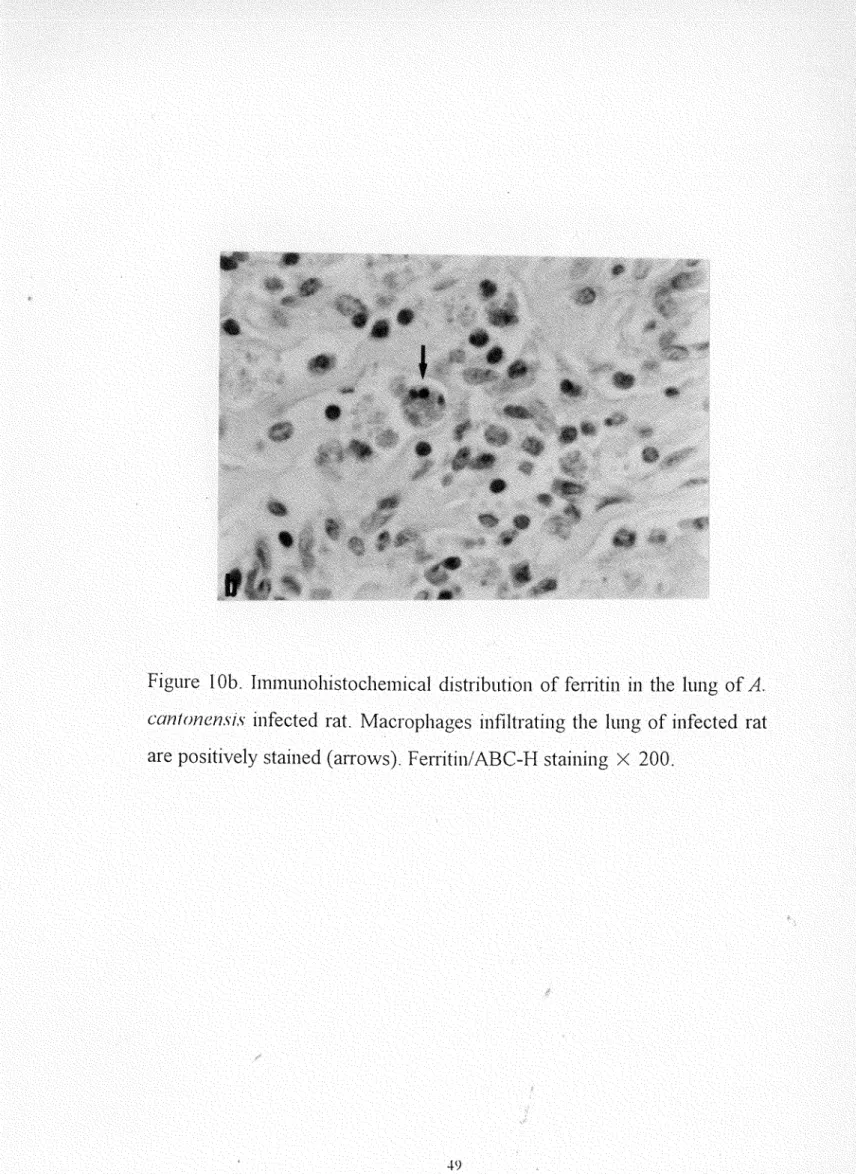
Figure 10b. lmmunohistochemical distribution of fenitin in the lung of A.
COl1 t仰的lS;S infected rat. Macrophages infiItrating the lung of infected rat are positively stained (a汀ows). Fenitin/ABC-H staining X 200.
-\.9
Figure 10c. lmmunohistochemical distribution of ferritin in the lung of nonnal rat. Macrophages in the lung of nonnal rat are not stained.(arrows).Ferritin/ABC-H staining X 200
50
參考文獻
Agu前, A. P., Grande, N. R. and Carvalho多五. (1991) 1nflammatory macrophages in the dog contaÎn high amounts of intravesicular ferritin and are associated with pouches of c01Ulective tissue fibers.
American Journal of Anatomy, 190( 1): 89-96.
Alica妞, J. E. (1962) (Nematoda: Metastronglidae) as a causative agent of eosinophilic meningitis of man in Hawaii and Tahiti. Can. ]. Zool.,
再0:5-8.
Alicata, J. H. (1965) Biology and distribution of the lungwonn An gi os t ro n f:,'Y! us ι﹒αntonens;s and its relation to eosinophilic meningitis and other. neurological disorders of man and animals.
1n: Adναnces m par的itο!06'Y Vol III. 1. B. Dawes (ed.) Academic Press Inc., London. New York. pp 223-248.
Ali侃侃, J. E. and Jindr法, K. (1970) Murine angiostrongylosis. 1n:
Anderson H.H.(ed) Angiostrongylosis in the Pacific and Southeast Asia. Thomas, Springfield, 1 口, pp 50-62
Ambnlso, D. R. and Johnsto11, R. B. (1981) Lactoferrin enha11ces hydroxyl radical productio11 by human neutrophils多 neutrophil
particulate fractio11s and enzymatic generating system. J. Clin.
1nvest., 41: 1628-36
Andrews, P. (1965) The gel-filtration behavior of proteins related to their molecular weights over a wide range. Biochem. J., 96: 595 個606.
51
Arosio多哎, Adelman, T. G. and Drysdale, J. W. (1978) On ferritin heterogeneity.主u抗her evidence for heteropolymers. 1. Biol. Chel咒,
253:445 卜4458.
Bacon, B. R., Tavi立, A. S., Brittenham歹 G. M., Park. C. H. and Reckagel, R. D. (1983) Hepatic lipìd peroxidation in vivo in rats with chronic íron over1oad. Joumal of Clinical Investigation, 71:
429-439.
Beck勻 M. 人 Cardina, T. M. and Alicata, J. E. (1980) Eosinophilic meningitis due to Angiostrongylus cantonensis in American Samoa.
Hawaii Med. J., 39: 254-257.
Benne缸, G. D. and Kay, M. M. B. (1981) Homeostatic removal òf senescent murine erythrocytes by splenic macrophages. Exp.
Hematol.亨 9: 297-307.
Bhaibulaya, M. (1975) Comparative stlldies of the life history of Angiostronf:,rylus m此kerras的 (Bhaibu1a戶 1968) and Angiostronf:,rylu,\' cαntonensis (Chen, 1935). Inter, J, Parasitol., 5: 7- 20.
Bomford, A., Conlon-Hollingshead多 C. and Munro ,日. N. (1981) Adaptive responses of rat tissue isoferritins to iron administration. J.
Biol. Chem., 256: 948-955.
Bottke, W. (1985) Electrophorestic and immlll1010gic stlldies on the stmcture of a mollusc fe訂itin. Comp. Biochem. Physiol., 81B: 325刪
334.
52
Bottke, \\丸, Burschyk, M. and Volmel\ J. (1988) On the origin of the yolk protein ferrìtin in snails. 訣。ux's Arch. Dev. Biol., 197(7): 377♂ 82.
Bowden, D. K. (1981) Eosinophilic meningitis in the N ew Hebrides: two outbreaks and 趴'0 deaths. Am J. Trop. Med. Hyg., 30: 1141-1143.
Chang多 P. K. and Cross, J. H. (1966) Eosinophilic meningoencepha1itis caused by Angiostrongylus cαntonensis: Report of a human infection and a new intermediate host. J. Fonnosan Med. Assoc. ,的:
637-638.
Chen,日. T. (1933) A preliminary report on a survey of animal Parasites of Canton, China, rats. Lingnan Sci. 1.多 12: 65-74.
Chen, E. R., Hsieh, H. C. and Shih, C. C. (1971) First repo討 onbiological study of Achαtinαfù/icα(an Intennediate host of A. cαntonensis) in south Taiwan. 1. Formosan Med. Assoc., 70: 364. (by title only) Chen, E.R. (1979) Angiostrongyliasis and eosinophilic meningitis on
Taiwan: A review." Studies ο 刀 An 且g♂J;I川fιο)λ}入川叭叫st圳s的:t訂iμr洲'01
正μωIηf刀1d A L.μIsl、Y吋;1力I舟花甘正αal叫/打lαa" edited by Cross, J .H. A special publication of the NAMRU-2, Taipei步 Taiwan. pp 57-73.
Chen, S. N. (1972) A Survey of Angiostron三ryluscantonensis in rodents and snails on Pescadores Islands (Peng-hu). Chinese J. Microciol., 5: 129.
Chen, S. N. , Lo, C. T.' Lee, S. Y‘ and Liu, K. H. (197 4b) Angiostrongylus
ι.'an{onensis infection in A ιhatina fúlica from Taiwan, Chinees J.
Microbio1., 7: 62間63.
53
Chiu, 1. K. (1964) Snail hosts of Angiosllηngylus c仰的nensis in Taíp凹,
Taiwan. BuU. 1nst. Zool. Acad. Sm., 3: 55-62.
Cross, 1. H. (1967) Review of angiostrongylíasis in Taiwan. Seminar on helminthiaises and eosinophilic meningitis. pp. 7, South Pacific COIrunission, N oumea, N ew Caledonia.
Cross, J. H. and Van Peenen, P. F. D. (1976) Angiostront,rylus canloncnsis in rats on 扎!faklll1ιPescadores Islands. Chinese J.
Microbiol., 9: 85“86.
Cuckler, A. C., Ege討on, J. R. and Alicata, J. E. (1965) Therapeutic effect of thiabendazole on Ang;oslrongy/us canlonensis infected in rats. 1.
Parasitol., 51: 392-396.
David, C.N. and Easterbrook, K. (1971) Ferritin in the fungus Phycomyces. 1. Cell Biol., 48: 15個28.
Dietzel, J., Hirzman, .1., Preís, D., Symmons, P. and Kunz,到'. (1992) Ferritins of Schislo,)'oma l71anson;: sequence comparison and expression in female and male wonns. Mol. Biochem. Parasito1., 50:245-254
Domer, M., Sìlverstone, A., Nishiya, K., de Sostoa, A., Munl1, C. G. and de Sousa合扎在. (1980) F erritin synthesis by hllman T lymphocytes.
Science., 209: 1019-21.
Domer, M. 缸, Sílverstone, A蝠, de Sostoa, A., MlInn, G., and de Sous扎扎在.
(1983) Relative subunit composition of the fe叮ìtin synthesized by selected human 1ymphomyeloid cell populations. Exp. Hematol., 11(9): 866-872.
54
Doughe討y, E. C. (1946) The genes Aelurostcont,rylus Cameron, 1927 (Nematoda: M說 as釘on草yloidea)實 its relatives~ with the description of Parafilaroid叭, gen. nov可 and Angiostrongylus
gubernaculatu爪 sp. nov. Proc. helminth Soc. Wash., 13: 16凶25.
Ersfe1d, K. and Craig, P. S. (1995) Cloning and ümnunological characterisation of Echinococcus grαnulosus ferritin. Parasito1. Res., 81: 382-387.
Fahmy, M., al1d YOUl1g, S. P. (1993) Modulatiol1 of iron metabo1ism in monocyte cel1 line U937 by il1flammatory cytokines; changes in tral1sferrin uptake, iron hal1d1ing and ferritin mRNA. Biochem. J., 1 296: 175 個 18 1.
Haber, F. and Wei郎, J. (1.934) The catalytic decomposition of hydrogen peroxide by iron sa1ts. Proc. Roy. Soc., LOl1don. Series A. 147:332- 351.
Hayas1泣,扎1. , Terada, M., Ishii, A. 1., Kino,日. and Sano多 M. (1982)
Studies on chemotherapy of parasitic he1minths (XIV). Anthelmintic effect of mebendazole on Angtο:.;trongylus cantonensis il1 rats. Jap.
J. Parasito1., 31: 575-580.
Heusterspreute,恥1. and Crichton, R. R. (1981) Amino acid sequel1ce of horse sp1eel1 apoferritin. FEBS Lett. 129, 322-327.
Hoy, T. G叮 al1d Jacobs, A. (1981) Char晦的 il1 the characteristics and distribution of ferritil1 in iron仆 oaded cell cultures. Biochem. J., 193:
87-92.
) )
Hsieh, H. C. (1959) Outline of Parasitic Zoonoses in Taiwan. Fonnosan Science, 13: 99輛 109.
日uang,到天旺, Cher嗯, K. H. and Chang, 1. H. (1964) The rat lungwonn多 Angiostronf::ylus cantonensis (Nematoda: Metastrongyloidea) found in human eye in Taiwan. J. Fonnasan Med. Assoc., 63: 598.
Hung, T. P. and Chen, E. R. (1988) Angiostronbryliasis (Ang;ostrongylus
canlon仰Si8), ln fI Handbook of c1inical neurology" Edited by Vinken, P. J., Bruyn, G. 明久, Klawans歹 H. L. P 545“562. Elsevier Science Publishing C缸, Inc. New York.
Hwar袍, K. P., Chen, E 氏, Yen, C. M., Hsieh, H. C. and Shih, C. C.
( 1986) An outbreak of angiostrongyliasis cantonensis in a family in Taiwan, Pro. Sino-Japanese Sym. Parasitic Zoonosis, 165-171.
HWaI嗯, K. P. and Chen ,五. R. (1988) Larvicidal e在ect of albendazole against Angioslron幻!/us canfonensis in mice. Am. Trop. Med. Hyg., 39(2): 191-195.
Ishii, A. 1., Terada雪恥1., Kill0,日 and Sano多扎在. (1983) Studies on chemotherapy of parasitic helminths: effects of avennectin BJu on
Angios的nf::,rylus' cantonensis in r剖s. 1nt. J. Parasitol., 13: 491-498.
Ishii, A. 1., Terada,孔1. and Sallo,恥1. (1985) Studies on chemotherapy of parasitic helminths (XXIII) effects of ivennectin on
Angiostrongyl此\' Cαntonensis in rats. Jpn. J. Parasito1., 34: 411-417.
Jindrak, K. (1968) Early mibrratiol1 and pathogenicity of Angiostron6rylus
cαntonensis in labratory rats. Ann. Trop. Med. Parsito1., 62: 506-517.
56