©2009
Landes
Bioscience.
Do not
distribute.
[Autophagy 5:4, 571-572; 16 May 2009]; ©2009 Landes Bioscience
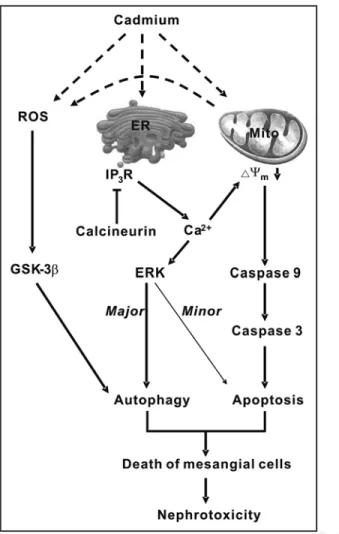
This study summarizes our most recent findings on the mecha- nisms underlying the cadmium-induced death of mesangial cells, which leads to nephrotoxicity. Multiple pathways participate in cadmium-induced nephrotoxicity. In the ROS-GSK-3β autophagy pathway, cadmium induces ROS most likely from the mito- chondria, and the ROS consequently activate GSK-3β leading to autophagic cell death. In the calcium-ERK autophagy and apoptosis pathway, cadmium stimulates calcium release from the endoplasmic reticulum, which activates ERK leading to predomi- nantly autophagic cell death and a minor level of apoptotic cell death. In the calcium-mitochondria-caspase apoptosis pathway, cadmium-induced elevation of calcium depolarizes the mitochon- drial membrane potential and then activates caspase signaling leading to apoptosis. A proposed model for cadmium-induced autophagy and apoptosis leading to nephrotoxicity is summarized in Figure 1.
Cadmium treatment causes calcium release from the endoplasmic reticulum, and the overload of intracellular calcium consequently activates ERK and depolarizes the mitochondrial membrane poten- tial, which in turn results in autophagy and apoptosis, respectively, leading to the death of mesangial cells. Application of cadmium to mesangial cells causes a fast increase in the intracellular calcium concentration. Pretreatment of mesangial cells with a calcium chelator, 1,2-bis (2-amino-phenoxy) ethane-N,N,N,N-tetraacetic acid (BAPTA-AM), suppresses ERK activation and attenuates apoptosis and autophagy, as manifested by the decreased formation of autophagosomes, reduced processing of microtubule-associated protein 1 light chain 3 (LC3)-I to LC3-II, and decreased acidic vesicular organelles (AVOs), further demonstrating that cadmium results in calcium-dependent autophagy and apoptosis.
*Correspondence to: Chwen-Ming Shih; Department of Biochemistry; College of Medicine; Taipei Medical University; 250 Wu-Hsing Street; Taipei 110 Taiwan; Tel.:
+886.2.27361661x3151; Fax: +886.2.27356689; Email: [email protected] Submitted: 02/06/09; Revised: 02/23/09; Accepted: 02/27/09
Previously published online as an Autophagy E-publication:
http://www.landesbioscience.com/journals/autophagy/article/8311
Punctum to: Wang SH, Shih YL, Kuo TC, Ko WC, Shih CM. Cadmium toxicity toward autophagy through ROS-activated GSK-3beta in mesangial cells. Toxicol Sci 2009;
108:124–31; PMID: 19126599; DOI: 10.1093/toxsci/kfn266.
Cadmium-induced calcium-dependent cell death comprises the calcium-ERK signaling pathway and the calcium-mitochon- dria-caspase signaling pathway. The expression of ERK increases significantly 1 hour following cadmium treatment, and pretreatment with PD 98059 (a MEK 1/2 inhibitor) suppresses cadmium-induced ERK expression and autophagy in mesangial cells. Similarly, pretreat- ment of mesangial cells with U0126 (another MEK 1/2 inhibitor) attenuates the cadmium-induced expression of ERK, autophagy and apoptosis. The results indicate that the cadmium-induced calcium- ERK signaling pathway leads predominantly to autophagy and to a lesser degree to apoptosis.
Cadmium can induce apoptosis via the calcium-mitochondria- caspase signaling pathway. In addition to the elevation of the intracellular calcium concentration, cadmium treatment depo- larizes the membrane potential of mitochondria, increases the expression of caspases 9 and 3 and induces cell death in mesangial cells. Pretreatment of mesangial cells with the calcium chelator BAPTA-AM suppresses the cadmium-induced depolarization of mitochondrial membrane potential and activation of caspases 9 and 3 and attenuates cadmium-induced cell death. These findings strongly suggest that the calcium-mitochondria-caspase signaling pathway contributes significantly to the cadmium-induced death of mesangial cells.
Cadmium results in the production of ROS (including hydrogen peroxide in mesangial cells), which in turn activate GSK-3β leading to autophagy and then cell death. Suppression of GSK-3β induction by the inhibitor SB216763 or small interfering RNA (siRNA) to GSK-3β attenuates cadmium-induced autophagy. Conversely, over- expression of GSK-3β via transfection aggravates cadmium-induced autophagy. These findings demonstrate that GSK-3β plays a critical role in cadmium-induced autophagy. Moreover, N-acetylcysteine (NAC, an ROS scavenger) pretreatment prevents the cadmium- induced activation of GSK-3β and attenuates cadmium-induced autophagy. Furthermore, NAC and the antioxidant vitamin E repress both the cadmium-induced elevation of ROS and autophagy. Taken together, these findings demonstrate that cadmium induces an ROS burst, which in turn potentiates the expression of GSK-3β leading to autophagic cell death.
ROS play a pivotal role in the pathogenesis of several kidney diseases, and cadmium might act on the mitochondria to induce the release of ROS, which in turn activate GSK-3β leading to cell death,
Autophagic Punctum
The cadmium-induced death of mesangial cells results in nephrotoxicity
Liang-Yo Yang,1,2,7,† Kuan-Hsun Wu,5,† Wen-Ta Chiu,4,6 Sheng-Hao Wang2 and Chwen-Ming Shih2,3,8,*
1Department of Physiology; College of Medicine; 2Graduate Institute of Medical Sciences; College of Medicine; 3Department of Biochemistry; 4School of Medicine; College of Medicine; Taipei Medical University; Taipei, Taiwan; 5Department of Pediatrics; Taipei Medical University-Wan Fang Hospital; Taipei, Taiwan; 6Department of Neurosurgery;
Taipei Medical University-Shuang Ho Hospital; Taipei, Taiwan; 7Neuroscience Research Center; 8Traditional Herbal Medicine Research Center; Taipei Medical University Hospital;
Taipei, Taiwan
†These authors contributed equally to this work.
Key words: cadmium, autophagy, apoptosis, calcium, GSK-3β
www.landesbioscience.com Autophagy 571
©2009
Landes
Bioscience.
Do not
distribute.
Cadmium-induced cell death in mesangial cells
572 Autophagy 2009; Vol. 5 Issue 4
Cadmium-induced cell death in mesangial cells
which causes nephrotoxicity. It remains unclear which organelles in the mesangial cells are the target of cadmium to generate ROS.
Mitochondrial dysfunction and oxidative stress play a crucial role in the pathogenesis of neurodegenerative diseases such as Alzheimer disease (AD), Parkinson disease (PD), Friedreich ataxia (FRDA), multiple sclerosis and amyotrophic lateral sclerosis (ALS). Thus, mitochondria are one of the most likely targets of cadmium to induce the generation of ROS. Moreover, oxidative stress contrib- utes significantly to the pathogenesis of several kidney diseases such as diabetic nephropathy, age-related ischemic kidney disease, and renal cancer. These findings together lead us to propose that the mitochondra-ROS-GSK-3β pathway leading to autophagic cell death is an important mechanism underlying cadmium-induced nephrotoxicity.
In addition to its potential effect on mitochondria, cadmium might act on the endoplasmic reticulum to induce nephrotoxicity via eleva- tion of the cytosolic calcium concentration, which in turn activates the calcium-ERK pathway and calcium-mitochondria-caspase pathway leading to autophagic and apoptotic cell death, respectively.
Endoplasmic reticulum stress and mitochondrial injury have been demonstrated to cause cell death in the glomerular and tubular epithelium of the kidney. Diabetes mellitus can often induce neph- ropathy. Evidence also indicates that diabetic rats show increased calcium-induced mitochondria depolarization, which is consistent with our finding that cadmium increases the intracellular calcium concentration and depolarizes the mitochondrial membrane poten- tial leading to activation of caspases 9 and 3. In our recent study, we find that cadmium activates the calcium-ERK-autophagy and calcium-ERK-apoptosis pathways leading to the death of mesangial cells. The above findings together guide us to propose that cadmium induces nephrotoxicity via the calcium-ERK pathway and calcium- mitochondria-caspase pathway leading to autophagic and apoptotic cell death. Thus, we propose that cadmium induces nephrotoxicity via multiple pathways.
Acknowledgements
This study was sponsored by grants from the Department of Health (Grant No. DOH-TD-B-111-002) and from Wan Fang Hospital (Grant No. 94TMU-WFH-214), Taipei, Taiwan
Figure 1. A proposed model for cadmium-induced nephrotoxicity. Cadmium induces nephrotoxicity via multiple pathways including the ROS-GSK-3β autophagy, calcium-ERK autophagy and apoptosis, and calcium-mitochon- dria-caspase apoptosis pathways. In the ROS-GSK-3β autophagy pathway, cadmium might act on the mitochondria to generate ROS, which in turn results in activation of GSK-3β leading to autophagic cell death. In the calcium-ERK autophagy and apoptosis pathway, cadmium acts on the endo- plasmic reticulum to induce elevation of the cytosolic calcium concentration, which in turn activates ERK leading predominantly to autophagic cell death and a minor level of apoptotic cell death. In the calcium-mitochondria- caspase apoptosis pathway, cadmium induces the release of calcium through the inositol-1,4,5-triphosphate receptor (IP3R) of the endoplasmic reticulum and then calcium depolarizes the membrane potential of mitochondria, which in turn activates caspases 9 and 3 leading to apoptotic cell death.
ER, endoplasmic reticulum; IP3R, inositol-1,4,5-triphosphate receptor; Mito, mitochondria; ΔYm, mitochondrial membrane potential.