The Combination of multiphoton and reflected confocal microscopy
for cornea imaging
Wei-Liang Chen
1, Wen Lo
1, Yen Sun
1, Sung-Jan Lin
2,3, Hsin-Yuan Tan
3,4#, Chen-Yuan Dong
1*1
Department of Physics, National Taiwan University, Taipei, Taiwan.
2
Institute of Medical Engineering, College of Medicine and College of Engineering, Taipei 100,
Taiwan
3
Department of Dermatology, National Taiwan University Hospital, Taipei 100, Taiwan
4Department of Ophthalmology, Chang Gung Memorial Hospital, Linko 333, Taiwan
ABSTRACT
The purpose of this work is to demonstrate the combination of reflective confocal microscopy and multiphoton microscopy and its application in imaging cornea. The difficulty of optically imaging the highly translucent cornea has prevented the development of an effective non-invasive system for the clinical monitoring of the physiological or pathological states of corneas. In this work, we combine reflective confocal microscopy with multiphoton microscopy to demonstrate the potential of our methodology in the minimally invasive imaging of the cornea. The visible reflection signals from cornea can provide structural information of interfaces of different refractive indices while the multiphoton signals generated from the use of near infrared excitation allows deep tissue penetration and reduced photo-damage. In multiphoton imaging, the second harmonic generation (SHG) signal is used to detect collagen in the stroma of the cornea, and the reflective confocal imaging allows detection of the cellular components located in the epithelium. The combination of reflective and multiphoton imaging can be used to reveal complementary structural information of the corneal architecture.. The system is first tested on porcine eye cornea. Assessment of the result on the porcine eye will be used to evaluate the potential of the system as a technique for in vivo clinical applications.
Keywords: multiphoton microscopy, second-harmonic generation, reflected confocal microscopy, cornea. Address correspondence to: #[email protected], *[email protected]
Ophthalmic Technologies XVI, edited by Fabrice Manns, Per G. Söderberg, Arthur Ho, Proc. of SPIE Vol. 6138, 61380M, (2006) · 1605-7422/06/$15 · doi: 10.1117/12.647225
1. Introduction
Real-time acquiring cornea structural information with no intervention and destruction may be crucial for investigating and diagnosing all corneal pathologies. Traditionally, the investigation of corneal pathologies relies heavily on histological procedures for probing the morphological alterations. However, the processing and fixating procedures can inevitably lead to morphological artifacts. The alternative non-invasive approach of confocal microscopy has been widely applied in corneal pathological imaging1-9, including infections, dystrophies, and various wound healing processes following surgical procedures. Cellular components, nerve, and certain pathogens may be visualized in vivo with reflected confocal microscopy. However, stromal collagen may not be easily imaged using reflected mode. Multiphoton microscopy is another new modality with minimal invasion that has been applied in ophthalmology10,11. Multiphoton microscopy using the nonlinear excitation from ultrafast, near-infrared light source provides several advantages for the application in biomedical sciences. The key features of multiphoton microscopy include the enhancement of axial depth discrimination, reduced photodamage, and increasing imaging depth12,13. In addition to multiphoton fluorescence excitation, the nonlinear optical effect of second harmonic generation (SHG) has also been provided for imaging biological structures that lack of an inversion symmetry. Several biological structures such as collagen, muscle fiber, and microtubules have been proven to be effective second harmonic generators14,15. We had previously demonstrate multiphoton autofluorescence and SHG imaging of normal porcine cornea11. The intracorneal cellular components can be visualized with multiphoton autofluorescence, and collagenous structures can be identified with SHG signals. In this work, we extend our investigation into the combination of reflected confocal microscopy with multiphoton microscopy for minimally invasive imaging of cornea. The use of near-infrared excitation source allows deeper tissue penetration and a lower potential for photodamage. Collagenous components within cornea can be identified with SHG signals, and the reflected confocal imaging allows detection of the cellular components. We provides this minimally invasive imaging technique for imaging porcine cornea ex vivo and our work demonstrates the feasibility of this optical technique as a clinical diagnostic and monitoring tool for imaging corneal pathologies.
2. Experimental method Specimen preparation
The porcine eyes used in this study were obtained directly from local market at the earliest possible time. The eyeballs with intact epithelium and optically clear media were selected for further processing. The corneal buttons were created with 8mm corneal trephine and placed in phosphate buffered solution.
Multiphoton microscopy and reflected confocal imaging
The imaging system used in this study is a commercial laser scanning microscope (Meta 510, Zeiss, Germany). For
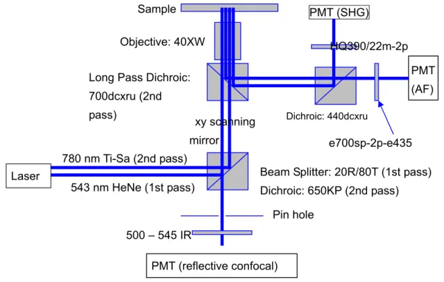
reflective microscopy, the 543 nm line from a visible source provided with the Meta 510 system was used. The multiphoton imaging of the porcine cornea specimen was achieved using the near-infrared excitation source from a titanium-sapphire laser (Tsunami, Spectra Physics, Mountain View, CA) pumped by a diode-pumped, solid state (DPSS) laser system (Millennia, Spectra Physics). The multiphoton excitation wavelength used was 780 nm and the we simultaneously acquired multiphoton autofluorescence and SHG signals (centered at 390 nm) from the cornea specimen. In our study, the reflected confocal and multiphoton images were separately acquired. The instrument set up of the reflective confocal and multiphoton microscope is shown in Fig. 1.
Fig. 1. The instrumentation setup of combined reflective confocal microscopy and multiphoton microscopy.
3. Results
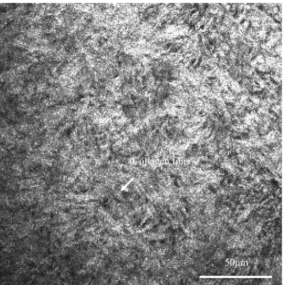
The separated reflective and multiphoton images are shown in Fig. 2 and 3. The wholelayered corneal structure can be imaged with multiphoton system. The corneal epithelium can be visualized with autofluorescence signals. In normal porcine corneal stroma, collagen fibers can be identified with SHG signals, while keratocytes cannot be easily detected with multiphoton autofluorescence excitation.
Laser
PMT (reflective confocal)
Pin hole
Beam Splitter: 20R/80T (1st pass) Dichroic: 650KP (2nd pass) Long Pass Dichroic:
700dcxru (2nd
pass) Dichroic: 440dcxru
PMT (AF) PMT (SHG) Sample Objective: 40XW 780 nm Ti-Sa (2nd pass) 543 nm HeNe (1st pass) e700sp-2p-e435 500 – 545 IR xy scanning mirror HQ390/22m-2p
Fig. 2. Reflected confocal image of normal porcine cornea at the approximate imaging depth of 10 µm.
50µm
keratocyte
Fig. 3. The corresponding second harmonic generation image of figure 2 at the same position of normal porcine conrea.
50µm
Collagen fiber
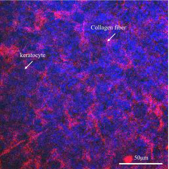
Fig. 4. The combined reflective and multiphoton imaging of normal porcine cornea at depth of 10µm below epithelium. Red color represents reflected signals while the blue color for SHG signals. Stromal collagen fibers are effective SHG generators, and keratocytes can be identified with reflective imaging. Notice that autofluorescence signals cannot be detectable here.
50µm
keratocyte
Collagen fiber
Fig. 5. The combined reflective and multiphoton imaging of normal porcine cornea at depth of 190µm below epithelium. Red color represents reflected signals while the blue color for SHG signals. Stromal collagen fibers are effective SHG generators, and keratocytes can be identified with reflective imaging. Notice that autofluorescence signals cannot be detectable here.
With the combined technique of multiphoton microscopy and reflective confocal microscopy, it can provide additional structural information. The corneal epithelium can be imaged with both the multiphoton autofluorescence excitation and reflective mode simultaneously. In the corneal stroma, collagenous structure can be identified with SHG signals (Fig. 4 and 5). As for the quiescent keratocytes, they can be identified in the reflected mode, but can be ineffective for generating autofluorescence. Therefore with the combination of SHG microscopy and reflective confocal microscopy as a minimally invasive imaging technique, both the cellular and extracelluar information can be visualized.
keratocyte
Collagen fibers
50µm
4. Conclusion
While we have demonstrated the ability of multiphoton imaging in resolving cornea structure, we anticipate the addition of reflected confocal imaging will widen the capability of cellular level corneal imaging. If only morphological information is needed, one can select the second harmonic generating wavelength such as not to excite cellular autofluorescence. In such a case, structural details of both the corneal epithelium and stroma can be obtained without minimal photodamage associated with autoflurescence excitation. This approach has potential application for in vivo imaging of cornea pathologies.
Acknowledgement
We like to acknowledge the support of National Research Program for Genomic Medicine, Taiwan (NSC 93-3112-B-002-034) for this work.
References
1. Tervo T, Moilanen. In vivo confocal microscopy for evaluation of wound healing following corneal refractive surgery. Prog Ret Eye Res. 2003;22:339-258.
2. Cavanagh HD, Petroll WM, Alizadeh AH. Clinical and diagnostic use of in vivo confocal microscopy in patients with corneal disease. Ophthalmology 1993; 100:1444-54.
3. Jalbert I, Stapleton F, Papas E, Sweeney DF, Coroneo M. In vivo confocal microscopy microscopy of the human cornea. Br J Ophthalmol 2003; 87:225-36.
4. Chew SJ, Beuerman RW, Assouline M, et al. Early diagnosis of infectious keratitis with in vivo real time confocal microscopy. CLAO J 1992; 18:197-201.
5. Winchester K, Mathers WD, Sutphin JE. Diagnosis of Aspergillus keratitis in vivo with confocal microscopy. Cornea 1997; 16:27-31.
6. Florakis GJ, Moazami G, Schubert H, et al. Scanning slit confocal microscopy of fungal keratitis. Arch Ophthalmol 1997; 115:1461-63.
7. Avunduk AM, Beuerman RW, Varnell ED, and Kaufman HE. Confocal microscopy of Aspergillus fumigatus keratitis. Br J Ophthalmol 2003; 87:409-10.
8. Pfister DR, Cameron JD, Krachmer JH, Holland EJ. Confocal microscopy findings of Acanthamoeba keratitis. Am J Ophthalmol 1996; 121:119-28.
9. Kaufman SC, Laird JA, Cooper R, Beuerman RW. Diagnosis of bacterial contact lens related keratitis with the white-light confocal microscope. CLAO J 1996; 22:274-7.
10. Yeh AT, Nassif N, Zoumi A, Tromberg BJ. Selective corneal imaging using combined second-harmonic generation and two-photon excited fluorescence. Optics Letters 2002; 27:2082-4.
11. Teng SW, Tan HY, Peng JL, et al. Multiphoton autofluorescence and second-harmonic generation (SHG) imaging of
ex-vivo porcine eye. Invest Ophthalmol Vis Sci 2006. In press.
12. Denk, W. et al. 2-Photon laser scanning fluorescence microscopy. Science 2006; 248, 73-76.
13. So, PTC. et al. Two-photon excitation fluorescence microscopy. Annu. Rev. Biomed. Eng. 2000;2, 399-429. 14. Zoumi, A. et al. Imaging cells and extracellular matrix in vivo by using second-harmonic generation and two-photon
excited fluorescence. P. Natl .Acad. Sci. USA. 2002; 99, 11014-11019.
15. Zoumi, A. et al. Imaging coronary artery microstructure using second-harmonic and two-photon fluorescence microscopy. Biophys. J. 2004; 87, 2778-2786.