Multiphoton fluorescence and second harmonic generation microscopy
for imaging keratoconus
Yen Sun
1, Wen Lo
1, Sung-Jan Lin
2,3, Wei-Chou Lin
4, Shiou-Hwa Jee
2,5, Hsin-Yuan Tan
3,6#,
Chen-Yuan Dong
1*1
Department of Physics, National Taiwan University, Taipei 106, Taiwan
2
Department of Dermatology, National Taiwan University Hospital, Taipei 100, Taiwan
3Institute of Medical Engineering, College of Medicine and College of Engineering, Taipei 100,
Taiwan
4
Department of Pathology, National Taiwan University Hospital, Taipei 100, Taiwan
5
Department of Dermatology, College of Medicine, National Taiwan University Hospital, Taipei 100,
Taiwan
6
Department of Ophthalmology, Chang Gung Memorial Hospital, Linko 333, Taiwan
ABSTRACT
The purpose of this study is to assess the possible application of multiphoton fluorescence and second harmonic generation (SHG) microscopy for imaging the structural features of keratoconus cornea and to evaluate its potential as being a clinical in vivo monitoring technique. Using the near-infrared excitation source from a titanium-sapphire laser pumped by a diode-pumped, solid state (DPSS) laser system, we can induce and simultaneously acquire multiphoton autofluorescence and SHG signals from the cornea specimens with keratoconus. A home-modified commercial microscope system with specified optical components is used for optimal signal detection. Keratoconus cornea button from patient with typical clinical presentation of keratoconus was obtained at the time of penetrating keratoplasty. The specimen was also sent for the histological examination as comparison. In all samples of keratoconus, destruction of lamellar structure with altered collagen fiber orientation was observed within whole layer of the diseased stromal area. In addition, the orientation of the altered collagen fibers within the cone area shows a trend directing toward the apex of the cone, which might implicate the biomechanical response of the keratoconus stroma to the intraocular pressure. Moreover, increased autofluorescent cells were also found in the cone area, with increased density as one approaches the apical area. In conclusion, multiphoton autofluorescence and SHG microscopy non-invasively demonstrated the morphological features of keratoconus cornea, especially the structural alternations of the stromal lamellae. We believe that in the future the multiphoton microscopy can be applied in vivo as an effective, non-invasive diagnostic and monitoring technique for
keratoconus.
Keywords: multiphoton fluorescence microscopy, second-harmonic generation, keratoconus
Address correspondence to: #[email protected],*[email protected]
1. Introduction
Keratoconus is a non-inflammatory ectatic disorder of cornea which is characterized by progressive corneal steepening and stromal thinning1. It affects young people in their second or third decade of lives. Clinically, it may lead to visual disturbance due to the progressive increase of irregular astigmatism and myopia1. Histologically, thinning of corneal stroma, break of Bowman’s membrane, and iron deposition in the basal epithelial layers represent the classical characteristics of keratoconus2,3. The etiology of this ectatic disorder remains unknown until now. Several predisposing factors have been proposed4-7. Atopy, genetics, and contact lens have all been proposed. It has been suggested that the disease may be originated from the epithelium8; however, the whole layer of cornea has been demonstrated to be altered morphologically during disease progress3,8-11.
In this study, we used multiphoton microscopy to characterize human keratoconus in ex vivo specimens. Multiphoton micrscopy using the nonlinear excitation from ultrafast, near-infrared light source can provide several advantages for the application in biomedical imaging, which include enhancement of axial depth discrimination, reduction of photodamage, and increase of imaging penetrating depth12,13. All these properties allow us to observe living cells and tissues without inducing detectable damages14-17. In addition, characteristic autofluorescence (AF) from various cells and components of extracellular matrix can help to analyze biological structures of interest18-20. In addition to fluorescence excitation imaging, another nonlinear optical effect of second harmonic generation (SHG), has also been proven to be useful for imaging certain biological structures lacking an inversion symmetry. Several biological materials such as collagen, muscle fiber, and microtubules have been shown to be effective second-harmonic generators21,22. Cornea may be a perfect target of interest for applying multiphoton fluorescence and SHG microscopy due to the main component of corneal stroma is constructed by regularly aligned type I collagen fiber bundles. In particular, since the transparent nature of cornea is believed to contribute to the unique regular three-dimensional arrangement of stromal collagen fibers23, SHGmicroscopy may be an effective tool for investigating the physiology of corneal stroma as well as visualizing abnormalities of the corneal stroma. We have previously demonstrated the application of multiphoton AF and SHG microscopy for imaging three-dimensional structure of normal porcine ocular surface.24 In these studies, the multiphoton generated fluorescence is effective for visualizing the cellular component within cornea, and the SHG signal derived from collagen can be used to image the collagen structure within the corneal stroma. In this work, we further extend our study for studying the structural alterations of the corneas due to keratoconus. In this disease, the morphological changes of the collagenous stroma may possibly play an important role during pathogenesis, and the
success of our demonstration can lead to potential clinical applications of multiphoton fluorescence and SHG imaging as an effective diagnostic and disease monitoring tool for keratoconus, and possibly, for the investigation of the pathogenesis of disease progression.
2. Experimental Method
The study protocol was approved by an institutional review board, and was kept to the Helsinki Declaration with respect to human subjects in biomedical research.
Specimen preparation
Corneal buttons of keratoconus were obtained from patients during penetrating keratoplasty. All patients were diagnosed as keratoconus according to clinical slitlamp findings and topographical data. All patients had been initially tried with conservative methods of vision correction including glasses and rigid contact lenses fitting. Penetrating keratoplasty was only indicated when conservative methods failed. The trephined corneal specimens were placed in balanced salt solution and sent for imaging immediately. The samples were kept within PBS buffer and covered with a standard No. 1.5 cover glass and mounted on the upright microscope for multiphoton microscopy to be performed. After images were acquired, the specimens were then placed in the fixation medium composed of formalin:PBS in 1:9 ratio (v/v). The specimens were then dehydrated and embedded in paraffin and then processed for hematoxylin and eosin staining. Histological images were then acquired and compared with the multiphoton results.
Multiphoton AF and SHG microscopy
The multiphoton AF and SHG microscopic system used in this study is similar to the experimental set up that was previously used for corneal studies24,25. It is a home-built laser scanning microscopic imaging system based on a commercial upright microscope (E800, Nikon, Japan). A titanium-sapphire (ti-sa) laser (Tsunami, Spectra Physics, Mountain View, CA) pumped by a diode-pumped solid state laser (Millennia X, Spectra Physics, Mountain View, CA) was used as the excitation source. The 760 nm of the ti-sa laser capable of inducing AF and SHG from cornea is scanned in the focal plane by a galvanometer-driven x-y mirror scanning system (Model 6220, Cambridge Technology, Cambridge, MA). The multiphoton images were acquired using an water-immersion objective (Fluor WI 40x, NA 0.8, Nikon). To ensure optimal focusing, the laser was beam expanded to ensure overfilling of the objective’s back aperture and a short-pass dichroic mirror (700DCSPRUV, Chroma Technology, Brattleboro, VT) was used to reflect the incident excitation laser source into the back aperture of the focusing objective. The MF and SHG signals are collected by the same focusing objective, and passed through the dichroic and additional filters before the signals reach the photo-detectors. Prior to reaching the detectors, the AF and SHG signals are separated by a secondary dichroic mirror (435DCSX, Chroma Technology). The SHG signal centered at 380nm was reflected by the secondary dichroic and filtered using a bandpass filter (HQ380/20, Chroma Technology), while the longer wavelength AF signal passes through
the dichroic mirror and an additional broad-band filter (E435LP, Chroma Technology) before being detected. Both AF and SHG signals were detected using single-photon counting photomultiplier tubes (R7400P, Hamamatsu, Japan). For large area scans of the corneal specimens, a two-dimensional stage scanning system (H101, Prior Scientific, UK) was used for specimen translation after each x-y scan. The overlapping images acquired in this fashion can then be assembled into a large area, high resolution map of the cornea.
3. Results
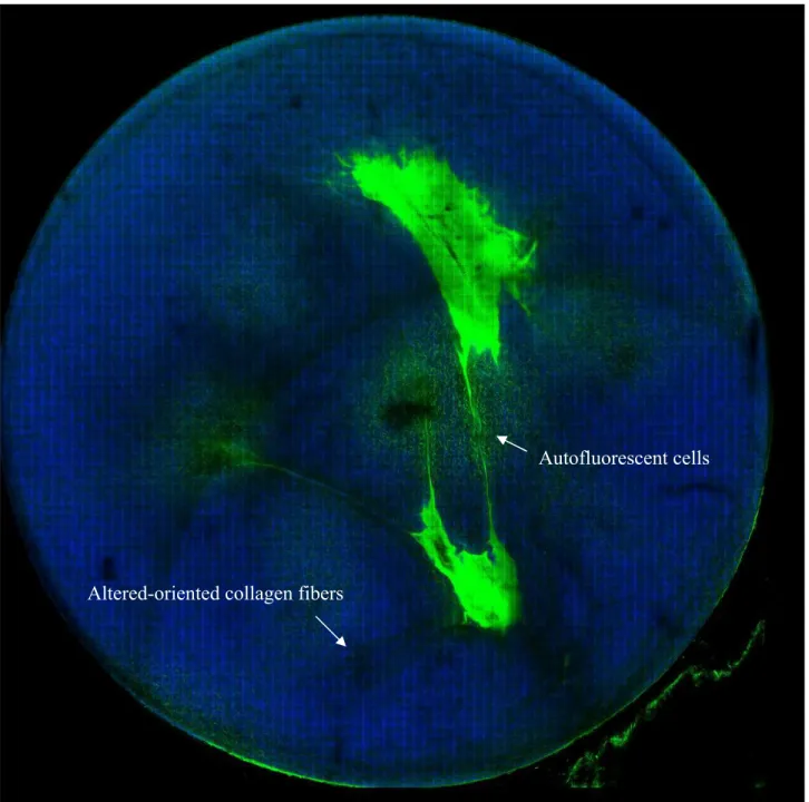
Multiphoton fluorescence and SHG imaging demonstrated morphological alterations in both cellular and extracellular matrix in the whole layer of cornea (Fig. 1). Within the pathological cone area of cornea, abnormal scattered spindle-shaped epithelial cells can been found. As the depth increase, numerous autofluorescent stellate cells can be identified, which may represent the activated keratocytes. For the stromal collagen, we can identify significant architectural alterations within cone area. In the anterior stroma of pathological cone area, in addition to the presence of normal interweave packing of collagen bundles, we can identify inhomogeneous alignment of collagen bundles with preferential orientation centripetally, directing toward the apex.As the depth increases, the alteration of stroma collagen lamellae becomes apparent. The normal stroma architecture of orthogonal packing of lamellae composed of parallel collagen fibers bundles has been displaced partially by parallel, unidirectional, and centripetally oriented collagen bundles according to the severity of disease. This modulation of collagen bundles direct toward apex may possibly implicate the biomechanical response of pathologically weakening corneal stroma to the intraocular pressure during the disease progression.
Fig. 1. The multiphoton autofluorescence and SHG imaging of the whole keratoconic corneal specimen. (diameter of corneal button: 8mm)
4. Conclusion
Multiphoton autofluorescence and SHG microscopy had been demonstrated as an useful technique for corneal imaging with no need of additional tissue processing24,26. The nonlinear fluorescence excitation from cellular component and the nonlinear optical effect of SHG from collagen allow us to image corneal structure with subcellular resolution. In this
Altered-oriented collagen fibers
work, we extend our previous work to demonstrate that the detail corneal structural alterations due to keratoconus can be effectively imaged, which can potentially provide important information for the underlying pathogenesis of disease per se.
Confocal microscopy and X-ray diffraction have been applied for imaging the morphological changes associated with keratoconus27-31. Confocal microscopy may demonstrate partially the morphological alterations of keratoconus in vivo, especially the cellular changes. Elongation of superficial epithelial cells and increase of reflectivity of keratocytes can be visualized as the characteristic findings of keratoconus. Yet the resolution for identifying collagenous morphological alteration is limited with the reflected technique27,28. X ray scattering is another imaging technique that had been applied for demonstrating the corneal collagenous structural alterations due to keratoconus29-31. Meek et al has demonstrated bulk information of significant shift of preferred orientation of collagen mass, especially around the apical area.31 However, individualized structural information of individual collagen bundle can not be obtained. To the best of our knowledge, the current study represents the first non-invasive demonstration of visualizing corneal structural alterations due to keratoconus. Both the cellular and collagenous morphological information in keratoconus corneas can be obtained. Elongation of superficial epithelial cells within the apical area which possibly implicit the biomechanical cellular response to the progress of corneal protusion can be demonstrated. In addition, we demonstrated the structural alterations of collagen lamellae with serial imaging of various depths in keratoconus. Instead of the homogenous respective interweaving and parallel alignment in the anterior and posterior stroma, centripetal collagen bundles directing toward the apex can be demonstrated to be uneven distributed within cone area, which may possibly be the consequence of the tissue modulation of pathological weakening collagenous stroma in response to the intraocular pressure.
In conclusion, we herein demonstrated using multiphoton autofluorescence and SHG microscopy for imaging the three-dimensional structural alterations in keratoconus cornea specimens under ex vivo conditions. We believe that this technique can potentially provide insights into the investigation of keratoconus pathogenesis and may be developed into an effective clinical diagnostic tool for keratoconus in the future.
ACKNOWLEDGMENT
We like to acknowledge the support of National Research Program for Genomic Medicine, Taiwan ( NSC 93-3112-B-002-034) for this work.
REFERRENCES
1. Rabinowitz YS. Keratoconus. Surv Ophthalmol. 1998;42:297-319.
2. Sherwin T. Brookes NH. Morphological changes in keratoconus: pathology or pathogenesis. Clinical & Experimental Ophthalmology. 2004;32(2):211-7.
4. Tuft SJ, Moodaley, LC, Gregory WM, Davison CR, Buckley RJ. Prognostic factors for the progression of keratoconus. Ophthalmology. 1994;101:439-447.
5. Harrison RJ, Klouda PT, Easty DL, Manku M, Charles J, Stewart CM. Association between keratoconus and atopy. Br J Ophthalmol. 1989;73:816-822.
6. Lindsay RG, Bruce AS, Gutteridge IF. Keratoconus associated with continual eye rubbing due to punctual agenesis. Cornea. 2000;19:567-569.
7. Hartstein J. Keratoconus that developed in patients wearing corneal contact lenses. Arch Ophthalmol. 1968;80:345-346.
8. Teng CC. Electron microscopic study of pathology of keratoconus. Am J Ophthalmol. 1963;55:18-47. 9. Scroggs MW, Proia AD. Histopathological variation in keratoconus. Cornea. 1992;11:553-559.
10. Sturbaum CW, Peiffer RIJ. Pathology of corneal endothelium in keratoconus. Ophthalmologica. 1993;206:192-208. 11. Sawaguchi S, Fukuchi T, Abe H, et al. Three-dimensional scanning electron microscopic study of keratoconus
corneas. Arch Ophthalmol. 1998;16:62-68.
12. Denk, W. et al. 2-Photon laser scanning fluorescence microscopy. Science 248, 73-76 (1990)
13. So, PTC. et al. Two-photon excitation fluorescence microscopy. Annu. Rev. Biomed. Eng. 2, 399-429 (2000). 14. Squirrell, JM. et al. Long-term two-photon fluorescence imaging of mammalian embryos without compromising
viability. Nat. Biotechnol. 17, 763-7(1999).
15. Miller, MJ. et al. Two-photon imaging of lymphocyte motility and antigen response in intact lymph node. Science 296, 1869-1873 (2002).
16. Masters, BR. et al. Multiphoton excitation fluorescence microscopy and spectroscopy of in vivo human skin. Biophys. J. 72, 2405-12 (1997).
17. Sun,Y. et al. Multiphoton polarization imaging of the stratum corneum and the dermis in ex-vivo human skin. Opt. Express. 11, 3377-3384 (2003).
18. Zipfel, WR. et al. (2003a) Nonlinear magic: multiphoton microscopy in the biosciences. Nat. Biotechnol. 21, 1369-77(2003).
19. Zipfel, WR. et al. (2003b) Live tissue intrinsic emission microscopy using multiphoton-excited native fluorescence and second harmonic generation. P. Natl .Acad. Sci. USA. 100, 7075-80 (2003).
20. Lin, SJ. et al. Evaluating cutaneous photoaging by use of multiphoton fluorescence and second harmonic generation microscopy. Opt. Lett. (2005) (in press)
21. Zoumi, A. et al. Imaging cells and extracellular matrix in vivo by using second-harmonic generation and two-photon excited fluorescence. P. Natl .Acad. Sci. USA. 99, 11014-11019 (2002).
22. Zoumi, A. et al. Imaging coronary artery microstructure using second-harmonic and two-photon fluorescence microscopy. Biophys. J. 87, 2778-2786 (2004).
23. Maurice DM. The structure and transparency of the cornea. J Physiol (Lond). 1957;136:263 286.
ex-vivo porcine eye. IOVS. 2005. In press.
25. Tan HY, Teng SW, Lo W, et al. Chracterizing the thermally induced structural changes to intact porcine eye, part 1: second harmonic generation imaging of cornea stroma. J. Biomed. Opt. 2005;10: 054019.
26. Yeh AT, Nassif N, Zoumi A, Tromberg BJ. Selective corneal imaging using combined second-harmonic generation and two-photon excited fluorescence. Opt. Lett. 2002;27:2082-2084.
27. Hollingsworth JG, Bonshek RE, Efron N. Correlation of the appearance of the keratoconic cornea in vivo by confocal microscopy and in vitro by light microscopy. Cornea. 2005;24:397-405.
28. Hollingsworth JG, Efron N, Tullo AB. In vivo corneal confocal microscopy in keratoconus. Ophthalmic & Physiological Optics. 2005;25:254-260.
29. Fullwood NJ, Tuft SJ, Malik NS, Meek KM, Ridgway AE, Harrison RJ. Synchrotron x-ray diffraction studies of keratoconus corneal stroma. Invest Oph Vis Sci. 1992;33:1734-1741.
30. Daxer A, Fratzl P. Collagen fibril orientation in the human corneal stroma and its implications in keratoconus. Invest Ophthalmol Vis Sci. 1997;38:121-129.
31. Meek KM, et al. Changes in collagen orientation and distribution in keratoconus corneas. Invest Ophthalmol Vis Sci. 2005;46:1948-1956.