Short Communication
Trisomy 7 mosaicism at amniocentesis: Interphase FISH, QF-PCR, and
aCGH analyses on uncultured amniocytes for rapid distinguishing of true
mosaicism from pseudomosaicism
Chih-Ping Chen
a,b,c,d,e,f,g,*
, Hsu-Kuang Huang
h, Yi-Ning Su
i, Schu-Rern Chern
c, Jun-Wei Su
b,j,
Chen-Chi Lee
b, Dai-Dyi Town
b, Wen-Lin Chen
b, Yu-Ting Chen
c, Wayseen Wang
c,kaDepartment of Medicine, Mackay Medical College, New Taipei City, Taiwan bDepartment of Obstetrics and Gynecology, Mackay Memorial Hospital, Taipei, Taiwan
c
Department of Medical Research, Mackay Memorial Hospital, Taipei, Taiwan d
Department of Biotechnology, Asia University, Taichung, Taiwan e
School of Chinese Medicine, College of Chinese Medicine, China Medical University, Taichung, Taiwan f
Institute of Clinical and Community Health Nursing, National Yang-Ming University, Taipei, Taiwan g
Department of Obstetrics and Gynecology, School of Medicine, National Yang-Ming University, Taipei, Taiwan h
Department of Obstetrics and Gynecology, Taiwan Adventist Hospital, Taipei, Taiwan i
Department of Medical Genetics, National Taiwan University Hospital, Taipei, Taiwan j
Department of Obstetrics and Gynecology, China Medical University Hospital, Taichung, Taiwan k
Department of Bioengineering, Tatung University, Taipei, Taiwan Accepted 25 October 2011
Abstract
Objective: To present prenatal diagnosis of true trisomy 7 mosaicism.
Materials, Methods and Results: A 36-year-old woman underwent amniocentesis at 18 weeks of gestation. Amniocentesis revealed a karyotype of 47,XY,þ7[20]/46,XY[9]. The parental karyotypes were normal. Repeated amniocentesis was performed at 20 weeks of gestation. Array comparative genomic hybridization (aCGH) analysis on uncultured amniocytes manifested a genomic gain in chromosome 7. Quantitative fluorescent polymerase chain reaction (QF-PCR) analysis on uncultured amniocytes showed a biparental diallelic pattern with a dosage increase in the maternal allele. Interphase fluorescence in situ hybridization (FISH) on uncultured amniocytes revealed three 7q-specific signals in 13 of 50 (26%) of the cells. The cultured amniocytes had a karyotype of 47,XY,þ7[12]/46,XY[14]. The ultrasound findings were unremarkable. The pregnancy was subsequently terminated, and a fetus was delivered with facial dysmorphisms. Postnatal tissue samplings revealed the mosaic trisomy 7 level of 37.5% (15/40), 30% (12/40), 42.5% (17/40), 82.5% (33/40), 52.5% (21/40), and 27.5% (11/40) in skin, liver, lungs, placenta, membrane, and cord, respectively. The cord blood had a karyotype of 46,XY. PEG1/MEST methylation-sensitive high-resolution melting PCR assay of cord blood showed no uniparental disomy for chromosome 7.
Conclusion: Interphase FISH, QF-PCR, and aCGH analyses on uncultured amniocytes are useful for rapid distinguishing of true mosaicism from pseudomosaicism for trisomy 7 at amniocentesis. Cord blood sampling for confirmation of fetal trisomy 7 mosaicism is not practical. CopyrightÓ 2012, Taiwan Association of Obstetrics & Gynecology. Published by Elsevier Taiwan LLC. All rights reserved.
Keywords: aCGH; amniocentesis; cord blood sampling; interphase FISH; QF-PCR; trisomy 7 mosaicism
Introduction
Genetic counseling of mosaic trisomy at amniocentesis is difficult because of the phenotypic variability associated with the condition with some fetuses exhibiting the typical * Corresponding author. Department of Obstetrics and Gynecology, Mackay
Memorial Hospital, 92, Section 2, Chung-Shan North Road, Taipei, Taiwan. E-mail address:[email protected](C.-P. Chen).
Taiwanese Journal of Obstetrics & Gynecology 51 (2012) 77e82
www.tjog-online.com
1028-4559/$ - see front matter CopyrightÓ 2012, Taiwan Association of Obstetrics & Gynecology. Published by Elsevier Taiwan LLC. All rights reserved. doi:10.1016/j.tjog.2012.01.015
phenotypic abnormalities while others appear normal [1]. Trisomy 7 mosaicism may present variable and non-specific clinical features ranging from normal to facial dysmorphisms, enamel dysplasia, sparse hair, hypomelanosis of Ito, pigmentary abnormalities, radial defects, Potter syndrome, Goldenhar syndrome, and Blaschkolinear malformation syndrome[2e8]. Trisomy 7 mosaicism at amniocentesis has been reported to be associated with a normal karyotype in the lymphocytes of the cord blood and peripheral blood [9]. Maternal uniparental disomy for chromosome 7 (UPD 7) may be observed in cases of trisomy 7 mosaicism with Silver-Russell syndrome (SRS)
[10e13]. Here, we report prenatal diagnosis of trisomy 7 mosaicism by amniocentesis using cultured and uncultured amniocytes in the second trimester. We demonstrate the application of molecular cytogenetic technologies in uncul-tured amniocytes for rapid confirmation of fetal mosaicism, searching for UPD, and distinguishing true mosaicism from pseudomosaicism.
Methods and results
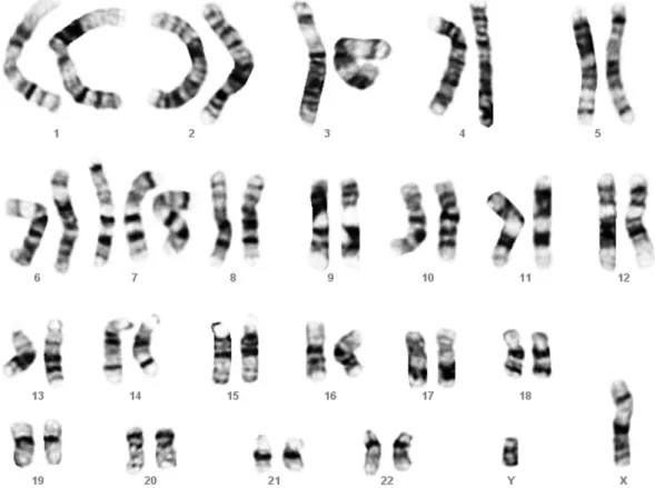
A 36-year-old, gravid 3, para 1 woman underwent amnio-centesis at 18 weeks of gestation at a local hospital because of advanced maternal age. There was no family history of congenital malformations. Her husband was 39 years old, and the couple had a 3-year-old healthy child. In 20 of 29 sepa-rated cultured amniocyte colonies, an abnormal karyotype of 47,XY,þ7 was found (Fig. 1), while the other nine colonies had a karyotype of 46,XY. The cytogenetic result of
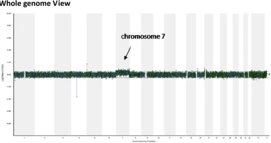
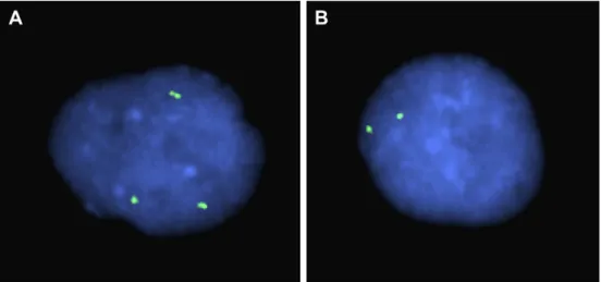
amniocentesis was 47,XY,þ7[20]/46,XY[9]. She was referred to the hospital at 19 weeks of gestation for genetic counseling and repeated amniocentesis. The parental karyotypes were normal. The fetal ultrasound findings were unremarkable. Molecular cytogenetic techniques such as array comparative genomic hybridization (aCGH), interphase fluorescence in situ hybridization (FISH), and quantitative fluorescent polymerase chain reaction (QF-PCR) were applied to the uncultured amniocytes. The aCGH investigation using CytoChip Oligo array (BlueGnome, Cambridge, UK) on uncultured amniocytes manifested a genomic gain in chromosome 7 (Fig. 2). Poly-morphic DNA marker analysis of the amniotic fluid sample with uncultured amniocytes using QF-PCR and microsatellite markers specific for chromosome 7 revealed a biparental dia-llelic pattern. The uncultured amniotic fluid sample had unequal biparental inheritance of chromosome 7 with a dosage increase in the maternal allele (Fig. 3). Interphase FISH anal-ysis on uncultured amniocytes using a 7q11.1-specific probe (RP11-432A1) showed three 7q-sepcific signals in 13/50 (26%) of uncultured amniocytes and two signals in 37/50 (74%) of uncultured amniocytes (Fig. 4). The molecular cytogenetic results were consistent with true mosaicism, and UPD 7 was excluded. Cytogenetic analysis of cultured amniocytes at repeated amniocentesis revealed a karyotype of 47,XY,þ7[12]/ 46,XY[14]. The parents decided to terminate the pregnancy. A 426-g male fetus was delivered with facial asymmetry, low-set ears, long philtrum, and hypertelorism. Cytogenetic analysis of the cord blood revealed a karyotype of 46,XY in 40/40 of lymphocytes. Fetal and extraembryonic tissue samplings
Fig. 1. A karyotype of 47,XY,þ7.
revealed a karyotype of 47,XY,þ7[15]/46,XY[25] in the skin, a karyotype of 47,XY,þ7[12]/46,XY[28] in the liver, a karyo-type of 47,XY,þ7[17]/46,XY[23] in the lungs, a karyotype of 47,XY,þ7[11]/46,XY[29] in the umbilical cord, a karyotype of 47,XY,þ7[21]/46,XY[19] in the amniotic membrane, and a karyotype of 47,XY,þ7[33]/46,XY[7] in the placenta. A PEG1/MEST methylation-sensitive high-resolution melting PCR assay was performed to identify the differential methyla-tion of the imprinted PEG1/MEST locus on 7q32 in the cord
blood. The result revealed biparental inheritance of chromo-some 7 in the cord blood (Fig. 5).
Discussion
The present case was associated with true mosaic trisomy 7 and dysmorphisms. Mosaic trisomy 7 at amniocentesis has been associated with pseudomosaicism [14]. We previously observed discrepancy between the cytogenetic results of Fig. 2. Oligonucleotide-based array comparative genomic hybridization (aCGH) analysis using CytoChip Oligo array on uncultured amniocytes shows a genomic gain in chromosome 7.
cultured amniocytes and the molecular results of uncultured amniocytes in mosaic trisomy 7 at amniocentesis [9]. In the present case, the first amniocentesis revealed 69% (20/29) mosaicism for trisomy 7 in cultured amniocytes, and the second amniocentesis revealed 46.2% (12/26) mosaicism for trisomy 7 in cultured amniocytes. However, the interphase FISH result
revealed only 26% (13/50) mosaicism for trisomy 7. This result provides evidence that a high level of trisomy 7 in cultured amniocytes might be derived from a cell culture effect from a low level of trisomy 7 mosaicism in uncultured amniocytes.
In this presentation, both QF-PCR and aCGH were able to detect 26% mosaicism for trisomy 7 in uncultured amniocytes. QF-PCR assay has been reported to detect mosaicism as low as 15% of the whole sample[15]. aCGH has been reported to detect mosaicism as low as 20% in the peripheral blood samples[16,17]or even the level of 10% in prenatal diagnosis
[18]. However, the detection rate of mosaic trisomy using aCGH on uncultured amniocytes is variable according to different products of array chips. For example, we previously failed to detect mosaicism in uncultured amniocytes in a case with 25% (5/20) mosaicism for trisomy 8 by aCGH using CMDX Oligo HD Scan array[19]. In another study, we failed to detect mosaicism in uncultured amniocytes in a case with 18% (9/50) mosaicism for trisomy 9 by aCGH using CMDX BAC aCGH chips [20]. However, we were able to detect mosaicism in uncultured amniocytes in two separate cases with 48% (12/25) mosaicism for trisomy 9 and 12% (6/50) mosaicism for trisomy 2, respectively, by aCGH using Cyto-Chip Oligo array[21,22].
The present case provides evidence for cytogenetic discrepancy between cord blood and other fetal and extra-embryonic tissues in prenatally detected mosaic trisomy 7. It is obvious that cord blood sampling for confirmation of mosaic trisomy 7 at amniocentesis is not useful. In this study, cyto-genetic analyses of the fetal tissues including skin, liver, and lungs revealed mosaic trisomy 7 in the mosaic levels of 37.5% (15/40), 30% (12/40), and 42.5% (17/40), respectively; and cytogenetic analyses of the extraembryonic tissues including placenta, membrane, and cord revealed mosaic trisomy 7 in the mosaic level of 82.5% (33/40), 52.5% (21/40), and 27.5% (11/40), respectively. However, the cord blood presented a normal karyotype in 40/40 cells examined. Our observation is in accordance with previous reports that most patients with trisomy 7 mosaicism have a normal karyotype in blood lymphocytes[5,8e12,23].
Fig. 3. Representative electrophoretograms of quantitative fluorescent poly-merase chain reaction (QF-PCR) assays at short tandem repeat markers specific for chromosome 7 in the amniotic fluid sample with uncultured amniocytes. The marker D7S794 (7q34) shows two peaks (166 bp: 170 bp; maternal allele: paternal allele) from two different parental alleles and a dosage increase in the maternal allele.
Fig. 4. Interphase fluorescence in situ hybridization (FISH) analysis on uncultured amniocytes using a bacterial artificial chromosome probe RP11-432A1 (7q11.1; spectrum green) shows (A) three green signals in an abnormal cell with trisomy 7 and (B) two green signals in a normal cell with disomy 7.
Prenatal diagnosis of trisomy 7 mosaicism by amniocentesis should alert intrauterine growth restriction, UPD 7, and SRS
[10e13]. Prenatal diagnosis of UPD 7 can be rapidly made either by QF-PCR assays by means of genotyping the fetus and the parents with microsatellite markers to identify the loss of one parental contribution, or by methylation-specific PCR by means of differential methylation of the imprinted PEG1/ MEST locus on 7q32. Because cord blood usually has a normal karyotype in cases with mosaic trisomy 7, it is a useful source for perinatal identification of maternal UPD 7.
In conclusion, interphase FISH, QF-PCR, and aCGH analyses on uncultured amniocytes are useful for rapid dis-tinguishing of true mosaicism from pseudomosaicism for trisomy 7 at amniocentesis. Cord blood sampling for confir-mation of fetal trisomy 7 mosaicism is not practical.
Acknowledgments
This work was supported by research grants NSC-97-2314-B-195-006-MY3 and NSC-99-2628-B-195-001-MY3 from the National Science Council, and MMH-E-100-04 from Mackay Memorial Hospital, Taipei, Taiwan.
References
[1] Chen C- P. Prenatal diagnosis and genetic counseling for mosaic trisomy 13. Taiwan J Obstet Gynecol 2010;49:13e22.
[2] Hodes ME, Gleiser S, DeRosa GP, Yune HY, Girod DA, Weaver DD, et al. Trisomy 7 mosaicism and manifestations of Goldenhar syndrome
with unilateral radial hypoplasia. J Craniofac Genet Dev Biol 1981;1: 49e55.
[3] Pflueger SMV, Scott Jr CI, Moore CM. Trisomy 7 and Potter syndrome. Clin Genet 1984;25:543e8.
[4] Kahler SG, McConkie-Rosell A, Iafolla AK, Lamman JT. Radial defects with other malformations e possible trisomy 7 mosaicism. Proc Greenwood Genet Center 1990;9:65e6.
[5] Jenkins D, Martin K, Young ID. Hypomelanosis of Ito associated with mosaicism for trisomy 7 and apparent ‘pseudomosaicism’ at amniocen-tesis. J Med Genet 1993;30:783e4.
[6] Magenis E, Webb MJ, Spears B, Opitz JM. Blaschkolinear malformation syndrome in complex trisomy-7 mosaicism. Am J Med Genet 1999;87: 375e83.
[7] Verghese S, Newlin A, Miller M, Burton BK. Mosaic trisomy 7 in a patient with pigmentary abnormalities. Am J Med Genet 1999;87:371e4. [8] Kivirikko S, Salonen R, Salo A, von Koskull H. Prenatally detected
trisomy 7 mosaicism in a dysmorphic child. Prenat Diagn 2002;22: 541e4.
[9] Chen C-P, Su Y-N, Chern S-R, Hwu Y-M, Lin S-P, Hsu C-H, et al. Mosaic trisomy 7 at amniocentesis: prenatal diagnosis and molecular genetic analyses. Taiwan J Obstet Gynecol 2010;49:333e40.
[10] Bilimoria KY, Rothenberg JM. Prenatal diagnosis of a trisomy 7/ maternal uniparental heterodisomy 7 mosaic fetus. Am J Med Genet 2003;118A:60e3.
[11] Font-Montgomery E, Stone KM, Weaver DD, Vance GH, Das S, Thurston VC. Clinical outcome and follow-up of the first reported case of Russell-Silver syndrome with the unique combination of maternal uniparental heterodisomy 7 and mosaic trisomy 7. Birth Defects Res A Clin Mol Teratol 2005;73:577e82.
[12] Flori E, Girodon E, Samama B, Becmeur F, Viville B, Girard-Lemaire F, et al. Trisomy 7 mosaicism, maternal uniparental heterodisomy 7 and Hirschsprung’s disease in a child with Silver-Russell syndrome. Eur J Hum Genet 2005;13:1013e8.
[13] Petit F, Holder-Espinasse M, Duban-Bedu B, Bouquillon S, Boute-Benejean O, Bazin A, et al. Trisomy 7 mosaicism prenatally misdiagnosed Fig. 5. PEG1/MEST methylation-sensitive high-resolution melting polymerase chain reaction (PCR) assay shows (A) a normal wild type with unmethylated and methylated alleles, (B) a positive control of maternal uniparental disomy for chromosome 7 with only the methylated maternal allele, and (C) the cord blood of the fetus with unmethylated and methylated alleles.
and maternal uniparental disomy in a child with pigmentary mosaicism and Russell-Silver syndrome. Clin Genet 2012;81:265e71.
[14] Chen C-P, Chern S-R, Chen L-F, Chen W-L, Wang W. Prenatal diagnosis of low-level mosaic trisomy 7 by amniocentesis. Prenat Diagn 2005;24: 1067e9.
[15] Donaghue C, Mann K, Docherry Z, Ogilvie CM. Detection of mosaicism for primary trisomies in prenatal samples by QF-PCR and karyotype analysis. Prenat Diagn 2005;25:65e72.
[16] Ballif BC, Rorem EA, Sundin K, Lincicum M, Gaskin S, Coppinger J, et al. Detection of low-level mosaicism by array CGH in routine diag-nostic specimens. Am J Med Genet 2006;140A:2757e67.
[17] Shaffer LG, Kashork CD, Saleki R, Rorem E, Sundin K, Ballif BC, et al. Targeted genomic microarray analysis for identification of chromosome abnormalities in 1500 consecutive clinical cases. J Pediatr 2006;149: 98e102.
[18] Cross J, Peters G, Wu Z, Brohede J, Hannan GN. Resolution of trisomic mosaicism in prenatal diagnosis: estimated performance of a 50K SNP microarray. Prenat Diagn 2007;27:1197e204.
[19] Chen C-P, Chen M, Pan Y-J, Su Y-N, Chern S-R, Tsai F-J, et al. Prenatal diagnosis of mosaic trisomy 8: clinical report and literature review. Taiwan J Obstet Gynecol 2011;50:331e8.
[20] Chen C-P, Lin M-H, Su Y-N, Chern S-R, Tsai F-J, Wu P-C, et al. Mosaic trisomy 9 at amniocentesis: prenatal diagnosis and molecular genetic analyses. Taiwan J Obstet Gynecol 2010;49:341e50.
[21] Chen C-P, Su Y-N, Lin S-Y, Chern S-R, Chen Y-T, Lee M-S, et al. Prenatal diagnosis of mosaic trisomy 2: discrepancy between molecular cytogenetic analyses of uncultured amniocytes and karyotyping of cultured amniocytes in a pregnancy with severe fetal intrauterine growth restriction. Taiwan J Obstet Gynecol 2011;50:390e3.
[22] Chen C-P, Hung F-Y, Su Y-N, Chern S-R, Su J-W, Lee C-C, et al. Prenatal diagnosis of mosaic trisomy 9. Taiwan J Obstet Gynecol 2011; 50:549e53.
[23] Hsu LYF, Yu M-T, Neu RL, Van Dyke DL, Benn PA, Bradshaw CL, et al. Rare trisomy mosaicism diagnosed in amniocytes, involving an autosome other than chromosomes 13, 18, 20 and 21: karyotype/phenotype correlations. Prenat Diagn 1997;17:201e42.