嘉南藥理科技大學專題研究計畫成果報告
計畫編號:CN9802
計畫名稱:多酚化合物活性及有效成分探討─ 子計畫二:川陳皮素 (nobiletin)及其代謝產物 3',4'-dihydroxy-5,6,7,8-tetramethoxyflavone 對 LDL 氧化及脂泡細胞形成之影響
執行期間:98 年 1 月 1 日至 98 年 12 月 31 日
■整合型計畫 □個別型計畫
計畫總主持人:楊朝成 計畫主持人:
子計畫主持人:
一、楊朝成 二、吳明娟 三、葉東柏
中華民國99 年 02 月 28 日
子計畫二:川陳皮素(nobiletin)及其代謝產物 3',4'-dihydroxy-5,6,7,8- tetramethoxyflavone 對 LDL 氧化及脂泡細胞形成之影響
子計畫二主持人: 吳明娟 ABSTRACT
There is accumulating evidence that LDL oxidation is essential for atherogenesis, and antioxidants that prevent oxidation may either decelerate or reduce atherogenesis.
Current study focused on the effect and mechanism of 3',4'-dihydroxy-5,6,7,8- tetramethoxyflavone (DTF), a major metabolite of nobiletin (NOB, a citrus
polymethoxylated flavone) on atherogenesis. We found DTF had stronger inhibitory activity than α-tocopherol on inhibiting Cu2+-mediated LDL oxidation measured by thiobarbituric acid-reactive substances assay (TBARS), conjugated diene formation and electrophoretic mobility. Monocyte-to-macrophage differentiation plays a vital role in early atherogenesis. DTF (10-20 μM) dose-dependently attenuated
differentiation along with the reduced gene expression of scavenger receptors, CD36 and SR-A, in both PMA- and oxidized low density lipoprotein (oxLDL)-stimulated THP-1 monocytes. Furthermore, DTF treatment of monocytes and macrophages lead to reduction of fluorescent DiI-acLDL and DiI-oxLDL uptake. In conclusion, at least three mechanisms are at work in parallel: DTF reduces LDL oxidation, attenuates monocyte differentiation into macrophage, and blunts uptake of modified LDL by macrophage. The effect is different from that of NOB, from which DTF is derived.
This study thus significantly enhanced our understanding on how DTF may be beneficial against atherogenesis.
Keywords: low-density lipoprotein (LDL); atherogenesis; scavenger receptor A (SR-A); CD36
INTRODUCTION
Atherosclerosis is an inflammatory disease characterized by monocyte recruitment and cytoplasmic lipid accumulation within cells, leading to lipid-laden foam cells beneath the aortic endothelium [1]. Under oxidative stress, both blood monocytes and plasma lipoproteins invade the arterial wall, where they are exposed to atherogenic modifications [2]. In intima, monocytes were subsequently converted to macrophages, which express high level of scavenger receptors that bind modified LDL [3]. These processes give rise to the arterial foam cell, a hallmark of the arterial lesion [1]. Among the scavenger receptors, CD36, SR-A, and lectin-like oxidized LDL receptor-1 (LOX-1) represent the principal receptors in the process of foam cell formation [4, 5].
CD36, an 88-kDa membrane glycoprotein, plays a quantitatively significant role in modified LDL binding to macrophages [6]. In addition to binding acetylated LDL (acLDL) and oxidatively modified LDL (oxLDL), CD36 binds thrombospondin, anionic phospholipids, long-chain fatty acids and collagen [7]. CD36 is highly expressed on lipid-laden macrophages in human atherosclerotic aorta [8], possibly as a result of a positive feedback loop mediated by oxLDL and its lipid content [9, 10].
SR-A is a trimer of 77 kDa that binds to a diverse array of macromolecules including modified lipoproteins (acLDL or oxLDL), bacterial surface lipids (endotoxin and lipoteichoic acid), proteins modified by advanced glycation (advanced glycation end products, AGE), and β-amyloid fibrils [11]. LOX-1, a 50-kDa type II membrane glycoprotein, is expressed at very low levels in healthy endothelium but is upregulated by oxLDL or proinflammatory cytokines [12]. LOX-1 is also highly expressed by macrophages and smooth muscle cells in the intima of human carotid atherosclerotic plaques [13]. The natural ligand of LOX-1 is oxLDL, but not acLDL
[14]. The direct evidences of the involvement of scavenger receptors in atherosclerosis have been demonstrated using knockout mice [15-20] and these findings suggest that CD36, SR-A and LOX-1 play pro-atherogenic roles in vivo.
The oxidative hypothesis of atherosclerosis development has attracted extensive investigation of a possible preventive role of antioxidants [21, 22]. Dietary antioxidants, such as α-tocopherol or polyphenolics, not only protect LDL from oxidation but also reduce the development of atherosclerotic lesions [23-26]. Lots of studies have described their anti-atherogenic effects attributed to down-regulation of scavenger receptor gene expression in addition to antioxidant activities [26-30].
Strong in vivo [31, 32] evidence now exists to indicate that citrus flavonoids could reduce the occurrence of cardiovascular disease. In vitro data have demonstrated that these citrus flavonoids could reduce hepatic production of cholesterol containing lipoproteins [33] and induce hepatic LDL receptor gene transcription [34], and hence decrease total plasma cholesterol concentrations. In addition to the noted cholesterol lowering potential, it was reported that nobiletin (NOB, 5,6,7,8,3',4'-hexamethoxyflavone, Figure 1), a citrus polymethoxylated flavone, also attenuated scavenger receptor expression in PMA-induced THP-1 cells [35] and inhibited SR-A-mediated metabolism of acLDL in J774A.1 macrophages [36]. The di-demethylated metabolite of NOB, 3',4'-dihydroxy-5,6,7,8- tetramethoxyflavone (DTF, 3',4'-didemethylnobiletin; Figure 1), was found to exhibit stronger anti-inflammatory [37, 38] and anti-monocyte differentiation effects than its parent compound [39].
In this research, we aim to better understand the possible antiatherogenic effects and mechanisms of DTF in several aspects: (1) Cu2+-induced LDL oxidation; (2) monocyte-to-macrophage differentiation; and (3) foam cell formation. The extent of
Cu2+-induced LDL oxidation was studied by measuring the formation of conjugated dienes [40], and various aldehydic products (thiobarbituric acid-reactive substances, TBARS) [41] as well as changes of the electrophoretic mobility of apoB100 [42].
Monocyte-to-macrophage differentiation was investigated using THP-1 monocytes treated with PMA (30 nM) alone or PMA (1.6 nM) in combination with oxLDL (25 μg/ml). Foam cell formation was assessed by measuring the expression of scavenger receptors and modified LDL uptake in THP-1-derived macrophages. Our results clearly demonstrate that the anti-atherogenic effect of DTF is different from that of NOB, from which DTF is derived. There are at least three mechanisms at work in parallel for the effect: DTF reduces LDL oxidation, attenuates monocyte differentiation into macrophage, and blunts uptake of modified LDL by macrophages.
MATERIALS AND METHODS
Materials. Nobiletin (NOB) and 3',4'-dihydroxy-5,6,7,8-tetramethoxyflavone (DTF, 3',4'-didemethylnobiletin) were purified and synthesized as described before [38]. The RPMI 1640 medium, PMA (phorbol 12-myristate 13-acetate) and other chemicals were purchased from Sigma-Aldrich Co. (St. Louis, MO, USA) unless otherwise stated. Fetal bovine serum was from Hyclone (Logan, UT, USA).
1,1'-dioctadecyl-3,3,3',3'-tetramethylindocarbocyanide perchlorate (DiI) was from Invitrogen Life Technologies (Carlsbad, CA, USA).
Preparation and oxidation of LDL. LDL (d 1.019–1.063) was prepared from the plasma of healthy donors by sequential ultracentrifugation [43]. Lipoprotein was desalted and concentrated by ultra-filtration (Centricon 4, Amicon, Beverly, MA) against PBS at 450 × g, 4oC for 120 min. The protein concentration was measured by the method of Bradford [44], using bovine serum albumin as a standard.
OxLDL was prepared by incubation of LDL (0.2 mg of LDL protein/ml) with 10
μM Cu2+ in PBS at 37°C for 24 h followed by addition of 0.24 mM EDTA. The degree of LDL oxidation was determined by measurement of TBARS formation as described below. Lipoproteins for cell culture were sterilized by filtration through a 0.45 μm pore size filter (Gelman Sciences, Ann Arbor, MI) and stored at 4oC.
Biochemical markers of lipid peroxidation.
Conjugated diene formation: Oxidation of LDL is accompanied by an increase
in absorbance at 234 nm, due to the formation of conjugated dienes in constituent polyenoic fatty acids [45]. The quantity of conjugated dienes in LDL was assessed by monitoring the change at A234 at indicated time point [40].
Thiobarbituric acid-reactive substances (TBARS) formation: The formation of
TBARS was determined based on the reaction of one molecule of malondialdehyde (MDA) with two molecules of thiobarbituric acid (TBA) to form a TBA-MDA adduct.
The TBARS concentration was measured at 532 nm and expressed as MDA equivalents [46].
Electrophoresis of LDL: The electrophoresis mobility of LDL was used as an
indication of protein oxidation and was measured using agarose gel. The Cu2+
mediated oxLDL was concentrated by ultra-filtration (Microcon YM-3, Amicon).
About 1-2 μL of each concentrated sample was loaded onto Titan Lipoprotein Gel (Helena Laboratories, Beaumont, TX, USA) and run at 80 V for 45 min. Gel was then dried and stained with Fat Red 7B according to the manufacturer’s instructions.
Relative electrophoretic mobility (REM) was calculated as the mobility of oxLDL relative to that of native LDL (nLDL).
Cell culture. The monocyte-like cell line THP-1 was obtained from Bioresource Collection and Research Center (Hsinchu, Taiwan). THP-1 cells were cultured in RPMI 1640 medium, which contained 0.3 g/L L-glutamine, 4.5 g/L glucose, 10 mM
HEPES, 1.0 mM sodium pyruvate and 10% fetal bovine serum. Cell cultures were maintained at 37oC in a humidified 5% CO2/95% air incubator.
For monocytic-to-macrophage differentiation, THP-1 monocytes were cultured in 6-well plates (1 × 10⁶ cells/well) in RPMI-1640 medium supplemented with 30 nM of PMA, which is known to induce the maturation of monocytes, or with 1.6 nM of PMA together with 25 μg of oxLDL [47], as specified in each experiment for 24 h.
To prepare THP-1-derived macrophages, THP-1 monocytes were cultured in 6-well plates (1 × 10⁶ cells/well) supplemented with PMA (200 nM) for 72 h.
Non-adherent cells were then removed by washing the wells twice with RPMI.
mRNA expression of CD11b, SR-A, CD36 and LOX-1. Total cellular RNA was prepared using Illustra RNAspin Mini RNA Isolation Kit (GE Healthcare, Buckinghamshire, UK). Reverse-transcription was carried out using 1 μg RNA and High-Capacity cDNA Archive kit (Applied Biosystems, Foster City, CA, USA).
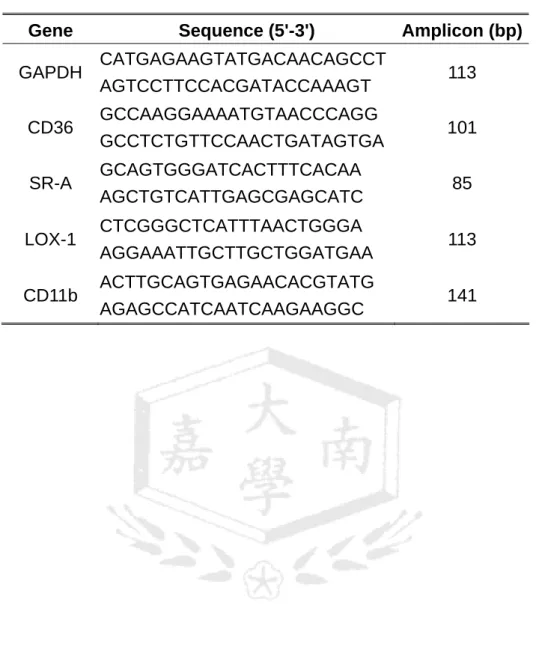
Quantitative PCR was performed with 2 μL cDNA obtained above in 25 μL containing 200 nM primers using iTaq™ SYBR Green Supermix with ROX (BioRad, Hercules, CA, USA). The primer sequences for GAPDH, CD11b and scavenger receptors were deduced from PrimerBank [48] and listed in Table 1. Amplification was conducted in an ABI Prism 7300 sequence detection system. PCR conditions were as follows: 95 °C for 2 min, 40 cycles at 95 °C for 15 s, and 60 °C for 45 s. The optimal concentrations of primers and templates used were established based on the standard curve created before the reaction and corresponded to approximately 100%
reaction efficiency. PCR results were then normalized to the expression of GAPDH in the same samples.
CD36 and SR-A phenotypic expression. Phenotypic expression of CD36 and SR-A were quantified by flow cytometry (FACScan, BD Biosciences, San Jose, CA,
USA). For detection of CD36 expression, cells (1 × 10⁶) were resuspended in 100 μL PBS. Twenty μl fluorescein (FITC)-conjugated murine anti-human CD36 (clone FA6.152, Immunotech, Beckman Coulter, Fullerton, CA, USA) was added and incubated on ice for 60 min before washing twice with PBS. For detection of SR-A expression, cells (5×10⁵) were resuspended in 100 μL PBS. Ten μL mouse anti-human SR-A/MSR1 antibody (25 μg/mL, clone 351615, R&D Systems, Minneapolis, MN, USA) was added and incubated at room temperature for 60 min. Cells were washed twice with PBS and then resuspended in 200 μL PBS. Ten μl FITC-labelled goat anti-mouse IgG antibodies (10 μg/mL, R&D Systems) were then added and incubated on ice for 60 min before washing twice with PBS. These cells were investigated in duplicates by flow cytometry (FACScan, BD Biosciences, San Jose, CA, USA). Data were acquired from 10,000 cells (events), and the fluorescent intensity was determined and expressed as the geometric mean fluorescence intensity (MFI).
Preparation of DiI-oxLDL and DiI-acLDL. LDL was incubated overnight at 37oC under nitrogen and light protection with 50 μL of DiI (3 mg/ml in DMSO) for each milligram of LDL protein. The LDL must be labeled before acetylation or oxidation [49]. For preparation of DiI-oxLDL, DiI-LDL (0.1 mg/ml) was incubated with 10 μM Cu2+ in PBS at 37oC in dark for 18 h [30]. Unbound dye and copper ions from the oxidation step described above which would otherwise be toxic to the cells were removed by passing through Sephadex G-25 (GE Healthcare). For preparation of DiI-acLDL, the method of Basu et al. [50] was employed. Unbound dye and acetate from acetylation step were then removed by passing through Sephadex-G25. Protein concentration of elute was determined by Bradford [44]. All lipoprotein preparations were stored at 4°C in sterile containers after filtration sterilization (0.45 μm).
Modified LDL uptake measurement by flow cytometry and fluorescent
microscopy. DiI-oxLDL or DiI-acLDL (10 μg/ml) was added into THP-1 monocytes or THP-1-derived macrophages and incubated for 24 h. The cells were washed with PBS and then examined with a fluorescence inverted microscope (IX-71, Olympus) followed by flow cytometry analysis (FACScan) using the FL2 emission filter. Data were acquired from 15,000 cells (events), and the DiI-oxLDL or DiI-acLDL uptake was determined and expressed as the geometric mean fluorescence intensity (MFI).
Statistical analyses. Student’s t-tests were used to assess significant differences in parameters measured between in the presence and absence of test substances. The level of significance was set at p<0.05. All experiments have been performed at least three times.
RESULTS
Inhibition of LDL oxidation by DTF. It is recognized that oxidative modified low density lipoproteins (oxLDL) play an important role in the generation and progression of the atherosclerotic plaque [51]. When LDL is oxidized by radical generating substances or by Cu2+ ions, three consecutive phases of the reaction, naming lag phase, propagation phase and the decomposition phase, can be observed in kinetic experiments by measuring compositional changes of LDL [52]. As shown in the top panel of Figure 2A, incubation of LDL (0.1 mg/mL) with Cu2+ (10 μM) resulted in a significant increase in conjugated diene formation, with maximum at 7 h oxidation without detectable lag phase. Loading LDL with DL-α-tocopherol (AT, 5 μM) increased the length of the lag phase to 1 h and therefore delayed onset of propagation. In comparison, loading LDL with DTF (5 μM) resulted in distinct kinetic conjugated diene formation which exhibited four successive oxidation phases: a longer lag phase (3 h), the first propagation phase, an inhibition phase with a conjugated diene formation rate slower than in the first propagation phase and the
second propagation phase [53]. However, LDL loaded with nobiletin (NOB) did not show significant difference from that of vehicle.
To better measure the antioxidant activity of DTF, LDL oxidation was performed with lower concentration of DTF (0.5 or 1 μM) or higher concentration of AT (10 or 20 μM) (bottom panel of Figure 2A). The conjugated diene formation of LDL supplemented with 1 μM of DTF or 20 μM of AT exhibited five successive phases (lag phase, the first propagation phase, an inhibition phase, the second propagation phase and a decomposition phase).
Incubation of LDL (0.1 mg/mL) with Cu2+ (10 μM) at 37oC for 3 h caused TBARS formation to increase from 0.95 ± 0.05 to 43.53±1.67 nmol MDA equivalent/mg LDL (Figure 2B). DTF treatment produced a dose-dependent reduction in TBARS formation, and IC50 was calculated as 1.14 ± 0.03 μM from linear regression curve. In parallel to the conjugated diene data, the antioxidant potency of DTF was significantly higher than positive control, AT, which had IC50 of 10.84 ± 0.95 μM.
To further investigate the protection of DTF towards electronegative charge modification of LDL-protein moiety (apoB) induced by Cu2+, agarose gel electrophoresis was carried out as described in Materials and Methods. Native LDL (nLDL, 0.1 mg/mL) treated with Cu2+ (10 μM) for 3 h increased REM to 3.03 (Figure 2C). Oxidation of LDL in the presence of 5 μM DTF resulted in decreased REM to 1.13. On the other hand, LDL treated with 5 μM NOB or AT exhibited less protective effect against Cu2+-mediated electronegative charge modification of apoB.
DTF inhibits monocyte-to-macrophage differentiation. The human monocytic cell line THP-1 was used as a model system to analyze gene expresssion and activity involved in monocyte-to-macrophage differentiation [54]. To evaluate the maximal
dosages of NOB and DTF could be used in THP-1 monocytes and macrophages, cell viability was analyzed using 3-(4,5-dimethylthiazol-2-yl)-2,5- diphenyltetrazolium (MTT) assay [55]. Our result demonstrates that 50 μM NOB and DTF exhibited significant (33~36%) cytotoxicity in THP-1 monocytes after 48 h incubation; while lower doses (10 and 20 μM) did not show significant adverse effect. A similar phenomenon was observed in THP-1 derived macrophages (data not shown). The differentiation-inducing dose of 30 nM PMA and working duration of 24 h was also determined in preliminary dose response experiments by analyzing differentiation marker CD11b expression and morphology change (data not shown).
THP-1 monocytes, which were originally in suspension, were treated with indicated concentrations of NOB or DTF 30 min prior to the addition of PMA (30 nM) for 24 h. The mRNA expression of differentiation marker, CD11b, and the major scavenger receptors, naming CD36, SR-A and LOX-1, measured by RT-Q-PCR normalized to the level of GADPH is shown in Figure 3. CD11b expression was increased by 17.0 ± 1.6 folds when THP-1 monocytes were treated with PMA (30 nM) for 24 h (Figure 3A). This is in a good agreement with the well-known theory that differentiation of monocytes into tissue macrophages results in an increase in mRNA and surface of CD11b/CD18 expression [56]. Addition of DTF (10 and 20 μM) in the process of monocyte differentiation dose-dependently inhibited CD11b mRNA expression, indicating the differentiation was attenuated. On the other hand, NOB had no influence on PMA-stimulated CD11b expression.
As expected, THP-1 monocytes exhibited very low level of LOX-1 mRNA and treated with PMA (30 nM) for 24 h induced more than 1000-fold increase (Figure 3B). Treatment of THP-1 cells with DTF or NOB strongly attenuated PMA-induced LOX-1 expression and the percentage of reduction was 81.4%, 98.8%, 58.9% and
79.4% for 10 μM DTF, 20 μM DTF, 10 μM NOB and 20 μM NOB, respectively, as compared with PMA-treated cells (p<0.01).
The impact of DTF and NOB on CD36 expression during PMA-stimulated monocyte differentiation is shown in Figure 3C. THP-1 monocytes exhibited little CD36 mRNA expression, while PMA-treated cells increased CD36 expression by 26.8 ± 0.4 folds. THP-1 monocytes treated with DTF significantly suppressed PMA-stimulated CD36 mRNA expression dose-dependently and 20 μM DTF can down-regulated CD36 mRNA to the level of monocytes. Surprisingly, NOB induced CD36 mRNA expression during THP-1 differentiation.
Figure 3D further demonstrates the effects of NOB and DTF on SR-A transcript expression in THP-1 monocytes treated with PMA (30 nM) for 24 h. It was found that PMA induced about 480-fold SR-A mRNA expression and addition of 10 μM and 20 μM DTF reduced PMA-induced SR-A expression by 56.8% and 98.6%, respectively.
However, no effect was observed for NOB-treated cells.
CD36 and SR-A are the principal receptors responsible for the binding and uptake of modified LDL in macrophages and together these two receptors account for 75 to 90% of the uptake and degradation of acetylated and oxidized LDL [57]. We thus further studied the effects of NOB and DTF on CD36 and SR-A surface protein level using flow cytometry as described in Materials and Methods. Data demonstrated that the expression of CD36 and SR-A surface protein increased by 6.3- and 1.9-fold, respectively (Figures 3E and F). Addition of 20 μM DTF could significantly reduced surface CD36 and SR-A protein expression by 80.5% and 41.8% (p<0.01 and 0.05), respectively, as compared with PMA-treated cells. On the other hand, treatment of THP-1 cells with NOB or lower dose of DTF did not have significant effect on CD36 or SR-A surface protein expression.
The LDL particle acquires a number of important biological activities as a result of oxidative modification in addition to the ability to bind scavenger receptors.
OxLDL is both a potent chemoattractant for circulating monocytes and a potent inhibitor of resident macrophage motility [58]. Previous studies have indicated that oxLDL plays a role in promoting differentiation of monocytes to macrophages [59, 60]. Recently, it has been reported that treatment of THP-1 monocytes with oxLDL plus 1.6 nM of PMA resulted in a dramatic synergistic effect on differentiation as compared with oxLDL alone [47]. By RT-Q-PCR analysis, we also observed that in the presence of 1.6 nM of PMA, CD36 mRNA expression of THP-1 monocytes increased with oxLDL concentration (5-25 μg/ml) (data not shown). mRNA transcripts for CD36, SR-A and LOX-1 were increased following cell treatment with oxLDL (25 μg/ml) by up to 1.5-, 1.7- and 2.2-fold, respectively, after 24 h culture as compared with PMA (1.6 nM) alone (Figure 4). DTF (20 μM) could significantly reduce the transcripts of CD36, SR-A and LOX-1 (p<0.01, 0.05 and 0.05, respectively). On the contrary, NOB did not exert any detectable inhibitory effect on any of the gene expression (data not shown).
It has been well-known that differentiated monocytes exhibit characteristic macrophage activity, i.e., increased cellular uptake of oxLDL. Our next approach was to examine whether NOB and DTF could block modified LDL uptake associated with monocyte differentiation. THP-1 monocytes were pre-treated with vehicle or indicated concentration of NOB or DTF for 30 min prior to exposure to PMA (30 nM) for 24 h. These cells were then incubated with DiI-acLDL (10 μg/mL) for 24 h.
Accumulation of DiI-acLDL into the cytoplasm was observed by fluorescence microscopy (vehicle of Figure 5A). Addition of DTF or NOB (10 and 20 μM) dose-dependently abolished acLDL uptake. In parallel, flow cytometry (Figure 5B)
shows that 10 and 20 μM DTF inhibited PMA-induced DiI-acLDL uptake by 61.5 and 86.2%, respectively. Lower concentration of NOB (10 μM) did not affect PMA-induced DiI-acLDL uptake, while higher concentration (20 μM) inhibited only 30.0% uptake.
Fluorescence microscopy (Figure 5C) also demonstrates that treatment of THP-1 monocytes with PMA (30 nM) for 24 h stimulated DiI-oxLDL uptake dramatically. In parallel, flow cytometry (Figure 5D) revealed PMA increased 25-fold DiI-oxLDL uptake as compared with monocytes. Treatment of THP-1 monocytes with DTF and NOB (10-20 μM) dose-dependently inhibited PMA-stimulated DiI-oxLDL uptake by 68.8-88.4% and 26.2-67.3%, respectively.
DTF inhibits the expression and activity of scavenger receptors in THP-1-derived macrophages. THP-1-derived macrophages have been extensively used as a model for studies of scavenger receptor expression and foam cell formation in response to various agents [61-63]. THP-1 monocytes were first allowed to differentiate into adherent macrophages by PMA (200 nM) for 3 days prior to exposure to test agent for 48 h. It has been shown that SR-A and CD36 are responsible for the preponderance of modified LDL uptake in macrophages and that other scavenger receptors do not compensate for their absence [57]. As a consequence, we investigated whether DTF or NOB treatment could inhibit foam cell formation by focusing on SR-A and CD36 expression and modified LDL uptake. Figure 6 shows that the mRNA expression of CD36 and SR-A of THP-1-derived macrophages was significantly repressed by DTF in a dose-dependent manner. About 46.1 ± 3.4 and 12.6 ± 2.8% of CD36 expression was detected in 10 and 20 μM DTF-treated THP-1 macrophages, respectively, relative to vehicle (Figure 6A). NOB (10-20 μM) was less potent and inhibited about 40% of CD36 expression with no clear dose response.
Likewise, SR-A expression in THP-1 macrophages was significantly inhibited by DTF (10-20 μM) by 68-92% dose-dependently. NOB inhibited SR-A expression by about 55% without dose-response relationship (Figure 6B).
DiI-oxLDL and DiI-acLDL uptake was studied to determine the effects of NOB and DTF on scavenger receptor activity. Fluorescence microcopy of Figure 6C shows that the uptake of DiI-oxLDL was highly enhanced in THP-1 macrophages and the addition of DTF or NOB significantly reduced the number of fluorescent cells. In parallel, flow cytometry of Figure 6D shows that treatment of THP-1 macrophages with DTF and NOB (10-20 μM) dose-dependentlyinhibitedDiI-oxLDL fluorescence signal by 45-80 and 34-68%, respectively. Similar effect was observed for DiI-acLDL uptake into macrophages (Figure 6E).
DISCISSION
Oxidative modification of LDL is believed to be an important event in atherogenesis [64], and several studies have reported on the antioxidant effect of flavonoids, that is, decreasing the susceptibility of LDL to oxidation [65-68]. To study the effect of 3',4'-dihydroxy-5,6,7,8-tetramethoxyflavone (DTF), as antioxidants in preventing copper-induced oxidation of LDL, three different approaches were employed to measure changes in parameters known to be associated with LDL oxidation: formation of conjugated dienes and thiobarbituric acid reactive substances (TBARS) during lipid peroxidation, as well as increase in the electrophoretic mobility of LDL due to apolipoprotein B100 modification [69]. In agreement with previous study [53], we found that artificial enhancement of antioxidant defenses by increasing α-tocopherol (AT) concentration (from 5 to 20 μM) invariably made LDL more resistant to peroxidative modifications as extended lag phase shown in Figure 2. Both LDL loaded with AT (20 μM) or DTF (1 μM) showed five successive oxidation
phases: lag phase, the first propagation phase, an inhibition phase, the second propagation phase, and a decomposition phase. It has been suggested that the delayed onset of the second propagation (Figure 2) may reflect the chain-breaking activity of ΑΤ to halt the progression of lipid peroxidation and the second inhibition phase is a period where ΑΤ is being consumed via peroxyl radical scavenging [53].
Consequently, a rise in the second inhibition phase for LDL enriched with ΑΤ (shown in bottom panel of Figure 2A) may be due to lipid hydroperoxides (LOOH) formation via the reaction with ΑΤ. Although no termination phase was observed for LDL enriched with DTF (5 μM) in top panel of Figure 2A, the oxidation would have stop if we had extended incubation longer. In addition to chain breaking activity, DFT might have a metal binding activity which would reduce the overall rate of initiating radical generation.
A key feature of LDL oxidation is the breakdown of polyunsaturated fatty acids to yield a broad array of smaller fragments, 3–9 carbons in length, including aldehydes and ketones [70]. As a result, besides evaluating the conjugated dienes that form following the Cu2+-triggered peroxidation, we also studied the accumulation of TBARS for LDL supplemented with DTF or ΑΤ after 3 h oxidation (Figure 2B). The data presented and obtained by utilizing these two different analytical procedures, clearly demonstrate that DTF has about 10- to 20-fold higher antioxidant activity as compared with ΑΤ; while NOB has negligible antioxidant activity.
It has been found that malondialdehyde (or other aldehydes) generated during oxidation can form Schiff bases with the ε-amino groups of lysine residues of apoB [71]. In this research, we also found that the relative electrophoretic mobility (REM) of LDL increased in the presence of Cu2+ and the addition of DTF (5 μM) could significantly protect apoB from oxidative modification. NOB had no detectable
antioxidant effect; while ΑΤ (5 μM) had limited inhibitory activity against conjugated diene formation (Figure 2A) and TBARS formation (Figure 2B). As a result, these characteristics might explain why there was no significant difference in REM assay between LDL enriched with AT or NOB and vehicle control (Figure 2C).
In atherosclerotic lesions, monocytes migrate into the vessel wall and differentiate into macrophages. Some macrophages become lipid-loaded after uptake of oxLDL. As a result, to mimic events in atherosclerotic lesions, both monocyte-to-macrophage differentiation and foam cell formation need to be studied [72]. In this study, we first used PMA- and oxLDL-stimulated THP-1 cells as models to study the effects of NOB and DTF on monocyte differentiation. The expression of scavenger receptors is upregulated during the differentiation of monocytes into macrophages, which is a key event in the process of atherosclerosis. It was found that THP-1 monocytes stimulated with PMA (30 nM) for 24 h significantly induced CD11b, LOX-1, CD36 and SR-A mRNA expression by ~17, >1000, ~27 and ~480 folds, respectively (Figures 3A-D). These levels were higher than those published by using semi-quantitative RT-PCR in combination with end-product gel electrophoretic analysis [35, 39, 47]. Phenotypic expression analyzed by flow cytometry further demonstrated that PMA (30 nM) induced CD36 and SR-A surface protein expression by 6.3- and 1.9-fold (Figures 3E and F). The protein induction was similar to those published in literature [47] but was much lower than that of mRNA. The discrepancy between induction of mRNA and surface protein expression might be caused by (1) the newly made mRNA does not completely contribute to the protein synthesis or the turnover rate of mRNA is relatively slow so that detected mRNA level is higher; (2) nascent protein does not completely translocate from the intracellular compartments to cellular surface so that surface protein level is lower [73]; (3) RT-Q-PCR has higher
detection sensitivity than flow cytometry.
Current result (Figure 3) also showed that DTF (10 and 20 μM) could attenuate monocyte-to-macrophage differentiation because the transcripts of CD11b, LOX-1, CD36 and SR-A, as well as surface CD36 and SR-A protein expression were significantly reduced in a dose-dependent manner. On the other hand, the effect of parent compound, NOB, on monocyte differentiation was distinct. In the mRNA expression, NOB had no effect on CD11b or SR-A; while decreased LOX-1 and increased CD36. These results are slightly different from those published by Eguchi et al. [35, 39], which demonstrated that both NOB and DTF could reduce LOX-1, SR-A, and CD36 mRNA levels. We reasoned that these discrepancies may be accounted by variations in THP-1 culture condition and primer design for the target gene or detection limit difference between the real-time PCR and traditional PCR [74].
It has been well-known that oxLDL, in contrast to native LDL, induces a differentiation process in monocytes, resulting in a more mature macrophage-like phenotype [60]. Stimulation of monocyte-to-macrophage differentiation was dependent on the extent of LDL oxidation, and required oxLDL internalization by the cells [47]. Current research (Figure 4) demonstrated that DTF, rather than NOB, downregulated oxLDL-stimulated scavenger receptor expression. Among these scavenger receptors, the expression of CD36 was the most sensitive to DTF treatment.
It has been indicated that there might be different pathways activated by oxLDL and PMA. PKB/PPARγ signaling pathways are thought to mediate CD36 expression in response to oxLDL [75]; while PKC/PPARγ is to mediate PMA-induced differentiation [75, 76]. From the current result, it is likely that DTF rather than its parent compound, NOB, interferes with both pathways.
The above results led us to examine whether DTF and NOB can affect
differentiation-associated scavenger receptor activity by measuring uptake of acLDL and oxLDL. AcLDL is known to be bound to and taken up mainly through SR-A;
while a less extent is through CD36. OxLDL is taken up, for the most part, through CD36; while through SR-A and LOX-1 in a less degree. Fluorescence microscope (Figures 5A and C) provided live-cell imaging of increased number of fluorescence cells in response to PMA. Flow cytometry (Figures 5B and D) revealed 6-fold DiI-acLDL and 25-fold DiI-oxLDL mean fluroscence (MFI) increase in PMA-treated cells as compared with monocytes. The increase is associated with PMA-induced expression and activities of scavenger receptors. Treatment of THP-1 cells with DTF (10-20 μM) during differentiation dose-dependently block PMA-stimulated uptake of modified LDL by 61-88% (Figure 5). This effect may result from the combined decrease of SR-A, LOX-1 and CD36 expression (Figure 3). In comparison, NOB decreased PMA-stimulated DiI-acLDL and DiI-oxLDL uptake, in less extent (Figure 5); although it did not suppress PMA-induced SR-A or CD36 mRNA or surface protein expression (Figure 3). This result was similar to the effect of NOB in murine J774A.1 macrophages [36]. We also found that NOB exhibited stronger inhibitory effect against DiI-oxLDL uptake than DiI-acLDL uptake and this may be associated with its inhibition on LOX-1 mRNA expression. Nevertheless, the attenuation of modified LDL uptake by NOB could not be completely explained by merely down-regulation of scavenger receptor expression, it is likely that NOB function downstream of scavenger receptor ligand binding as well.
We further questioned whether DTF and NOB also inhibited foam cell formation in THP-1-derived macrophages. RT-Q-PCR (Figures 6A and B) indicated that addition of DTF and NOB markedly inhibited the expression of SR-A and CD36, which were responsible for the majority of modified LDL processing in macrophages
[57]. To elucidate whether there are any functional consequences for DTF- or NOB-mediated inhibition of SR-A and CD36 expression, quantitative analysis of DiI-acLDL and DiI-oxLDL uptake was performed in THP-1-derived macrophages.
Both DTF and NOB significantly inhibited DiI-acLDL as well as DiI-oxLDL uptake by THP-1 macrophages dose-dependently. It appears that both DTF and NOB exert dual effects in inhibiting SR-A and CD36 activities in macrophages. By comparing data from Figure 3 to Figure 6, it is apparent that the effect of NOB on scavenger expression and activity is more evident in macrophages than during monocyte-to-macrophage differentiation.
It has been generally accepted that catechol compounds which possess two adjacent OH-groups in the benzene ring can be further oxidized to reactive semiquinone, which may bring about the pharmacological or toxicological activities [77]. Similarly, DTF has been reported stronger anti-inflammatory and antitumor activity than NOB [37]. In this research we further demonstrated that DTF exerts a stronger protective effect against atherogenesis than NOB because of its stronger antioxidant activity against LDL oxidation and its potent inhibitory effect against monocyte-to-macrophage differentiation and foam cell formation. This observation, if confirmed in vivo, might have important clinical implications in the prevention and treatment of atherosclerosis.
ACKNOWLEDGEMENTS
Critical reviews and suggestions from Professor Verne N. Schumaker and Professor Chenbei Chang are very much appreciated. This research was supported by Grants NSC-96-2320-B-041-006-MY3 (to Dr. M-J Wu) and NSC-98-2313-B-041 -002-MY3 (to M.-C. Kou) from the National Science Council, Taiwan and Campus Integrated Project Grant (to Dr. M-J Wu) from Chia Nan University, Taiwan.
Table 1. Primer pairs used in RT-Q-PCR.
Gene Sequence (5'-3') Amplicon (bp)
GAPDH CATGAGAAGTATGACAACAGCCT
AGTCCTTCCACGATACCAAAGT 113
CD36 GCCAAGGAAAATGTAACCCAGG
GCCTCTGTTCCAACTGATAGTGA 101
SR-A GCAGTGGGATCACTTTCACAA
AGCTGTCATTGAGCGAGCATC 85
LOX-1 CTCGGGCTCATTTAACTGGGA
AGGAAATTGCTTGCTGGATGAA 113
CD11b ACTTGCAGTGAGAACACGTATG
AGAGCCATCAATCAAGAAGGC 141
Figure 1. Chemical structures of nobiletin and 3',4'-dihydroxy-5,6,7,8- tetramethoxyflavone.
Figure 2. Effects of 3',4'-dihydroxy-5,6,7,8-tetramethoxyflavone (DTF), nobiletin (NOB), and DL-α-tocopherol (AT) on inhibiting Cu2+-induced LDL oxidation. Native LDL (nLDL) (0.1 mg/ml) was oxidized with 10 μM Cu2+ in the presence of vehicle (0.1% DMSO) or indicated compound at 37oC. (A) The formation of conjugated diene was measured by change in absorbance at 234 nm (ΔΑ234) after oxidation for the indicated period. (B) TBARS formation was measured after oxidization for 3 h as described in Materials and Methods. (C) Electrophoretic mobility was measured in the presence of 5 μM of indicated compound as described in Materials and Methods.
Figure 3. Effects of 3',4'-dihydroxy-5,6,7,8-tetramethoxyflavone (DTF) and nobiletin (NOB) on the mRNA expression of CD11b (A), LOX-1 (B), CD36 (C) and SR-A (D);
as well as surface protein expression of CD36 (E) and SR-A (F) during
monocyte-to-macrophage differentiation. THP-1 monocytes were treated with vehicle (0.1% DMSO), DTF or NOB for 30 min prior to 30 nM PMA addition and incubated for 24 h. To measure mRNA expression, total cellular RNA was prepared and the expression of mRNA was analyzed as described in Materials and Methods. The data were normalized with reference to the expression levels of the corresponding GAPDH mRNAs. Data represent mean ratio± SEM of three independent experiments relative to the value of the monocytes (A-D). Cell surface CD36 protein expression was measured by incubation cells with fluorescein (FITC)-conjugated murine anti-human CD36 (clone FA6.152, Immunotech, Beckman Coulter). Cell surface SR-A expression was measured by incubation cells with mouse anti-human SR-A/MSR1 antibody
(clone 351615, R&D Systems) followed by FITC-labelled goat anti-mouse IgG
antibodies. The level of fluorescence was investigated in duplicates by flow cytometry (Coulter EPICS XL, Beckman Coulter) as described in Materials and Methods (E and F). *, p<0.05 and **, p < 0.01 represents significant differences compared with
vehicle control of 30 nM PMA-treated cells.
Figure 4. Effects of 3',4'-dihydroxy-5,6,7,8-tetramethoxyflavone (DTF) on the mRNA expression of CD36, SR-A and LOX-1 in oxLDL-induced monocyte-to-macrophage differentiation. THP-1 monocytes were treated with vehicle (0.1% DMSO), NOB for 30 min prior to 1.6 nM PMA and 25 μg/mL oxLDL treatment and incubated for 24 h.
Total cellular RNA was prepared and the expression of mRNA was analyzed as described in Materials and Methods. The data were normalized with reference to the expression levels of the corresponding GAPDH mRNAs. Data represent the mean ratio ± SEM of three independent experiments relative to the value of the PMA (1.6 nM)-treated cells. *, p<0.05 and **, p<0.01 represent significant differences compared with cells treated with PMA (1.6 nM) plus oxLDL (25 μg/mL).
Figure 5. Effects of 3',4'-dihydroxy-5,6,7,8-tetramethoxyflavone (DTF) and nobiletin (NOB) on DiI-acLDL and DiI-oxLDL uptake in PMA-stimulated THP-1 monocytes.
THP-1 monocytes were treated with vehicle (0.1% DMSO), DTF or NOB for 30 min prior to PMA (30 nM) addition and incubated for 24 h. DiI-acLDL (A and B) and DiI-oxLDL (C and D) (10 μg/mL) was added to cells and incubated for 24 h. Cell association of DiI-modified LDL was observed under fluorescence microscopy (A and C). Cells were then analyzed by flow cytometry (B and D) as described in
Material and Methods. Data represent the mean ± SEM of three independent experiments. *, p<0.05 and **, p<0.01 represent significant differences compared with vehicle control of 30 nM PMA-treated cells.
Figure 6. Effects of 3',4'-dihydroxy-5,6,7,8-tetramethoxyflavone (DTF) and nobiletin (NOB) on the mRNA expression of CD36 (A) and SR-A (B) and DiI-modified LDL uptake (C-E) in THP-1-derived macrophages. THP-1-derived macrophages were treated with vehicle (0.1% DMSO), DTF or NOB for 48 h. To measure mRNA expression level, total cellular RNA was prepared and analyzed as described in Materials and Methods. The data were normalized with reference to the expression levels of the corresponding GAPDH mRNAs and were represented as the mean ratio
± SEM of three independent experiments relative to the value of the vehicle-treated cells (A and B). To measure modified LDL uptake, DiI-oxLDL or DiI-acLDL (10 μg/ml) was added to cells and incubated for another 24 h. Cell association of DiI-oxLDL was observed under fluorescence microscopy (C) and the uptake of DiI-modified LDL was quantitated by flow cytometry (D and E). Data represent the mean ± SEM of three independent experiments.*, p<0.05 and **, p < 0.01 represent significant differences compared with vehicle control.
O O
OCH3
OCH3
OCH3
H3CO
H3CO
OCH3
Nobiletin
O O
OH OH
OCH3
H3CO
H3CO
OCH3
3',4'-dihydroxy-5,6,7,8-tetramethoxyflavone
(Figure 1)
Incubation Time (h)
0 2 4 6 8 10 12 14 16 18 20 22 24 26
ΔA234
0.0 0.5 1.0 1.5 2.0 2.5
Vehicle DTF 5 μM NOB 5 μM AT 5 μM
Incubation Time (h)
0 5 10 15 20 25
ΔA234
0.0 0.2 0.4 0.6 0.8 1.0 1.2 1.4
Vehicle DTF 0.5 μM DTF 1 μM AT 10 μM AT 20 μM
A Conjugated Diene Formation
(Figure 2A)
B
TBARS Assay
n-LDL vehicle 0.8 0.9 1 1.1 1.2
nmol MDA/mg LDL
0 10 20 30 40 50
+ CuSO4
DTF concentration (μM)
+ CuSO4
n-LDL vehicle 5 10 15 20
nmol MDA/mg LDL
0 10 20 30 40 50
AT concentration (μM)
(Figure 2B)
C
Relative Electrophoretic Mobility
_ o +
_ o + _
o +
_ o +
REM 3.03 2.81 1.13 2.94 1.00
Origin
NOB
Vehicle DTF AT n-LDL
_ o +
_ o + _
o +
_ o +
REM 3.03 2.81 1.13 2.94 1.00
Origin
NOB
Vehicle DTF AT n-LDL
(Figure 2C)
concentration (μM)
monocytes vehicle 10 20
CD11b mRNA expression (folds of monocytes )
0 5 10 15 20
DTF NOB
+ PMA (30 nM) A
*
*
monocytes vehicle 10 20
LOX-1 mRNA expression (folds of monocytes)
0 200 400 600 800 1000 1200 1400
DTF NOB
concentration (μM) + PMA (30 nM)
**
**
**
**
**
**
B
(Figure 3 A,B)
+ PMA (30 nM) C
concentration (μM) + PMA (30 nM) D
concentration (μM)
monocytes vehicle 10 20
CD36 mRNA expression (folds of monocytes)
0 10 20 30 40 50 60
DTF NOB
**
**
**
**
**
monocytes vehicle 10 20
SR-A mRNA expression (folds of monocytes)
0 100 200 300 400 500 600
DTF NOB
**
** **
(Figure 3 C,D)
E
concentration (μM)
monocytes vehicle 10 20
Mean fluorescence intensity of CD36 (MFI)
0 50 100 150 200 250
DTF NOB
+ PMA (30 nM)
**
(Figure 3E)
vehicle monocytes
20 μM DTF 20 μM NOB
+PMA
F
monocyte Vehicle 10 20
0 2 4 6 8 10 12 14
DTF NOB
Mean flourescence intensity of SR-A (MFI)
concentration (μM)
+ PMA (30 nM)
*
(Figure 3F)
+ PMA 1.6 nM
monocyte vehicle 10 20
CD36 mRNA expression (fold of monocyte)
0.0 0.2 0.4 0.6 0.8 1.0 1.2 1.4 1.6 1.8
DTF (μM) +oxLDL (25 μg/ml)
**
*
monocyte vehicle 10 20
SR-A mRNA expression (fold of monocyte)
0.0 0.2 0.4 0.6 0.8 1.0 1.2 1.4 1.6 1.8 2.0
+ PMA 1.6 nM +oxLDL (25 μgl/ml)
DTF (μM)
*
**
monocyte vehicle 10 20
LOX-1 mRNA expression (fold of monocyte)
0.0 0.5 1.0 1.5 2.0 2.5 3.0
+ PMA 1.6 nM +oxLDL (25 μgl/ml)
DTF (μM)
*
**
*
(Figure 4)
A
B
**
**
*
#
#
concentration (μM)
monocytes vehicle 10 20
24 h DiI-acLDL uptake (MFI)
0 20 40 60 80 100 120
DTF NOB
+ 30 nM PMA
**
**
**
**
(Figure 5AB)
a monocyte
b vehicle
c DTF 10 μM
d DTF 20 μM
e NOB 10 μM
f NOB 20 μM
C
D
concentration (μM)
monocytes vehicle 10 20
DiI-oxLDL uptake (MFI)
0 100 200 300 400
DTF NOB
+ PMA (30 nM)
**
**
**
**
*
(Figure 5CD)
a monocyte
b vehicle
c DTF 10 μM
d DTF 20 μM
e NOB 10 μM
f NOB 20 μM
concentration (μM)
vehicle 10 20
CD36 mRNA expression (fold of macrophages)
0.0 0.2 0.4 0.6 0.8 1.0 1.2 1.4
DTF NOB
**
**
* *
A
concentration (μM)
vehicle 10 20
SR-A mRNA expression (fold of macrophages)
0.0 0.2 0.4 0.6 0.8 1.0 1.2
DTF NOB
**
**
** **
B
(Figure 6 A and B)
C
(Figure 6 C)
a blank control b vehicle
c DTF 10 μM d DTF 20 μM
e NOB 10 μM f NOB 20 μM
vehicle 10 20
DiI-oxLDL uptake (MFI)
0 50 100 150 200 250 300 350
DTF NOB
concentration (μM)
D
E
**
**
**
**
concentration (μM)
vehicle 10 20
DiI-acLDL uptake (MFI)
0 20 40 60 80 100 120 140 160
DTF NOB
**
** **
**
(Figure 6 D and E)
[1] P. Libby, Inflammation in atherosclerosis, Nature 420 (2002) 868-874.
[2] M. Kaplan, M. Aviram, Oxidized low density lipoprotein: atherogenic and proinflammatory characteristics during macrophage foam cell formation. An inhibitory role for nutritional antioxidants and serum paraoxonase, Clin. Chem.
Lab. Med. 37 (1999) 777-787.
[3] R. Ross, Atherosclerosis--an inflammatory disease, N. Engl. J. Med. 340 (1999) 115-126.
[4] Y. Yamada, T. Doi, T. Hamakubo, T. Kodama, Scavenger receptor family proteins: roles for atherosclerosis, host defence and disorders of the central nervous system, Cell. Mol. Life Sci. 54 (1998) 628-640.
[5] W.J. de Villiers, E.J. Smart, Macrophage scavenger receptors and foam cell formation, J. Leukoc. Biol. 66 (1999) 740-746.
[6] G. Endemann, L.W. Stanton, K.S. Madden, C.M. Bryant, R.T. White, A.A.
Protter, CD36 is a receptor for oxidized low density lipoprotein, J Biol Chem 268 (1993) 11811-11816.
[7] Y. Zeng, N. Tao, K.N. Chung, J.E. Heuser, D.M. Lublin, Endocytosis of oxidized low density lipoprotein through scavenger receptor CD36 utilizes a lipid raft pathway that does not require caveolin-1, J. Biol. Chem. 278 (2003) 45931-45936.
[8] A. Nakata, Y. Nakagawa, M. Nishida, S. Nozaki, J. Miyagawa, T. Nakagawa, R. Tamura, K. Matsumoto, K. Kameda-Takemura, S. Yamashita, Y.
Matsuzawa, CD36, a novel receptor for oxidized low-density lipoproteins, is highly expressed on lipid-laden macrophages in human atherosclerotic aorta, Arterioscler Thromb Vasc Biol 19 (1999) 1333-1339.
[9] D.P. Hajjar, M.E. Haberland, Lipoprotein trafficking in vascular cells.
Molecular Trojan horses and cellular saboteurs, J. Biol. Chem. 272 (1997) 22975-22978.
[10] J. Han, D.P. Hajjar, M. Febbraio, A.C. Nicholson, Native and modified low density lipoproteins increase the functional expression of the macrophage class B scavenger receptor, CD36, J. Biol. Chem. 272 (1997) 21654-21659.
[11] N. Platt, S. Gordon, Is the class A macrophage scavenger receptor (SR-A) multifunctional? - The mouse's tale, J. Clin. Invest. 108 (2001) 649-654.
[12] N. Kume, T. Murase, H. Moriwaki, T. Aoyama, T. Sawamura, T. Masaki, T.
Kita, Inducible expression of lectin-like oxidized LDL receptor-1 in vascular endothelial cells, Circ. Res. 83 (1998) 322-327.
[13] H. Kataoka, N. Kume, S. Miyamoto, M. Minami, H. Moriwaki, T. Murase, T.
Sawamura, T. Masaki, N. Hashimoto, T. Kita, Expression of lectinlike
oxidized low-density lipoprotein receptor-1 in human atherosclerotic lesions, Circulation 99 (1999) 3110-3117.
[14] H. Moriwaki, N. Kume, T. Sawamura, T. Aoyama, H. Hoshikawa, H. Ochi, E.
Nishi, T. Masaki, T. Kita, Ligand specificity of LOX-1, a novel endothelial receptor for oxidized low density lipoprotein, Arterioscler. Thromb. Vasc.
Biol. 18 (1998) 1541-1547.
[15] M. Febbraio, E.A. Podrez, J.D. Smith, D.P. Hajjar, S.L. Hazen, H.F. Hoff, K.
Sharma, R.L. Silverstein, Targeted disruption of the class B scavenger
receptor CD36 protects against atherosclerotic lesion development in mice, J.
Clin. Invest. 105 (2000) 1049-1056.
[16] H. Suzuki, Y. Kurihara, M. Takeya, N. Kamada, M. Kataoka, K. Jishage, O.
Ueda, H. Sakaguchi, T. Higashi, T. Suzuki, Y. Takashima, Y. Kawabe, O.
Cynshi, Y. Wada, M. Honda, H. Kurihara, H. Aburatani, T. Doi, A.
Matsumoto, S. Azuma, T. Noda, Y. Toyoda, H. Itakura, Y. Yazaki, T.
Kodama, et al., A role for macrophage scavenger receptors in atherosclerosis and susceptibility to infection, Nature 386 (1997) 292-296.
[17] V.R. Babaev, L.A. Gleaves, K.J. Carter, H. Suzuki, T. Kodama, S. Fazio, M.F.
Linton, Reduced atherosclerotic lesions in mice deficient for total or macrophage-specific expression of scavenger receptor-A, Arterioscler.
Thromb. Vasc. Biol. 20 (2000) 2593-2599.
[18] H. Sakaguchi, M. Takeya, H. Suzuki, H. Hakamata, T. Kodama, S. Horiuchi, S. Gordon, L.J. van der Laan, G. Kraal, S. Ishibashi, N. Kitamura, K.
Takahashi, Role of macrophage scavenger receptors in diet-induced atherosclerosis in mice, Lab. Invest. 78 (1998) 423-434.
[19] J.L. Mehta, N. Sanada, C.P. Hu, J. Chen, A. Dandapat, F. Sugawara, H. Satoh, K. Inoue, Y. Kawase, K. Jishage, H. Suzuki, M. Takeya, L. Schnackenberg, R.
Beger, P.L. Hermonat, M. Thomas, T. Sawamura, Deletion of LOX-1 reduces atherogenesis in LDLR knockout mice fed high cholesterol diet, Circ. Res.
100 (2007) 1634-1642.
[20] C. Hu, J. Chen, A. Dandapat, Y. Fujita, N. Inoue, Y. Kawase, K. Jishage, H.
Suzuki, D. Li, P.L. Hermonat, T. Sawamura, J.L. Mehta, LOX-1 abrogation reduces myocardial ischemia-reperfusion injury in mice, J. Mol. Cell. Cardiol.
44 (2008) 76-83.
[21] M. Aviram, Macrophage foam cell formation during early atherogenesis is determined by the balance between pro-oxidants and anti-oxidants in arterial cells and blood lipoproteins, Antioxid. Redox. Signal. 1 (1999) 585-594.
[22] M.N. Diaz, B. Frei, J.A. Vita, J.F. Keaney, Jr., Antioxidants and atherosclerotic heart disease, N. Engl. J. Med. 337 (1997) 408-416.
[23] B. Fuhrman, N. Volkova, R. Coleman, M. Aviram, Grape Powder Polyphenols Attenuate Atherosclerosis Development in Apolipoprotein E Deficient (E0) Mice and Reduce Macrophage Atherogenicity, J. Nutr. 135 (2005) 722-728.
[24] B. Fuhrman, M. Aviram, Anti-atherogenicity of nutritional antioxidants, IDrugs 4 (2001) 82-92.
[25] B. Fuhrman, M. Aviram, Flavonoids protect LDL from oxidation and attenuate atherosclerosis, Curr. Opin. Lipidol. 12 (2001) 41-48.
[26] A. Munteanu, J.M. Zingg, R. Ricciarelli, A. Azzi, CD36 overexpression in ritonavir-treated THP-1 cells is reversed by alpha-tocopherol, Free Radic. Biol.
Med. 38 (2005) 1047-1056.
[27] Y. Kawai, T. Nishikawa, Y. Shiba, S. Saito, K. Murota, N. Shibata, M.
Kobayashi, M. Kanayama, K. Uchida, J. Terao, Macrophage as a target of quercetin glucuronides in human atherosclerotic arteries: implication in the anti-atherosclerotic mechanism of dietary flavonoids, J. Biol. Chem. 283 (2008) 9424-9434.
[28] T.W. Lian, L. Wang, Y.H. Lo, I.J. Huang, M.J. Wu, Fisetin, morin and myricetin attenuate CD36 expression and oxLDL uptake in U937-derived macrophages, Biochim. Biophys. Acta 1781 (2008) 601-609.
[29] A. Munteanu, M. Taddei, I. Tamburini, E. Bergamini, A. Azzi, J.M. Zingg, Antagonistic effects of oxidized low density lipoprotein and alpha-tocopherol on CD36 scavenger receptor expression in monocytes: involvement of protein kinase B and peroxisome proliferator-activated receptor-gamma, J. Biol.
Chem. 281 (2006) 6489-6497.
[30] S. Devaraj, I. Hugou, I. Jialal, Alpha-tocopherol decreases CD36 expression in human monocyte-derived macrophages, J. Lipid Res. 42 (2001) 521-527.
[31] E.M. Kurowska, J.A. Manthey, Hypolipidemic effects and absorption of citrus polymethoxylated flavones in hamsters with diet-induced
hypercholesterolemia, J. Agric. Food Chem. 52 (2004) 2879-2886.
[32] J.M. Roza, Z. Xian-Liu, N. Guthrie, Effect of citrus flavonoids and tocotrienols on serum cholesterol levels in hypercholesterolemic subjects, Altern. Ther. Health Med. 13 (2007) 44-48.
[33] L.J. Wilcox, N.M. Borradaile, L.E. de Dreu, M.W. Huff, Secretion of
hepatocyte apoB is inhibited by the flavonoids, naringenin and hesperetin, via reduced activity and expression of ACAT2 and MTP, J. Lipid Res. 42 (2001) 725-734.
[34] B. Morin, L.A. Nichols, K.M. Zalasky, J.W. Davis, J.A. Manthey, L.J.
Holland, The Citrus Flavonoids Hesperetin and Nobiletin Differentially Regulate Low Density Lipoprotein Receptor Gene Transcription in HepG2
Liver Cells, J. Nutr. 138 (2008) 1274-1281.
[35] A. Eguchi, A. Murakami, H. Ohigashi, Nobiletin, a citrus flavonoid,
suppresses phorbol ester-induced expression of multiple scavenger receptor genes in THP-1 human monocytic cells, FEBS Lett. 580 (2006) 3321-3328.
[36] S.C. Whitman, E.M. Kurowska, J.A. Manthey, A. Daugherty, Nobiletin, a citrus flavonoid isolated from tangerines, selectively inhibits class A scavenger receptor-mediated metabolism of acetylated LDL by mouse macrophages, Atherosclerosis 178 (2005) 25-32.
[37] C.S. Lai, S. Li, C.Y. Chai, C.Y. Lo, S. Dushenkov, C.T. Ho, M.H. Pan, Y.J.
Wang, Anti-inflammatory and antitumor promotional effects of a novel urinary metabolite, 3',4'-didemethylnobiletin, derived from nobiletin, Carcinogenesis 29 (2008) 2415-2424.
[38] S. Li, S. Sang, M.H. Pan, C.S. Lai, C.Y. Lo, C.S. Yang, C.T. Ho,
Anti-inflammatory property of the urinary metabolites of nobiletin in mouse, Bioorg. Med. Chem. Lett. 17 (2007) 5177-5181.
[39] A. Eguchi, A. Murakami, S. Li, C.T. Ho, H. Ohigashi, Suppressive effects of demethylated metabolites of nobiletin on phorbol ester-induced expression of scavenger receptor genes in THP-1 human monocytic cells, Biofactors 31 (2007) 107-116.
[40] H. Esterbauer, G. Striegl, H. Puhl, M. Rotheneder, Continuous monitoring of in vitro oxidation of human low density lipoprotein, Free Radic. Res. Commun.
6 (1989) 67-75.
[41] G. Knipping, M. Rotheneder, G. Striegl, H. Esterbauer, Antioxidants and resistance against oxidation of porcine LDL subfractions, J. Lipid Res. 31 (1990) 1965-1972.
[42] L. Lanningham-Foster, C. Chen, D.S. Chance, G. Loo, Grape extract inhibits lipid peroxidation of human low density lipoprotein, Biol. Pharm. Bull. 18 (1995) 1347-1351.
[43] A.M. Gotto, Jr., H.J. Pownall, R.J. Havel, Introduction to the plasma lipoproteins, Methods Enzymol 128 (1986) 3-41.
[44] M.M. Bradford, A rapid and sensitive method for the quantitation of
microgram quantities of protein utilizing the principle of protein-dye binding, Anal Biochem 72 (1976) 248-254.
[45] H.A. Kleinveld, H.L. Hak-Lemmers, A.F. Stalenhoef, P.N. Demacker, Improved measurement of low-density-lipoprotein susceptibility to copper-induced oxidation: application of a short procedure for isolating low-density lipoprotein, Clin Chem 38 (1992) 2066-2072.
[46] M. Hermann, B. Gmeiner, Altered susceptibility to in vitro oxidation of LDL
in LDL complexes and LDL aggregates, Ann N Y Acad Sci 683 (1993) 363-364.
[47] B. Fuhrman, A. Partoush, N. Volkova, M. Aviram, Ox-LDL induces monocyte-to-macrophage differentiation in vivo: Possible role for the macrophage colony stimulating factor receptor (M-CSF-R), Atherosclerosis 196 (2008) 598-607.
[48] X. Wang, B. Seed, A PCR primer bank for quantitative gene expression analysis, Nucl. Acids Res. 31 (2003) e154.
[49] D. Teupser, J. Thiery, A.K. Walli, D. Seidel, Determination of LDL- and scavenger-receptor activity in adherent and non-adherent cultured cells with a new single-step fluorometric assay, Biochimica et Biophysica Acta (BBA) - Lipids and Lipid Metabolism 1303 (1996) 193-198.
[50] S.K. Basu, J.L. Goldstein, G.W. Anderson, M.S. Brown, Degradation of cationized low density lipoprotein and regulation of cholesterol metabolism in homozygous familial hypercholesterolemia fibroblasts, Proc. Natl. Acad. Sci.
U. S. A. 73 (1976) 3178-3182.
[51] U.P. Steinbrecher, H.F. Zhang, M. Lougheed, Role of oxidatively modified LDL in atherosclerosis, Free Radic Biol Med 9 (1990) 155-168.
[52] P.M. Abuja, H. Esterbauer, Simulation of lipid peroxidation in low-density lipoprotein by a basic "skeleton" of reactions, Chem. Res. Toxicol. 8 (1995) 753-763.
[53] O. Ziouzenkova, A. Sevanian, P.M. Abuja, P. Ramos, H. Esterbauer, Copper can promote oxidation of LDL by markedly different mechanisms, Free Radic.
Biol. Med. 24 (1998) 607-623.
[54] J. Auwerx, The human leukemia cell line, THP-1: a multifacetted model for the study of monocyte-macrophage differentiation, Experientia 47 (1991) 22-31.
[55] J. Carmichael, W.G. DeGraff, A.F. Gazdar, J.D. Minna, J.B. Mitchell, Evaluation of a tetrazolium-based semiautomated colorimetric assay:
assessment of chemosensitivity testing, Cancer Res. 47 (1987) 936-942.
[56] M.A. Arnaout, Structure and function of the leukocyte adhesion molecules CD11/CD18, Blood 75 (1990) 1037-1050.
[57] V.V. Kunjathoor, M. Febbraio, E.A. Podrez, K.J. Moore, L. Andersson, S.
Koehn, J.S. Rhee, R. Silverstein, H.F. Hoff, M.W. Freeman, Scavenger receptors class A-I/II and CD36 are the principal receptors responsible for the uptake of modified low density lipoprotein leading to lipid loading in
macrophages, J. Biol. Chem. 277 (2002) 49982-49988.
[58] M.T. Quinn, S. Parthasarathy, L.G. Fong, D. Steinberg, Oxidatively modified