行政院國家科學委員會專題研究計畫 期中進度報告
Fas-L 影響腫瘤組織結構及腫瘤免疫反應的機制(1/3)
計畫類別: 個別型計畫
計畫編號: NSC93-2320-B-006-060-
執行期間: 93 年 08 月 01 日至 94 年 07 月 31 日 執行單位: 國立成功大學微生物學科(所)
計畫主持人: 楊倍昌
計畫參與人員: 吳怡靜, 張雅南
報告類型: 精簡報告
處理方式: 本計畫可公開查詢
中 華 民 國 94 年 5 月 26 日
Title: Fas-L 影響腫瘤組織結構及腫瘤免疫反應的機制
中文摘要:
當 Fas-ligand (CD95-L; Fas-L)與細胞表面上它的受體 Fas (CD95)結合後,會激活 Fas 的 訊號傳遞,最後導致細胞凋亡(apoptosis)。以前我們發現 Fas/Fas-L 的交互作用會誘發 T 細 胞產生 IL-10。特別是人類的腦瘤細胞在裸鼠身上長成腫瘤時,Fas-L 會讓腫瘤的組織結構 較為密實、與正常組織之間的界線分明,浸潤的免疫細胞集中在組織外圍。然而,Fas-L 表 現量低的腦瘤細胞所形成的腫瘤則容易擴散侵入肌肉層,而且浸潤的免疫細胞分散。顯然 在腫瘤生長的過程中,Fas/Fas-L 傳遞的訊號可能不止於造成細胞凋亡。它可能刺激細胞激 素的產生而調節免疫反應,並且影響腫瘤的組織結構而增強轉移能力或逃避免疫偵測。
我們由小鼠黑色素瘤細胞建立表現不同程度的 Fas 及 Fas-L 細胞株。這些細胞轉移到 小鼠肺臟的能力各有不同。特別的是抑制了 Fas-L 的表現會增加黑色素瘤細胞轉移到肺臟 的能力。此外,利用三度空間培養方法將腫瘤細胞長成 tumor spheroid (腫瘤球)。這種體外 三度空間培養形成的腫瘤球和腫瘤細胞在小鼠體內長成的腫瘤組織很相像。腫瘤球內細胞 基因的表現和二度平面培養細胞差異極大。利用 ribozyme 抑制細胞 Fas-L 的表現量後導至 細胞在二維空間培養下的移形 (migration) 能力增加。抑制了細胞 Fas-L 的表現後三度空間 培養形成的腫瘤球內細胞的回貼能力也增加,但是不會改變 caspases 的基礎活性,而且使 用 caspases 的抑制劑也不會改變細胞移形能力。雖然 FAK 以及 MMP (gelatinase)的表現不 受 Fas-L 影響,抑制了細胞 Fas-L 的表現會增加 Paxillin。我們認為 Fas-L 影響到腫瘤細胞 在動物體內的轉移可能是與 MMP2/9 無關。
關鍵詞:Fas-ligand,腫瘤球,細胞移形
英文摘要:
Tumor cells escaping from immune surveillance and surviving in a new tissue environment are important for tumor metastasis. The mechanisms involving in these processes are still not completely understood. Fas/FasL mediated apoptosis is one of the strategies for immune escape.
Some FasL-expressing tumor cells induce apoptosis in cytotoxic lymphocytes (CTLs) by direct
“ Fas conterattack”. In order to investigate how Fas/FasL affects tumor metastasis, we established B16F10-derived cells expressing different levels of Fas or FasL and tested in a pulmonary
metastatic animal model. We found that down-regulation of FasL obviously increased lung
metastasis of B16F10 in B6 mice. Besides, up-regulation of Fas, but not up-regulation of FasL, reduced lung metastasis. Down regulation of FasL enhanced the cell migration in 2-dimensional culture system as well as in tumor spheroid-reattachment assay. We further explored if the Fas/FasL signaling might alter metalloproteinase activities to increase metastasis ability. A multicellular tumor spheroids system (MTS) was applied to measure cell migration and
matrixmetalloproteinases (MMPs) secretion. Tumor spheroids were grown on agar-medium based 96-well plates. MMPs activities in condition medium were detected by gelatinase-zymography assay. We found that the MMPs activities of tumor cells were not correlated with the expression of Fas and FasL. In summary, Fas/FasL system regulates tumor metastasis via an
MMP2/9-independent mechanism.
keywords:Fas-L, tumor spheroid, migration
Introduction:
Cancer is a kind of disease with significant impact on a society having long life-span, as in Taiwan. Understanding how tumor copes with host immune system can be valuable in designing an effective therapeutic strategy in tumor management. A novel aspect for the role of Fas-L on tumor surveillance, tumor escape, has been suggested in the last 5 years. Although it is still controversial about the effect of tumor Fas-L on immunity, study by dealing with only one molecule, here Fas-L, seems to be a straightforward action and easier to manipulate. Fas/Fas-L interaction can play multiple roles in addition to cell death. The study model established here provides a simple way to understand the complicate tissue microenvironment and immune response, which is critical and complimentary to current knowledge on tumor surveillance. As mentioned, Fas/FasL mediated apoptosis is one of the strategies for immune escape. Some FasL-expressing tumor cells induce apoptosis in cytotoxic lymphocytes (CTLs) by direct “Fas counter-attack.” In order to investigate how Fas/FasL affects tumor metastasis, we established B16F10-derived cells expressing different levels of Fas or FasL and tested in a pulmonary metastatic animal model. We further explored if the Fas/FasL signaling might alter metalloproteinase activities to increase metastasis ability. A multicellular tumor spheroids system (MTS) was applied to measure cell migration and matrixmetalloproteinases (MMPs) secretion.
Overall, this study tries to link the intracellular signal of tumor cells with interaction of extracellular matrix/ligand in the context of Fas/Fas-L signaling.
Results and discussion
The expression of Fas and FasL in B16F10 cells. To establish cells having different levels of Fas or FasL, the murine melanoma cells B16F10 were transfected with
pEGFP-N1 vector, FasLribozyme-carrying pEGFP-N1,
Fas-carrying pEGFP-N1, or FasL-carrying pEGFP-N1. As shown in Figure 1, we have established a panel of cells expressing different levels of Fas and FasL. A. The Fas mRNA detected by RT-PCR in B16F10, vector control (V), and Fas overexpression (oFas) cell lines. B.
The FasL mRNA detected by RT-PCR in B16F10, vector control (V), FasLribozyme (R), and FasL overexpression (oFasL) cell lines.
We injected the B16-F10 derived cells into B6 mice through tail vain to evaluate their metastatic ability to the lung. 5×10⁵ cells were inoculated into mouse through tail vein. Lung was harvested to observe tumor nodules at 14 days post-inoculation. V: vector control cells. R: FasLribozyme carrying cells. We observed an enhanced experimental pulmonary metastasis in B16F10 cells by down-regulating FasL expression, and reduced by up-regulating Fas expression (Figure 2).
Figure 1
Fig 2. Fas: Fas overexpression cells. FasL: FasL overexpress cells (bar = 1 cm)
Fig 3. A. Spheroids formation on an agar-covered plate. After 3 days, both cell lines could aggregate together to form single spheroid (bar = 200 mm). B. The formula v = 0.4ab2 was used to calculate the spheroids volume (a: largest; b: smallest diameter of the spheroid).
Fig 4. Morphological appearance of melanoma cell spheroids.
5×10³ cells were seeded into 96-well plate coated with a layer of
To establish a tumor spheroid model, spheroids were generated by the liquid overlay technique. We grew cells on the top of 0.5-2% Nobel agar to prevent cell attachment. Melanoma cells formed aggregate spheroids. The expression of FasL did not obviously influence the spheroid formation and growth rate
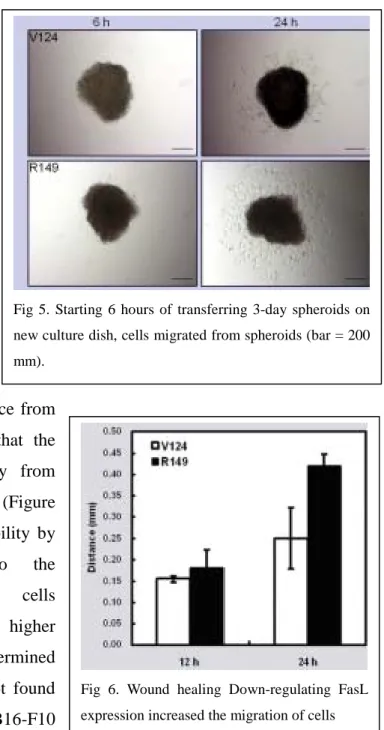
(Figure 3). We further characterized the structure of tumor spheroids by Hematoxylin/Eosin stain. Tumor spheroids formed by V124 (FasLhigh) and R149 (FasLlow) showed similar structures. Cell reattachment, mimicking the early process of tumor metastasis were evaluated by single cell migration distance from spheroid. When spheroid placed on normal culture plate, cells started to move apart from spheroid in 6 h. By counting those cells moved apart
from spheroid and determining the distance from the center of spheroid, we concluded that the FasLlow cells were easily to get away from spheroid and had higher migration ability (Figure 5). We further analyzed the migration ability by wound healing assay. Similar to the spheroid-reattachment assay, FasLlow cells migrated faster than those cells having higher FasL. Besides, MMPs activity were determined by gelatin zymography assay. We did not found obvious expression of MMP2/9 by B16-F10 derived cells.
In conclusion, suppression of FasL in melanoma cells by ribozyme for FasL enhanced lung metastasis in mice. Up-regulation of Fas, but not FasL, reduced lung metastasis. Tumor cells in spheroids might have differentiation property. Down-regulating FasL enhanced cell attachment and migration upon re-adherence to the solid surface. MMP-2/9 activities of tumor cells were not correlated with the expression of FasL. Thus, Fas/FasL system regulates tumor metastasis via an MMP2/9-independent mechanism, that involves enhanced cell motility.
Fig 5. Starting 6 hours of transferring 3-day spheroids on new culture dish, cells migrated from spheroids (bar = 200 mm).
Fig 6. Wound healing Down-regulating FasL expression increased the migration of cells