國立臺灣大學牙醫專業學院口腔生物科學研究所 碩士論文
Graduate Institute of Oral Biology School of Dentistry
National Taiwan University Master Thesis
配妥西菲林以及雷公藤內酯醇 對免疫細胞調控的機轉研究
Study on the Effects of Pentoxifylline and Triptolide on Immune Effector Cells
研究生:程玉如 Yu-Ju Cheng
指導教授:江伯倫 博士 Dr. Bor-Luen Chiang
中華民國 九十八 年 八 月
August, 2009
配
S
物科
國
配妥西菲
tudy on
本論文 科學所完 列考
國立臺 口
菲林以及
n the Eff
文係程玉如 完成之碩士
考試委員
臺灣大 口試委
及雷公藤
fects of P Immune
如君(r9 士學位論 員審查通過
大學碩士 委員會
藤內酯醇 研究 Pentoxi e Effect
9645001 論文,於 過及口試
士學位 會審定書
醇對免疫
fylline a tor Cells
13)在國 於民國 98
試及格,
位論文 書
疫細胞調
and Trip s
國立臺灣大 8 年 07 月
特此證明
文
調控的機
ptolide o
大學口腔 月 23 日承
明。
機轉
on
腔生
承下
誌謝
等寫致謝這一刻等很久了!不是說我一直趕著要畢業,而是一路上受到太多 的幫忙,心中一直很感激。
首先我要謝謝我的指導教授–江伯倫老師,有您建立了這麼棒且充滿自由風 氣 的 實 驗 室 , 才 能 讓 我 待 在 這 學 習 。 常 常 在 meeting 上 叮 嚀 我 們 唯 有 良 好 personality 才是成功的關鍵,這些話我也謹記在心。兩年來在實驗上對我的指導,
包容我大大小小的錯誤,培養我不同的人生觀,對我有很深的影響力。我也要謝 謝我的口試委員莊雅惠老師以及周涵怡老師,一直用笑容陪伴我,不僅在實驗上 的教導或是心靈上的鼓勵,都是讓我繼續走下去的動力。
待在這間實驗是最幸福的事情莫過於有好多親切有趣又熱心的學長姐們。首 先我要謝謝才華洋溢的演忠學長、充滿母愛的麒玲學姊、溫柔的郁里學姊、甚麼 都很神的心穎學姊、常哈哈笑的威君學姊、大方的俊億學姐,在我碩一時常常要 很耐心地回答我一些很蠢的問題,碩二時又要陪著我一起看一些奇妙的 data,而 你們也讓我學習到,越厲害就要越謙虛,你們的笑口常開也讓我學會要用樂觀的 態度來面對問題。在兩年當中,每當我實驗遇到困難而沮喪的時候,身邊總是會 出現一些如小天使般的羽涵學姊、開朗的芷菱學姐、氣質思瑋學姊、歌神般的 yoyo 學姊、High 到最高點文然學姐、cysospin 神童天才(銘)珈、奔放的以娜學姐、動 作神速的逸璉學姐、博學多聞的昆輝學長、還有條件最優的于珊,你們總像大太 陽般笑笑的鼓勵我,不厭其煩對著我說加油,並刻畫著未來美好的生活給我看,
總是逗得我開心大笑。常常在實驗室看到妳們的笑容,都讓我告訴自己不要被問 題給打倒,也不要讓實驗奪走了我的笑容。謝謝小舜在平日對我在生活上大大小 小的幫助,也謝謝岳倫學姊、妙妙醫師、陳幸津醫師、七共的惠萍學姊、蔡澤、
衣鈞,以及兒醫親切的純榮學姊、冠驊學姊、巧絨學姊、方瑜學姐、慧姚學姐、
黛萍學姊、欣妙學姐以及其他很厲害的學姐們,你們熱心的幫忙以及加油打氣,
都是支撐著我向前走的力量。
再來我要感謝一起趕畢業的好夥伴:靈氣四溢的昭妍、頂著獅子頭芊卉以及 吊嘎神坤珀,在拼了命的趕 data 時互相拍拍肩膀打氣,一起為一個實驗問題而沉 思不已,常常三個人面對面癡呆的發笑,有你們讓我找到實驗當中的樂趣,也因 為有你們的參與,才讓我覺得奮鬥中不孤單。還有三個小學妹:傻呼呼的昀穎、
鬼點子多的季萍、天然呆的思縈,你們真的很貼心也很可愛,畫滿圖畫的紙條及 卡片,每每看到都會令我嘴角不自覺上揚。
最後,我要感謝我親愛的媽媽及哥哥,常常擔心我的身體,帶無數食物和補 品給我,並一直鼓勵著我要我堅持下去。謝謝我爸爸,一定是你一直祝福著我,
讓我可以完成學業,並遇到很多貴人的幫助。還有謝謝舒涵、坦弟、阿茱、李屁、
阿萍、耀謙,一同陪我度過很多低潮期,並接收我很多壓力。希望我有讓你們感 到很驕傲!
玉如 于
民國九十八年八月十七日
I
中文摘要
現今有許多的疾病都被證實是由體內產生過度的免疫反應所造成,例如移植 體對宿主反應(GVHD)、氣喘(asthma),或是自體免疫方面的疾病,例如:類風濕 性關節炎(RA)、多發性硬化症(MS)、及紅斑性狼瘡(SLE)等等。目前市面上很多免 疫抑制藥物可供治療使用,卻也伴隨著副作用的產生。配妥西菲林(pentoxifylline) 和雷公藤內酯醇(triptolide)這兩種藥物在臨床上已經被使用在治療許多不同的疾病 上,也顯示出具有某種程度上能調節免疫的功能,但是當中詳細的機制還沒有完
全了解清楚。因此在我們的實驗當中,我們利用T 細胞接受器基因轉殖小鼠的 CD4
T 細胞以及 BALB/c 小鼠的樹突細胞來研究配妥西菲林(pentoxifylline)和雷公藤內 酯醇(triptolide)的免疫調節功能。實驗中我們針對藥物影響細胞分泌細胞激素的量、
細胞表面上標誌分子的表現、以及細胞增生的情形來觀察。實驗結果發現,配妥 西菲林(pentoxifylline)和雷公藤內酯醇(triptolide)兩種藥物都能有效的抑制 CD4 T
細胞活化,不管是在介白素 2 的分泌上或者是細胞增生情況都有顯著被抑制的情
形。同時實驗也發現這兩種藥物並不會去誘發T 細胞表現類似調節性 T 細胞的特
性,例如產生Foxp3 分子表現或者是增加分泌介白素 10 的分泌量。而在藥物處理
樹突細胞的實驗結果,發現配妥西菲林(pentoxifylline)會促進樹突細胞表現 CD86
分子,並且會促進樹突細胞分泌介白素12。而雷公藤內酯醇(triptolide)並不會對樹
突細胞表面上分子的表現量有影響,但是可以有效的抑制樹突細胞分泌介白素12。
在本次實驗當中我們可以了解到,配妥西菲林(pentoxifylline)以及雷公藤內酯醇 (triptolide)可以藉由抑制 CD4 T 細胞活化來抑制免疫反應,但是卻不會透過誘導調 節性T 細胞產生。然而,配妥西菲林(pentoxifylline)和雷公藤內酯醇(triptolide)藉由 不同機轉調節樹突細胞的功能,也能夠說明這兩種藥物可以各自用在各自不同的 疾病治療上。
II
ABSTRACT
Many diseases are well known to be caused by excessive and unexpected immune responses such as graft-versus-host diseases (GVHD), asthma, and other severe autoimmune diseases including rheumatoid arthritis (RA), multiple sclerosis (MS), and systemic lupus erythematosus (SLE). Up to the present time, many immunosuppressive drugs have been used to treat these diseases effectively to treat but with many side-effects. Pentoxifylline (PTX) and triptolide (TPT) are used in different kinds of diseases for a long time and show some immune regulatory functions, but the mechanisms are still not completely understood. In this study, we isolated CD4 T cells from DO11.10 mice and DCs from BALB/c mice. We treated these immune cells with PTX or TPT and analyzed their phenotype and function including cytokine secretion, cell surface markers expression, and cell proliferation. We found that both PTX and TPT inhibited CD4 T cells activation including IL-2 secretion and cell proliferation;
however, they did not induce Foxp3 expression and IL-10 secretion, which are the important characteristics of regulatory T cells. On the other hand, we found that PTX induced the expression of CD86 on DCs and promoted IL-12 secretion, while TPT did not affect the surface marker expression on DCs and inhibited the IL-12 secretion. In the present study, we suggest that both PTX and TPT inhibit CD4 T cell activation, and their immunosuppressive functions might not mediate through regulatory T cell
III
induction. In addition, these two drugs have different effects on dendritic cell functions, and these results show that they have the potential to be applied in different diseases.
IV
CONTENTS
摘要 --- I Abstract ---II Contents ---IV
Chapter I Introduction ---1
Pentoxifylline ---2
Triptolide ---3
Dendritic cells ---6
CD4 T cells ---13
Regulatory T cells ---16
Study Aims ---20
Chapter II Materials & Methods ---21
Materials Animals---22
Cell line---22
Cell culture reagents---22
Drugs---24
ELISA assay ---24
Flow cytometry---25
Magnetic cell sorting---26
Methods Isolation of naïve CD4 T cells---26
Cell toxicity test---27
Culture of bone narrow-derived dendritic cells---27
V
Drug treatment on CD4 T cells---28
Drug treatment on bone marrow-derived dendritic cells---29
Proliferation assay---30
Measurement of cytokine production---30
Flow cytometry analysis---31
Chapter III Results ---33
Pentoxifylline The cell toxicity test of PTX---34
PTX can inhibit CD4 T cell proliferation and IL-2 secretion upon stimulation----34
PTX do not have the regulatory T cell induction function---35
PTX can not affect the marker expression on BMDCs but selectively increase cytokine secretion---36
Inhibition of NF-κB prevent the IL-12p40 secretion changes induced by PTX----37
Triptolide The cell toxicity test of TPT---38
TPT can inhibit CD4 T cell proliferation and IL-2 secretion upon stimulation---38
TPT do not induce regulatory T cell generation---39
TPT do not affect the surface marker expression on BMDCs but selectively increase cytokine secretion---39
Inhibition of ERK and p38 pathways prevent the IL-12p40 secretion affected by TPT---40
Chapter IV Discussions & Conclusion---42
Pentoxifylline---43
Triptolide---46
Conclusion---48
VI
Figures ---49 References ---68
VII
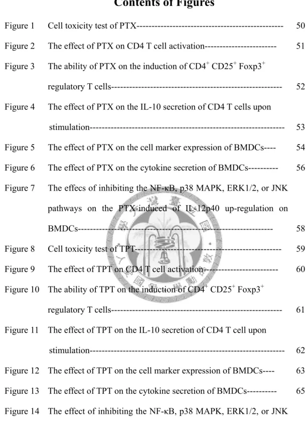
Contents of Figures
Figure 1 Cell toxicity test of PTX--- 50 Figure 2 The effect of PTX on CD4 T cell activation--- 51 Figure 3 The ability of PTX on the induction of CD4+ CD25+ Foxp3+
regulatory T cells--- 52 Figure 4 The effect of PTX on the IL-10 secretion of CD4 T cells upon
stimulation--- 53 Figure 5 The effect of PTX on the cell marker expression of BMDCs---- 54 Figure 6 The effect of PTX on the cytokine secretion of BMDCs--- 56 Figure 7 The effecs of inhibiting the NF-κB, p38 MAPK, ERK1/2, or JNK
pathways on the PTX-induced of IL-12p40 up-regulation on BMDCs--- 58 Figure 8 Cell toxicity test of TPT--- 59 Figure 9 The effect of TPT on CD4 T cell activation--- 60 Figure 10 The ability of TPT on the induction of CD4+ CD25+ Foxp3+
regulatory T cells--- 61 Figure 11 The effect of TPT on the IL-10 secretion of CD4 T cell upon
stimulation--- 62 Figure 12 The effect of TPT on the cell marker expression of BMDCs---- 63 Figure 13 The effect of TPT on the cytokine secretion of BMDCs--- 65 Figure 14 The effect of inhibiting the NF-κB, p38 MAPK, ERK1/2, or JNK
pathways on the TPT-suppressed the IL-12p40 production on BMDCs--- 67
1
CHAPTER I
INTRODUCTION
2
Pentoxifylline
Pentoxifylline (PTX), 3,7-dimetyl-1(5’-oxo-fexyl) xanthine is a non-specific type 4 phosphodiesterase inhibitor and widely used in therapy for peripheral vascular disease at first (Ward and Clissold, 1987). It could not only efficiently increase red blood cell deformability and reduce blood viscosity but also decrease the potential for platelet aggregation or thrombus formation. It is also used for other conditions with insufficient regional microcirculation (Moher et al., 2000; Samlaska and Winfield, 1994) and has been available on the market for approximately 30 years. In recent years, many researches focused on its anti-inflammatory properties, such as the protection of the endotoxin in animal model as well as beneficial effects in septic shock patients (Law et al., 1992; Mandi et al., 1995; Wu et al., 1999). Moreover, PTX has been demonstrated to suppress irritant and contact hypersensitivity reactions and used to treat some immune-mediated disorders, such as rheumatoid arthritis and multiple sclerosis despite the mechanism is still not completely understood (Maksymowych et al., 1995;
Rieckmann et al., 1996; Schwarz et al., 1993).
Some researches suggested that PTX have some immune modulatory functions. It could inhibit the synthesis of pro-inflammatory cytokines, such as TNF-α, IL-12, and IL-18 (Moller et al., 1997; Samardzic et al., 2001; Whitehouse, 2004), and selectively suppress T helper (Th) type 1 (IFN-γ, IL-2) cytokine production. Furthermore, this
3
drug was found to suppress human T cell activation and proliferation in vitro (Gonzalez-Amaro et al., 1998). In the case of DC, PTX could decrease the capacity of differentiation, maturation and antigen uptake upon stimulation (Vukanic et al., 2007;
Zhang et al., 2007). Previous studies (Mahnke et al., 2002) demonstrated that immature DCs might play some important roles in the maintenance of peripheral tolerance through the induction of anergic T cells or other T cells with regulatory properties. A report demonstrated that PTX could somehow induce regulatory T cells (Aricha et al., 2006). All of these research showed that PTX is a potent immune-suppressive and regulatory reagent.
Triptolide (TPT)
Tripterygium wilfordii Hook. f (TWHf), commonly known as thunder god vine, is a perennial vine-like member of the Celastracease plant family. This plant is native to many areas including Eastern and Southern China, Korea, Japan, and Taiwan. It has a long history of use in traditional Chinese Medicine (TCM) for the treatment of swelling, fever, chills, sores, joint pain, inflammation (Tao et al., 1991), and also for some autoimmune diseases including systemic lupus erythematosus (SLE) (Qin et al., 1981), rheumatoid arthritis (RA) (Lipsky and Tao, 1997), discoid lupus erythematosus (Qin et al., 1983), and pyoderma gangerenosum (Li, 2000). Tripterygium has been shown to
4
contain a vast array of natural molecules with strong biological activities, which may explain the multiple uses of this plant in traditional and allopathic medicine (Brinker et al., 2007).
Triptolide (TPT, also designated as PG-490), a highly oxygenated diterpenoid trieposide, has been identified as one of the main pharmacological active component purified from the root of TWHf (Gu et al., 1995; Tao et al., 1995; Tao et al., 1998). Even though it exhibits potent anti-inflammatory and immunosuppressive properties and has been used widely to treat a variety of autoimmune diseases (Jiang, 1994; Kupchan et al., 1972; Tao et al., 1989), the exact immunosuppressive mechanism of TPT has not been delineated fully.
To date, some immunosuppressive activities and immune regulatory function of TPT have been demonstrated in vitro. It is a strong inhibitor of NF-AT and NF-κB mediated transcription, which leads to down regulation of some gene products necessary for the immune responses (Lee et al., 1999; Liu et al., 2000; Qiu et al., 1999).
It also suppresses T-lymphocyte activation, including inhibition of lymphocyte proliferation, IL-2 expression at transcription level (Qiu and Kao, 2003), IL-2 receptor expression (Yang et al., 1994), and interferon-γ (IFN-γ) production (Chan et al., 1999), and it could also induce T cell apoptosis in a caspase-dependent and Bcl-2 -regulated manner (Yang et al., 1998). Some of these effects are even stronger than cyclosporine
5
(Lu et al., 1999) and FK-506 (Chan et al., 1999). In addition, TPT have some effects on DCs. For example, this drug can impair DC migration by suppressing the expression of CCR7 and COX-2, which are responsible for the migration of DCs to secondary lymphoid organ to initiate immune responses (Chen et al., 2005; Liu et al., 2007a).
Overall, these studies showed that TPT is a very powerful drug on immune regulation.
Immune activation & Immune tolerance
There are two main immune responses. One is immune activation, which means to develop appropriate and qualitatively different immune responses to protect the body from harmful pathogens including bacteria, fungi, viruses, and parasites. Another is immune tolerance. In contrast to immune activation, immune tolerance means to suppress harmful immune responses. Therefore, some excessive and self-destructive immune responses would be limited and some non-essential reactions and autoimmunity would be prevented. A balanced immune system is essential to the health and well-being. If the immune balance is broken, there comes to many severe diseases such as autoimmune diseases and so on. To achieve this balance, many immune cells play not only positive but also negative role in the immune system. Here we want to focus on two immune cells, DCs and CD4 T cells, to illustrate their role in the immune-regulation.
6
Dendritic cells
Dendritic cells (DCs) play an important role in mammalian immune system. They are identified by their dendritic morphology and unique phenotype. Without a doubt, DCs have been proved to be the most potent antigen presenting cells (APCs) among other APCs such as macrophages and B cells (Colonna et al., 2006; Liu et al., 2001;
Reid et al., 2000). Specialized in antigen presentation, they can initiate and regulate both cellular and humoral immune responses.
Subsets of DCs
Although all DCs are developed from hematopoietic progenitor cells in the bone marrow, they can be divided into two subsets in human. One is plasmacytoid DCs (pDCs) which evolve from lymphoid precursors. They have plasma cell-like morphology and express CD123 and blood DC antigen4+ (BDCA-2/CD303 、 BDCA-4/CD304) on the cell surface. The major function of pDCs is to secrete type I interferon against infections. They can detect viral and bacterial nucleic acid such as ssRNA and CpG DNA through TLR7 and TLR9 receptors, and secret large amount of type I interferon including IFN-α and IFN-β (Banchereau et al., 2000; Colonna et al., 2004; Novak and Bieber, 2008). The other subset of DCs is myeloid DCs (mDCs). In contrast to pDCs, mDCs evolve from myeloid precursors and express CD11c on the cell
7
surface. The mDCs are the major stimulators of T cells. They can sense pathogen by TLR2 and TLR4, secrete high amount of IL-12, which promote T cell differentiation and activation (Banchereau and Steinman, 1998).
Immune-stimulatory DCs Phenotype
The immune-stimulatory DCs often exhibit mature phenotype. Generally we can define this type of DCs by several features: (a) losing endocytic/ phagocytic receptors such as MMR (Kato et al., 2000); (b) upregulating costimulatory and signaling molecules such as CD80/B7-1, CD86/B7-2, TNFR-families, and CD40; (c) enhancing major histocompatibility complexes class II (MHC class II) expression (Banchereau et al., 2000; Banchereau and Steinman, 1998). (d) secreting high level of pro-inflammatory cytokines including interferon-alpha (IFN-α), tumor necrosis factor-alpha (TNF-α), IL-1, IL-6, IL-7, IL-12, and IL-15 (Banchereau and Steinman, 1998).
Functions
One unique and critical function of DCs is to prime naïve CD4 T cells in vivo and
in vitro. While sensing pathogens through their pathogen recognition receptors such as
8
TLRs (Gallucci and Matzinger, 2001; Iwasaki and Medzhitov, 2004), DCs not only uptake the pathogen but also process it for presentation. After that, they migrate to lymphoid tissues such as spleen and lymph nodes when they encounter naïve CD4 T cells. Upon the contact with antigen-specific CD4 T cells, the immune-stimulatory DCs would activate them. The DCs present high level of MHC class II-peptide complexes to T cell receptor (TCR) complexes on T cell surface and provide “signal one” to T cells.
Costimulatory molecules on DCs like CD80, CD86, and CD40 would also interact with CD28 and CD40L on the T cells. During these interactions, DCs can provide “signal two” to T cells. When receiving these two activation signals, CD4 T cells become fully activated and have the capacity to regulate other immune effector cells, including Ag-specific CD8 T cells and antibody secreting B cells. In addition, DCs also could influence T cell differentiation by providing “signal three”, which is the cytokine. For instance, they can drive the development of IFN-γ producing Th1 immune response by IL-12 secretion (Macatonia et al., 1995). In brief, the immune-stimulatory DCs are not only antigen presenting cells but also crucial effector cells in inducing adaptive immune responses.
Tolerogenic DCs Phenotype
9
Tolerogenic DCs, in contrast to immune-stimulatory DCs, usually present antigen with low MHC class II molecules and low/no costimulatory molecules including CD80/B7-1 and CD86/B7-2. Instead of pro-inflammatory cytokines, they can secrete higher level of immunosuppressive cytokines, such as IL-10, to affect other immune cells. In addition, some DCs can express Fas ligand (FasL) (Suss and Shortman, 1996), or secrete Indoleamine 2,3-dioxygenase, an amino acid digesting enzyme (Mellor and Munn, 2004), to fulfill their immune suppressive function.
Functions
These kinds of DCs are less potent to initiate adaptive immune responses. Despite so, these DCs are still very important in balancing the immune system. The most evident example is that they can induce effective peripheral tolerance. Studies suggested that central tolerance is essential, but not sufficient. The combination of peripheral and central tolerance is needed to prevent the body from harmful reaction. In the periphery, DCs could uptake and present peripheral self-antigens to auto-reactive T cells escaping from the central selection in thymus, and then induce tolerance to self-antigen (Steinman et al., 2000). Other peripheral tolerances could be seen in some organs such as lung, skin, and other mucosa areas (Novak and Bieber, 2008). In these tissues, tolerogenic DCs uptake non-self antigen but fail to induce further immune response
10
under anti-inflammatory environment.
DCs cannot induce tolerance by themselves. They need to work together with T cells. There are three major ways that DCs can affect T cells to suppress immune responses: anergy, apoptosis, and regulatory T cell induction.
T cell anergy
As describe above, T cell activation requires two activation signals from APCs:
MHC class II/peptide-TCR and CD80/86-CD28 signaling. When T cells encounter
“non-professional” APCs lacking costimulatory molecules, they recognize the peptide/MHC complexes but could not be fully activated. Under this situation, they would impair their effector functions and become anergic. That is, these T cells do not proliferate and secrete IL-2 upon re-stimulation, even in the presence of B7 costimulation (Gimmi et al., 1993).
T cell apoptosis
It has been demonstrated that after T cell activation, they would express Fas, which is also called death receptor. Many reports mentioned that Fas/FasL ligation is sufficient to induce T cell apoptosis. Some specialized subsets of DCs can express high level of FasL, such as CD8+ DCs in mouse. These DCs could suppress immune
11
responses by inducing activated CD4 effector T cells undergo apoptosis (Suss and Shortman, 1996). Another function of tolerogenic DCs to inhibit T cell response is to secret indoleamine 2,3-dioxygenase (IDO), a tryptophan degrading enzyme.
Because tryptophan is an essential amino acid for cell proliferation, the T cell response would be inhibited by IDO (Mellor and Munn, 2004; Munn et al., 2002).
Furthermore, some reports demonstrated that the metabolites of tryptophan by IDO could selectively induce Th1 cell apoptosis (Fallarino et al., 2002).
Regulatory T cell induction
Many papers have demonstrated that DCs could be an immune regulator through the induction of regulatory T cells. For example, DCs, which have immature phenotype and express low pro-inflammatory cytokines, can prime regulatory T cells and consequently play an important role in maintaining tolerance to self-antigens in the body (Banchereau and Steinman, 1998; Steinman and Nussenzweig, 2002; Wilson et al., 2004). Moreover, these DCs could induce Treg cells specific for foreign peptide or alloantigens (Dhodapkar et al., 2001; Jonuleit et al., 2000). On the other hand, there is another group of DCs which expresses high level of MHC class II (signal 1) and costimulatory molecules (signal 2), and secrete no pro-inflammatory cytokines (signal 3) or secrete IL-10 instead. These DCs are
12
known as semi-mature DCs, and they have been proved to induce antigen specific IL-10 producing CD4 Treg cells (Lundqvist et al., 2005).
Induction of tolerogenic DCs
Many types of tolerogenic DCs have been identified in vivo. Some of them are potent in inducing immune tolerance without any other stimulation. For example, the immature DCs under steady state can constantly uptake and present self-Ag from apoptotic cells or other harmless foreign antigens but fail to activate T cells effectively.
Because of their partial maturation, they can migrate to lymphoid tissue to induce tolerance (Hawiger et al., 2001; Steinman et al., 2000; Wilson et al., 2004). However, some DCs might be activated and become tolerogenic in special circumstances. For example, DCs secrete high amount of IL-10 to impair T cell responses and lead to the prolong existence of viral infection. (Brooks et al., 2006).
In addition, many papers have demonstrated that there are some methods to modulate DCs to become tolerogenic in vitro. For instance, we can induce these special DCs in specific culture conditions like low dose of GM-CSF (Lu et al., 1995). Besides, we can culture dendritic cell under the influence of IL-10 (Steinbrink et al., 1997), VEGF (Oyama et al., 1998), Glucocorticosteroid (Piemonti et al., 1999), and 1,25 (OH)2
vitamin D3 (Penna and Adorini, 2000; Piemonti et al., 2000) induce tolerogenic DCs.
13
Therefore, these induced tolerogenic DCs might be a useful therapy for immune diseases.
CD4 T cell
CD4 T cells in adaptive immunity
CD4 T cells are important members of adaptive immunity. As soon as the innate immunity induced, Ag-uptake APCs would further induce adaptive immune responses.
The first cells in contact with those APCs are CD4 T cells. These CD4 T cells could evoke further immunity through cytokines secretion. While in contact with APCs carrying different kinds of antigens and under the influence of different cytokines, CD4 T cells may differentiate into different types of effector cells that produce distinctive sets of cytokines with different functions. The functions of CD4 T cells are to influence innate and adaptive immunity. For example, if the activated CD4 T cells differentiate into Th1 cells and secrete IFN-γ, they could induce the activation of macrophage to become phagocytic and capable of killing the phagocytosed microbes (Nathan et al., 1983; Raveh et al., 1998). This CD4 T cells could also help B cells to secrete neutralizing antibody and subsequently eliminate pathogens such as viruses (Mahon et al., 1995). On the other hand, if activated CD4 T cells differentiate into Th2 cells secreting IL-4 or IL-5, they may activate other innate immune cells such as mast cells
14
and eosinophils to destruct pathogens like parasites (Finkelman et al., 1991; Gauchat et al., 1992; Gould et al., 2003; O'Byrne et al., 2001). Moreover, CD4 T cells could help the activation and differentiation of antigen-specific CD8 T cells and to promote cell-mediated immunity (Schoenberger et al., 1998; Shedlock and Shen, 2003).
The activation of CD4 T cells
After maturation in the thymus, CD4 T cells express a unique surface molecule called TCR. The TCR molecule and CD4 co-receptor work together to recognize the peptide epitope on MHC class II molecule, which provide a signal for activation. In addition to TCR-MHC interaction, there are some other molecules involved in the T cell response to the antigen. For instance, B7.1/B7.2 molecules expressed on APCs interact with CD28 expressed on T cells, which also provide an essential signal for T cell activation (Reiser et al., 1992). T cells would increase the avidity of integrin such as LFA-1 to bind with ICAM-1 on APCs in order to increase the binding stability between APCs and T cells for the activation (Dustin and Springer, 1989).
When T cells recognize the antigen, it will induce a number of intracellular signaling pathways. For example, several transmembrane proteins including Lck may be activated and then phosphorylate other adapter proteins such as ZAP70. These proteins would further phosphorylate other various enzymes or adapter proteins and
15
activate transcription factors such as NFAT, NF-κB, and AP-1 (Danielian et al., 1992) (Chan et al., 1995). These transcription factors would stimulate the transcription of some genes and new protein synthesis. The first cytokine made predominantly after T cell activation is IL-2, which is also called as T cell growth factor. After being secreted, the IL-2 will bind to the same T cells and induce the expression of high affinity IL-2 receptor alpha chain, CD25. The main function of IL-2 is to promote T cell proliferation, which means to promote T cells entering cell cycle and dividing (Toribio et al., 1989).
In this way, it will cause the expansion of antigen-specific T cells. Compared to CD8 T cells, the clonal expansion of antigen-specific CD4 T cells is probably only 100-fold to 1000-fold, but it is enough for them to secrete cytokines to activate other effector cells and induce the adaptive immune response.
When naïve CD4 T cells are activated, they would differentiate into effector T cells.
Many studies have demonstrated that some cytokines may play an important role in dictating CD4 T cell lineage. For example, IL-12 and IFN-γ could drive naïve T cells differentiation into Th1 cells, which predominantly secret IFN-γ and IL-2, and enhance the clearance of intracellular pathogens (Jacobson et al., 1996; Lighvani et al., 2001).
Another example is IL-4, which would drive naïve T cells differentiation into Th2 cells (Swain et al., 1990). These cells could secret IL-4, IL-5, and IL-13 and enhance the clearance of parasites. The third cytokine worth mentioning is IL-6. Combined with
16
TGF-β, IL-6 has been shown to play a critical role in Th17 development (Bettelli et al., 2006). In addition to effector cells, naïve CD4 T cells may have the opportunity to differentiate into regulatory T cells, and are very important in maintaining the immune homeostasis and the tolerance.
Regulatory T cell
The main function of our immune system is to protect the host from a variety of pathogenic microorganism infections. It has developed a very delicate immune system mediated by T and B cells. Here comes a question: how could this powerful immune response not responding to self-antigens but controlled so well in avoiding the damage to the host. There are two mechanisms that could answer this question. First one is the selection of lymphocytes in the primary lymphoid organs. In this stage, some self-reactive lymphocytes would be eradicated or educated. Another answer to this question is the T cell-mediated immune suppression.
There is a particular subpopulation of T cells generated in normal immune system called regulatory T cells. Regulatory T cells are specialized in immune suppression and thereby maintain immune homeostasis, self-tolerance, and also control excessive immune response to foreign antigens. Moreover, the disruption of regulatory T cells has been proved to be related to some autoimmune diseases and inflammatory diseases.
17
Currently, regulatory T cells are divided into two main subtypes: one is nature occurring regulatory T cells derived from thymus and well known as CD4+ CD25+ Foxp3+ regulatory T cells; the other one is inducible regulatory T cells induced by antigen or under some tolerogenic condition in the periphery.
Inducible regulatory T cells
In contrast to natural occurring regulatory T cells, inducible regulatory T cells are generated from naïve T cells in the periphery by immunosuppressive cytokines, such as TGF-β (Chen et al., 2003), by stimulation with low dose of antigen (Apostolou and von Boehmer, 2004), or some APCs (Jonuleit et al., 2000; Wakkach et al., 2003). The two most commonly studied inducible regulatory T cells are type 1 regulatory T cells (Tr1) and TGF-β induced Th3 cells
Tr1 cells
It has been described that when naïve CD4T cells are activated in the presence of high dose of exogenous IL-10, it will generate a special T cell clone with the cytokine production profile distinct from Th1 and Th2 cells. Because they produce interferon-γ (IFN-γ) at the level at least one log lower than Th1 cells, and low level of IL-2 and IL-4, we called these type of cells as Tr1 cells (Groux et al., 1997;
18
Roncarolo et al., 2006). Tr1 cells are differentiated from naïve CD4 T cells, which require the exposure of high level of IL-10 for the generation in vivo and in vitro (Roncarolo et al., 2006). Unlike nature occurring Treg, Tr1 cells do not express high level of CD25 and Foxp3. Antigen-specific Tr1 cells need to be activated via their TCR in the presence of IL-10. They show low proliferation capacity but secrete immunosuppressive cytokines such as IL-10 and TGF-β (Bacchetta et al., 2002). In addition, the secreted cytokines could inhibit both Th1 and Th2 cell-mediated immune response and modulate APCs in an Ag non-specific manner (Groux et al., 1997). In other words, Tr1 cells mediate their suppressive function in a cell-contact independent manner. Some papers also showed that the generation of Tr1 cells could be induced by immature DCs (Dhodapkar et al., 2001; Mahnke et al., 2003). In human, Tr1 cells are involved in the maintenance of immune tolerance to alloantigens (Bacchetta et al., 1994); moreover, they may play a role in modulating immune response to self-antigens (Yudoh et al., 2000), allergens (Cavani et al., 2000;
Reefer et al., 2004; Saloga et al., 1999), and non-harmful foreign antigens such as gliadin, an immunogenic element of gluten (Battaglia et al., 2004).
TGF-β induced Th3 cells
Th3 cells are first discovered in an experiment designed for the investigation of oral
19
tolerance (Chen et al., 1994). These cells are generated from gut lymphoid tissue and are capable of secreting TGF-β primarily and also helping the production of IgA in mucosa immunity (Weiner, 2001). Just like Tr1 cells, Th3 cells can be generated from naïve CD4 T cells exclusively under the stimulation of TGF-β. While being stimulated, in contrast to Tr1 cells, Th3 cells up-regulate their expression of CD25 and Foxp3. In addition, they secrete high level of TGF-β, which is the main suppressive agent of Th3 cells. TGF-β has been demonstrated to play an important role in the peripheral tolerance, such as inhibiting the activation of lymphocytes and monocyte-derived phagocytes (Prud'homme and Piccirillo, 2000). Many paper also showed that the homeostasis of T cell immunity could not be achieved without the existence of TGF-β (Lucas et al., 2000; Shull et al., 1992). Therefore, Th3 cells could control unwanted immune responses and prevent immune diseases through TGF-β secretion. In addition, some paper showed that purified CD4 T cells stimulated and cultured in the presence of TGF-β can generate a population of CD4 T cells which express Foxp3 and secrete TGF-β (Fantini et al., 2004). It was also suggested that these inducible regulatory T cells may be a potent population for the immunotherapy of autoimmune and inflammatory diseases (Fantini et al., 2006;
Huter et al., 2008).
20
Hypothesis
The mechanisms of PTX and TPT are through inhibiting the immune cell activation or inducing the regulatory function.
Study Aims
To date, many immune disorders are well defined to be caused by excessive and unexpected immune responses, such as graft-versus-host disease, asthma, and other autoimmune diseases including rheumatoid arthritis, multiple sclerosis, and systemic lupus erythematosus. Although many immunosuppressive drugs have been commonly used to treat patients with these diseases such as steroid, they are very effective but also come with many side-effects. In order to cure the immune diseases without extra side-effects, we need to find out other new substitutions for the treatment.
In my study, I chose PTX and TPT as the experimental targets. Both drugs are used to treat different kinds of diseases and show some regulatory functions in the immune system. However, the mechanisms underlying their actions are not very clearly defined.
We want to study the immune regulatory functions of these two drugs on CD4 T cells and DCs in vitro. Especially, PTX and TPT have been used as clinical drug for a long time, so the application of these drugs in the future would have less concern on their safety.
21
CHAPTER II
MATERIALS & METHODS
22
Materials
Animals
Female DO11.10 OVA-specific TCR-transgenic (Tg), BALB/c, and C57BL/6 mice were bred and maintained in the Animal Center of the College of Medicine, National Taiwan University. All mice were used at 4-5 week of age throughout the studies. All procedures were approved by the institutional Animal Care and Use Committee.
Cell line EL4 cells
This cell line is a C57BL/6N mouse lymphoma. The appropriate medium used to maintain this cell line is complete Dulbecco’s modified Eagle’s medium with 10% FBS.
Cell culture reagents RPMI-1640 (10% FBS)
RPMI-1640 (Hyclone, USA) 1L
Fetal Bovine Serum (Hyclone, USA) 10%
L-glutamine (GIBCO BRI, MD, USA) 1%
PSA (GIBCO BRI, MD, USA) 1%
23
HEPES (GIBCO BRI, MD, USA) 1%
Phosphate Buffered Saline (1L)
NaCl (Amersco, USA) 8g
Na2HPO4 (Sigma, USA) 2.871g
KCl (Merck, Germany) 0.2g
KH2PO4 (Ferak, Germany) 0.2g
ACK (RBC lysis buffer)
NH4Cl (Wako, Japan) 0.15M
KHCO3 (Wako, Japan) 10mM
EDTA (Sigma, USA) 0.1mM
Hank’s buffer
Hank’s Balanced Salts (Sigma, USA)
Dissolved the powder in 1L ddH2O and adjusted to pH 7.2~7.4 by NaHCO3
Cytokine
IL-2 (PeproTech Inc, USA) IL-4 (eBioscience, USA)
GM-CSF (PeproTech Inc, USA) TGF-β (R&D, USA)
24
Others
Anti-mouse CD3ε (Biolegend, USA) Anti-mouse CD28 (Biolegend, USA)
Drugs
pentoxifylline (Sigma, USA)
Dissolved the powder in complete medium and stocked at -20℃
Triptolide (Sigma, USA)
Dissolved the powder in DMSO and stocked at -20℃
ELISA assay (R&D System, USA) Wash buffer (1L)
Tween 20 (Sigma, USA) 0.05%
added Tween 20 in PBS and adjusted to pH 7.2~7.4 Reagent diluents
IL-2 0.1% BSA, 0.05% Tween 20 in Tris-buffered saline, pH 7.2~7.4 IL-10 1% BSA in PBS, pH 7.2~7.4
IL-12p40 1% BSA in PBS, pH 7.2~7.4
25
IL-12p70 1% BSA in PBS, pH 7.2~7.4 Blocking buffer
IL-2 1% BSA in PBS with 0.05% NaN3, pH 7.2~7.4 IL-10 1% BSA in PBS, pH 7.2~7.4
IL-12p40 1% BSA in PBS, pH 7.2~7.4
IL-12p70 1%BSA, 5% Sucrose in PBS, pH 7.2~7.4 Substrate solution
NelA-Blue tetramethylbenzidine substrate (TMB) (Clinical, USA) Stop solution
2N H2SO4 (Wako, Japan)
Flow cytometry
FACScan buffer (1L)
1X FACScan PBS 980ml
Fetal bovine serum (Hyclone, USA) 20ml
NaN3 1g
Foxp3 Fix/Perm Buffer Set (Biolegend)
(4X) Foxp3 Fix/Perm buffer (Biolegend, USA) (10X) Foxp3 Perm buffer (Biolegend, USA)
26
Diluted the above buffer to 1x working solution with PBS
Magnetic cell sorting
CD4 (L3T4) MicroBeads, mouse (Miltenyi Biotec, Germany) autoMACS running buffer
Bovine serum albumin (Sigma, USA) 0.5%
EDTA (Sigma, USA) 2mM
Dissolved the above powder in 1X PBS then sterilized and filtrated with 0.2μm filter
autoMACS rinsing buffer
EDTA (Sigma, USA) 2mM
Dissolved powder in 1X PBS then sterilized
Methods
Isolation of naïve CD4T cells
Spleens were harvested from female BALB/c DO11.10 OVA-specific TCR-transgenic mice or C57BL/6 mice, and then grinded it into single-cell suspensions in Hank’s buffer. After centrifuging at 1500 rpm for 5 minutes, large tissues were removed and RBCs were depleted by adding 1 ml of ACK lysis buffer and washed these
27
cells two to three times with Hank’s buffer. After determining the total cell number, spleen cells were prepared for the separation of CD4 T cell by CD4 T Cell Isolation Kit in Magnetic cell sorting machine. At first, spleen cells were re-suspended in 40μL per 1
× 107 cells of MACs running buffer and then add Biotin-Antibody Cocktail 10μL per 1
× 107 cellswas added, then mix well and incubate at 4℃~8℃ for 10 minutes. Cells were added with MACs running buffer 30μL per 1 × 10⁷ cells and Anti-Biotin MicroBeads 20μL per 1 × 10⁷ cellsand incubated at 4~8℃ for 15 minutes. Stained cells were washed with MACs running buffer and re-suspended with MACs running buffer in appropriate volume to get CD4T cells by auto-MACS.
Cell toxicity test
The EL-4 cell line was cultured in DMED with 10% FBS and in the concentration of 2 × 10 5 cells/ml; in addition, the cell line were treated with serial diluted dosages of PTX and TPT and used cell only as control. After cultured for 48 hours, the cells were collected from each condition, evaluated for cell viability using trypan blue staining.
The cell viability was shown in the percentage of alive cells in 500 cells per well.
Culture of bone marrow-derived dendritic cells (BMDCs)
The femurs and tibias were collected from mice, washed with 75% alcohol for 5
28
seconds, and transferred to HBSS buffer. The bone marrow cells were flushed out with 5ml needle and red blood cells were lysed by ACK. After washed with HBSS, cells were re-suspended in RPMI-1640 medium and adjusted to the concentration of 1 × 10⁶ cells/ml. Cells were cultured with 10% FBS-RPMI with recombinant murine granulocyte-macrophage colony-stimulating factor (GM-CSF, 500 U/ml) and interleukin-4 (IL-4, 1000 U/ml, Pepro Tech Inc., Rocky Hill, NJ ). On day 2, day 4, and day 6, the medium was removed by aspiration and lymphocytes were cultured in fresh medium containing GM-CSF and IL-4 in the same concentration as the first day. The percentages of bone marrow-derived DCs were evaluated by FACSort cell analyzer (Becton-Dickinson) on day 6.
Drug treatment on CD4T cells IL-2 secretion
Purified CD4 T cells were adjusted to the concentration of 1 × 10⁶ cells/ml, seeded in the 48-well plate, and stimulated with plate-coated anti-CD3 and anti-CD28 at 2μg/ml. Cells were also added with different concentration of PTX and TPT. After 48 hours, the cultured supernatant was collected for cytokine measurement.
29
3H-thymidine incorporation assay
Purified CD4 T cells were adjusted to the concentration of 1 × 10⁶ cells/ml, seeded in the 96-well round-bottom plate (Nunc, Roskilde, Dermark), and stimulated with plate-coated anti-CD3 and anti-CD28 at 2μg/ml. Cells were also added with different concentration of PTX and TPT. After 72 hours, cells were prepared for thymidine incorporation assay.
Regulatory T cell induction
Purified CD4 T cells were adjusted to the concentration of 2 × 10⁶ cells/ml, seeded in the 24-well plate, and stimulated with plate-coated anti-CD3 and anti-CD28 at 2μg/ml. IL-2 was added at 100U/ml in each well. TGF-β was added at 10ng/ml as positive control. After 72 hour of cultured, these cells were harvested to analyze the surface marker expression, and the cultured supernatant was collected for cytokines measurement.
Drug treatment on bone marrow-derived dendritic cells
The DCs, which were already cultured for 6 days, were adjusted to the concentration of 1 × 10⁶cells/ml, and also treated with different concentration of PTX and TPT. The 100 ng/ml of LPS were used to stimulate DCs. After 48 hours, the DCs
30
were harvested to analyze surface marker (CD11c, CD80, CD86, MHC class II) expression, and the cultured supernatant was collected for cytokines (IL-10, IL-12p40, IL-12p70) measurement.
Proliferation assay
The cultured CD4 T cells were pulsed with 1.0 μCi/ml of 3H-thymidine (Amersco, USA) and further incubated in 37℃ and 5% CO2 incubator for 16~18 hours. After incubation, these cells were harvested to glass fiber filter (Packard Instrument Co., Meriden, CT) with semi-automated device (Harvester, Packard, Filtermate 196). 3H- thymidine incorporation was quantitated by using Direct Beta Counter (Packard Instrument Co., Meriden, CT). The results were presented in counts per minute (CPM).
Measurement of cytokine production
Supernatants were collected from cell culture well to measure cytokine concentration by ELISA. The protocol was followed by Duoset, R&D Systems ( Minneapolis, MN, USA). First, ELISA plates (Nunc, Roskilde, Denmark) were coated with diluted capture antibody, 100 μL per well, at room temperature overnight . The next day, the plate were washed three times and blocked with blocking buffer. After washing, sample and standards were added at 100 μL per well and incubated 2 hours at
31
room temperature. Plates were washed and incubated with 100μL per well of diluted detection antibody. After two hours at room temperature, diluted streptavidin-HRP were added (100 μL per well) and then 1:1 mixture of H2O2 and Tetramethylbenzidine were used as the substrate. At last, H2SO4 were used as stop solution (50 μL per well). The OD450 and OD540 were read by using a microplate reader (Anthosreaser 2010, Austria), and the cytokine levels were determined by appropriate cytokine-specific standard curve.
Flow cytometry analysis
CD25 & Foxp3 expression of T cell
The cultured CD4 T cells were harvested and washed twice with FACS buffer, and then they were stained with anti-CD25 antibody (1μL /106 cells) at 4℃ for 30 minutes.
After washing to remove extra antibodies with FACS buffer, cells were resuspended with 1X FOXP3 Fix/Perm solution 1ml, and incubated at room temperature in the dark for 20 minutes. After removing the supernatant, the cells were washed once with FACS buffer and also with 1X FOXP3 Perm solution, then re-suspended with 1X FOXP3 Perm solution 1ml per tube, and incubated at room temperature in the dark for 15 minutes. Afterward, the supernatants were removed and added appropriate amount of anti-Foxp3 antibody (20μL /106 cells) and incubate at room temperature in the dark for
32
30 minutes. After washing twice with FACS buffer, cells were re-suspended in appropriate volume of FACS buffer for analyzing on FACSort cell analyzer (Becton-Dickinson).
MHC class II & CD11c & CD80 & CD86 expression of DC
Cultured DCs were harvested on day 8. In order to remove the culture medium, cells were washed twice with FACS buffer. Antibodies (anti-MHC class II, anti-CD11c, anti-CD80, anti-CD86, 1μL/106 cells) were added for staining. After incubated at 4℃
for 30 minutes, cells were further washed twice with FACS buffer and re-suspended in appropriate volume with FACS buffer for analyzing on FACSort cell analyzer (Becton-Dickinson).
33
CHAPTER III
RESULTS
34
Part I. Pentoxifylline (PTX) The cell toxicity test of PTX
First, we need to define a safety concentration range of PTX to make sure that the PTX do not damage the normal cells on its own. In order to find out the appropriate concentration range of PTX, we used EL4 cell line, a mouse lymphoma, to do this test.
We cultured EL4 cells with two-fold serial dilutions of PTX for 48 hours, and the cell viability was counted by trypan blue assay. The result showed that when cultured with 500μg/ml PTX, the cell viability had a significant difference compared with untreated group (P<0.05; Fig. 1).The EL4 cells with the concentrations of PTX lower than 500μg/ml seemed to have no cell toxicity with the viability close to 100%.
PTX can inhibit CD4 T cell proliferation and IL-2 secretion upon stimulation It is reported that CD4 T cells play an important role in adaptive immune response, and has some influence on innate immune response. We want to find out if PTX can directly inhibit CD4 T cells activation. CD4 T cells were stimulated with plate-coated anti-CD3 and anti-CD28 at 2μg/ml and treated with five-fold serial dilution of PTX.
After 48 hours, the supernatants were collected and analyzed by ELISA; in addition, the CD4 T cell proliferation was analyzed by 3H-thymidine incorporation assay. Our data showed that the CD4 T cell proliferation level were significantly decreased when
35
cultured with 50μg/ml and 250μg/ml of PTX (P<0.05; Fig. 2A). In addition, the IL-2 secretion from activated CD4 T cells were also inhibited by PTX at 50μg/ml and 250μg/ml (P<0.05; Fig. 2B).
PTX do not have the regulatory T cell induction function
In this experiment, we analyzed the percentage of CD4+CD25+Foxp3+ T cells, a kind of cell with immunosuppressive function, and the IL-10 level from cultured supernatant. IL-10 is an immunosuppressive cytokine and has some relationship with another regulatory T cells, Tr1 cells. Here, CD4 T cells were treated with 100U/ml of IL-2 together with 250μg/ml or 25μg/ml of PTX for 72 hours. The percentage of regulatory T cells was analyzed by flow cytometry. Since TGF-β has been proved to induce CD4+CD25+Foxp3+ T cell generation (Fantini et al., 2004), so we used TGF-β (10ng/ml) as the positive control in this experiment. The results showed that when CD4 T cells were treated with PTX, compared with TGF-β treatment (Fig. 3A), the percentage of CD4+CD25+Foxp3+ T cells was slightly but not significantly increased (Fig. 3B). The IL-10 level was significantly decreased when the CD4 T cells were cultured with 250μg/ml of PTX (P<0.05; Fig. 4).
We also want to investigate if PTX may have synergistic effect on TGF-β-induced Treg cells. In order to answer this question, we treated CD4 T cells with 250μg/ml or
36
25μg/ml of PTX together with TGF-β for 72 hours, and analyzed the percentage of CD4+CD25+Foxp3+ T cells. The results showed that when combined with PTX, the percentage of CD4+CD25+Foxp3+ T cells was lower than that of TGF-β treatment along (Fig. 3C).
PTX can not affect the marker expression on BMDCs but selectively increase cytokine secretion
As described before, some surface markers, such as MHC class II, CD80, and CD86 are very important for DC to regulate the immune response. Here we want to exam the effect of PTX on DCs for their surface marker expression and cytokine secretion. BMDCs were cultured with five-fold serial dilution of PTX (250, 50, 10μg/ml) with or without LPS (100 ng/ml) for 48 hours. The surface marker expression level was analyzed by flow cytometry. We first gated on CD11c+ cells and further analyzed individual surface marker expression. Our data showed that when BMDCs cultured with PTX, the expression of MHC class II, CD80, and CD86 had no significant difference compared with control group both either with or without LPS treatment (Fig. 5A-C). In addition, we also detected the cytokine secretion. We found that when BMDCs cultured with PTX and LPS, IL-12p40 secretion was significantly increased under 250μg/ml of PTX (P<0.05), and IL-12p70 secretion was increased
37
under 250μg/ml and 50μg/ml of PTX (P<0.05; Fig. 6A-B). In contrast, the IL-10 secretions were not different between control group and PTX treatment groups (Fig.
6C).
Inhibition of NF-κB prevent the IL-12p40 secretion changes induced by PTX
Since our data shows that PTX-treated BMDCs produce higher IL-12p40 and IL-12p70 (Fig. 6A-B), we want to further investigate whether the PTX-induced IL-12 secretion would be affected by inhibitors of NF-κB, p46/50 JNK, p42/44 ERK, and p38 MAPK.
BMDCs were first treated with Helenalin (NF-κB inhibitor), JNK inhibitor II (JNK pathway inhibitor), PD98059 (ERK pathway inhibitor), or SB203580 (p38 MAPK inhibitor) for 1 hours, and then incubated with PTX (250μg/ml) for 24 hours. The level of IL-12p40 in the supernatant was determined by ELISA. Our data showed that when BMDCs were first treated with Helenalin, the secretion IL-12p40 secretion was significantly decreased compared with PTX treatment group (P<0.05). On the other hand, when pre-treated with JNK inhibitor II, the level of IL-12p40 was significantly increased compared with PTX treatment group (P<0.05). In contrast, PD98059 and SB203580 seemed to have no effects on IL-12 secretion induced by PTX (Fig. 7).
38
Part II. Triptolide (TPT) The cell toxicity test of TPT
To find out the appropriate working concentration of TPT, which has no toxicity to normal cells, we treated EL4 cells with two-fold serial dilution of TPT for 48 hours, and then calculate cell viability by trypan blue assay. From our results, when the EL4 cells cultured with TPT at the concentration of 5000pg/ml, the cell viability had a significant difference compared to the cell only group (P<0.05; Fig. 8). However, when the concentration of TPT was lower than 5000pg/ml, TPT seemed to cause no significant cell toxicity.
TPT can inhibit CD4 T cell proliferation and IL-2 secretion upon stimulation
In order to find out if TPT can directly inhibit the CD4 T cell activation, we isolated CD4 T cells, stimulated them with plate-coated anti-CD3 and anti-CD28 at 2μg/ml and then treated with a five-fold serial dilution of TPT. After 48 hours, the supernatants were collected and IL-2 secretion was determined by ELISA. In addition, the CD4 T cell proliferation was analyzed by 3H-thymidine incorporation assay. Our data showed that CD4 T cell proliferation was significantly inhibited by TPT at concentration of 2500pg/ml (P<0.05; Fig. 9A). Moreover, IL-2 secretion was also inhibited under the same concentration of TPT (P<0.05; Fig. 9B).
39
TPT do not induce regulatory T cell generation
We also want to know whether TPT could induce CD4 T cell differentiation into regulatory T cells. CD4 T cells were treated with 100U/ml of IL-2 and with 2500pg/ml or 250pg/ml of TPT for 72 hours. The percentage of regulatory T cells was analyzed by flow cytometry. We used TGF-β (10ng/ml) as the positive control in this experiment.
The results showed that when CD4 T cells treated with TPT, compared with TGF-β treatment, the percentage of CD4+CD25+Foxp3+ T cells did not increased (Fig. 10A-B).
In addition, the levels of IL-10 in the culture supernatant show no significant difference between control group and TPT treatment group (Fig. 11).
In order to examine if TPT may have synergistic effect on TGF-β inducing Treg cells, we treated CD4 T cells with 2500 pg/ml or 250pg/ml of TPT together with TGF-β for 72 hours. The results showed that when combined with high concentration of TPT, the percentage of CD4+CD25+Foxp3+ T cells was slightly lower than TGF-β along (Fig.
10C).
TPT do not affect the surface marker expression on BMDCs but selectively increase cytokine secretion
In order to examine the effect of TPT on surface marker expression and cytokine secretion, BMDCs were cultured with five-fold serial dilution of TPT (2500, 500,
40
100pg/ml) in combination with or without LPS (100 ng/ml) for 48 hours. The surface marker expression was analyzed by flow cytometry. We first gated on CD11c+ cell and further analyzed the expression level of individual of surface marker. In our data, we could see that when cultured with TPT, the expression level of MHC class II, CD80, and CD86 on BMDCs had no significant difference compared with control group either with and without LPS treatment (Fig. 12A-C).
Moreover, we detected the cytokine level in culture supernatant. The data showed that the IL-12p40 levels were decreased when BMDCs were cultured with 2500pg/ml of TPT with or without LPS treatment (P<0.05; Fig. 13A), and IL-12p70 was decreased when cultured with 2500 pg/ml of TPT with LPS (P<0.05; Fig. 13B). The IL-10 level was not different between control group and TPT treatment group (Fig. 13C).
Inhibition of ERK and p38 pathways prevent the IL-12p40 secretion affected by TPT
Our data showed that TPT-treated BMDCs produced lower level of IL-12p40 and IL-12p70 (Fig. 13A-B). Here we want to further investigate whether this suppressive effect on the secretion of IL-12 would be affected by the inhibitors of NF-κB, p46/50 JNK, p42/44 ERK, and p38 MAPK. BMDCs were pre-treated with Helenalin (NF-κB inhibitor), JNK inhibitor II (JNK pathway inhibitor), PD98059 (ERK pathway inhibitor),
41
or SB203580 (p38 MAPK inhibitor) for 1 hours, and then incubated with TPT (2500pg/ml) for 24 hours. The IL-12p40 levels in the supernatant were detected by ELISA. Our data showed that when BMDCs were pre-treated with PD98059 and SB203580, the IL-12p40 secretion levels were significantly increased compared with TPT-treated group (P<0.05); In contrast, Helenalin and JNK inhibitor II seemed to have no effect on IL-12 secretion induced by TPT (Fig. 14).
42
CHAPTER IV
DISCUSSION & CONCLUSION
43
Many human diseases are well known to be caused by uncontrolled immune responses. Although nowadays a lot of drugs have been discovered to suppress the unexpected immunity potently and have been used for the treatments, they are still not as good as we expected. In other words, they would bring some side effects and further worsen a patient’s condition. PTX and TPT, the two drugs we focused here, are used as clinical drugs for a long time. PTX is used for the treatment of peripheral vascular disease; however, it’s immune regulatory functions are being focused in recent years.
TPT is a Chinese herb commonly used to treat some inflammatory diseases and autoimmune diseases despite the exact mechanism are still unclear. In this study, we provided some explanation on their immune effects on CD4 T cells and DCs.
Part I. Pentoxifylline (PTX)
Firstly, we tested the cell toxicity of PTX and found that PTX has very low toxicity to cells. Even under high concentration of this drug, the target cells are still alive.
To explore the effect of PTX on the activation of T cells, we analyzed the effect of this drug on the secretion of IL-2 and cell proliferation. Our results showed that PTX inhibited the IL-2 secretion and proliferation of CD4 T cells upon stimulation. It is well known that IL-2 can induce the expression of CD25, which is a high affinity IL-2 receptor (Depper et al., 1985). We found that when the T cells were cultured with PTX
44
and exogenous IL-2, the up-regulation of CD25 was inhibited. Previous studies showed that the signals delivered by the TCR/CD3 complex and accessory molecules induce the expression of IL-2 receptor and the secretion of IL-2. Furthermore, the signaling through CD25 in response to IL-2 is required for the T cell proliferation (Cantrell and Smith, 1984). Here, we proposed that PTX is a strong inhibitor for CD4 T cell activation.
To further investigate the immunomodulatory function of PTX, we tested the potency of this drug on the induction of regulatory T cells, which have been mentioned to be important in controlling immune responses. However, we found that the expression of Foxp3 was not induced by PTX, and this drug had no synergistic effect in conjunction with TGF-β, a Treg inducing cytokine, on Treg induction. A previous study demonstrated that the mRNA level of Foxp3 in total spleen cells was significantly increased after i.p injection of PTX in to the mice (Aricha et al., 2006). But in our data, we could not see the expression of Foxp3 increased by PTX treatment. It is possible that the inducible effect of PTX on Foxp3 is at the mRNA level, but not at protein level.
Additionally, PTX seemed to inhibit the generation of Treg cells by TGF-β. It has been reported that IL-2 is essential for TGF-β to induce CD25+Foxp3+ regulatory T cells (Zheng et al., 2007). In our data, we can see that PTX could inhibit the CD25 expression on CD4 T cells. Therefore, we propose that PTX might interfere with the
45
generation of Treg cells by TGF-β due to decreasing IL-2 signaling. Apart from Foxp3 expression, IL-10 secretion is one important function of regulatory T cells. So we also detected the IL-10 secretion level from CD4 T cells and found that the IL-10 level was not increased by PTX. Therefore, in our study, we could not identify the ability of PTX on induction of regulatory T cells.
In addition to CD4 T cells, DCs also play an important role in modulating immune responses. The ways of DCs to influence T cells are through the ligation of costimulatory molecules and cytokine secretion. In our data, we found that the expression level of CD86 was significantly increased by PTX, but the levels of MHC class II and CD80 were not affected. On the other hand, PTX increased IL-12 secretion by DCs, and this effect existed in both PTX only or combined with the LPS treatment.
We also identified that the induction of IL-12 secretion of DCs by PTX is through NF-κB pathway, since the induction phenomenon was disappeared when the NF-κB pathway was blocked. It is well known that IL-12 is involved in Th1 differentiation (Jacobson et al., 1996). It is possible that PTX promotes Th1 differentiation by inducing IL-12 secretion from DCs, but this hypothesis requires further examination. However, there is a confliction between our data and previous reports. They showed that in the presence of PTX and LPS, the expression of CD86 on the human DCs were significantly decreased; and moreover, IL-12 secretion from DCs was also inhibited by
46
this drug (Vukanic et al., 2007). The time point of PTX treatment was different between our experimental designs and theirs. They treated DCs with PTX on the day when they isolated CD14+ cells from PBMCs and also maintained these cells in the presence of PTX during the whole culture period. On the contrary, we treated DCs with PTX after six days of culture in the medium containing IL-4 and GM-CSF, therefore, these treated DCs were already differentiated. In the same paper they indicated that PTX impaired the differentiation of human DCs. It is possible that PTX has different effects on DCs at different differentiation stages.
Part II Triptolide (TPT)
TPT is extracted from Tripterygium wilfordii Hook. f, which is a toxic plant. After tested the cell toxicity, we found that the safety dose of this drug is at about ng/ml. This drug has been mentioned to be a special drug with narrow therapeutic window, Therefore, this drug requires more attention on the safety usage.
We explored the effect of TPT on the activation of T cells and analyzed the secretion of IL-2 and cell proliferation. Our data showed that under non-toxic concentration, TPT inhibited CD4 T cell activation including lower IL-2 secretion and cell proliferation. In our study, we could not demonstrate the relationship between its immunosuppressive function of TPT and regulatory T cell induction. We found that the
47
TPT did not induce Foxp3 expression; in addition, it has no synergy between TPT and TGF-β, which has been proved to have the potential for Foxp3 induction on CD4 T cells.
Additionally, we not only detected the molecule specialized for regulatory T cells but also examined whether the TPT was able to induce IL-10-secreting cell, which were one important kind of inducible regulatory T cells. Our result showed that TPT did not induce the IL-10-secreting CD4 T cell production. In this part, we suggested that the immunosuppressive function of TPT on CD4 T cells is to inhibit the activation but not to induce regulatory T cells production.
We further explored the effects of TPT on DCs and analyzed the expression of costimulatory molecules and the cytokine secretion. Our data showed us that in the present of TPT or combined with 100ng/ml of LPS, the expression of MHC class II on DCs was not affected by this drug. Also, the expression levels of costimulatory molecules including CD80 and CD86 were not influenced by TPT treatment either.
Although a published paper suggested that the percentages of CD80 and CD86 expressing cells in total DC population were decreased by TPT under 10ng/ml LPS stimulation (Liu et al., 2007b). There was another paper showed that when DCs were treated with TPT and stimulated by 1μg/ml of LPS, the expression level of CD80 and CD86 were not affected (Liu et al., 2004). LPS can activate DCs through Toll-like receptor 4 and induce costimulatory molecules expression. It is possible that the effects
48
of TPT on costimulatory molecules expression might be diminished under high LPS stimulation.
The cytokine secretion is also an important function of DCs. We showed that TPT significantly inhibited the secretion of IL-12 by DCs, both cultured in drug treatment only or combined with LPS stimulation, and the suppressive effect might through ERK and p38 MAPK pathway. As described before, IL-12 is involved in the differentiation of Th1 cells. In our data, we identified that the effect of TPT on DCs is through the cytokine secretion instead of the expression of costimulatory molecules, which may further regulating the differentiation of CD4 T cells.
Conclusion
PTX and TPT have been widely used in clinical treatment of autoimmune diseases and other immune disorders, but the mechanisms are still unclear. In our study, we showed that PTX is an inhibitor of CD4 T cell activation, and it could promote DCs to secrete IL-12. As for TPT, we identified that this drug could inhibit the activation of CD4 T cells and the secretion of IL-12 by DCs. The different effects of these two drugs on DCs suggested that they can be used to treat different diseases.