中藥純化成份 stevioside 降血壓作用機轉之研究
The Mechanism of Antihypertensive Effect of Stevioside isolated from the Herb Stevia
陳保羅 Paul Chan
台北醫學大學萬芳醫院內科部心臟內科
Department of Medicine, Taipei Medical University -Wan Fang Hospital; Taipei, Taiwan
Running title:Antihypertensive effect of stevioside on dogs
Correspondence to: Dr. Paul Chan, MD, Ph.D
Department of Medicine, Taipei Medical University – Wan Fang Hospital, 111, Sec. 3, Sing – Lung Road , Wen-Shan, Taipei 117, Taiwan
Tel: +886-2-2930-7930 ext. 2817, Fax: +886-2-2933-9378 E-mail: [email protected]
Abstract
Stevioside is a sweet-tasting glycoside isolated from the leaves of Stevia rebaudiana. It has been used as a non-caloric sugar substitute in Japan and Brazil for decades. Previous studies have shown that it lowered blood pressure in spontaneously hypertensive rats by intravenous injection. This study was designed to evaluate the hypotensive effect of stevioside on dogs and to define the underlying mechanism. After nasogastric feeding with stevioside powder (200 mg/kg), blood pressure of healthy Mongrel dogs began to decrease significantly at 60 min and returned to baseline level at 180 min. The reduction of blood pressure was more rapid (at 5-10 min) and was effective under intravenous injection. However, no significant change of blood pressure was noted after injection though left vertebral artery, implicating that the hypotensive effect was not related to central nervous system. Stevioside also showed significant hypotensive effect on renal hypertensive dogs in a dose-dependent manner. In cultured rat aortic smooth muscle cells (A7r5 cell line), stevioside can dose-dependently inhibit the stimulatory effect of vasopressin and phenylephrine on intracellular Ca2+ under calcium-containing medium. However, no intracellular Ca2+ inhibitory effect was observed in calcium-free medium, implicating that stevioside may inhibit Ca2+ influx from extracellular fluid. Our present data show that stevioside did not influence calcium ionophore (A23187) induced Ca2+ influx, showing the antagonistic effect was through Ca2+ channels. This study confirmed that stevioside is an effective antihypertensive natural product, and its hypotensive mechanism may be probably due to inhibition of Ca2+ influx.
Key Words:Stevioside, Hypotension, Anesthetized dogs
Introduction
Hypertension is one of the most common cardiovascular disease causing major mortality and morbidity in many epidemiological studies [1]. However, inadequate blood pressure control still persists as a major public health problem [2]. Compliance of patients to antihypertensive treatment may be an important barrier to improve blood pressure control, since antihypertensive drugs commonly have a negative effect on quality of life [3]. Diuretics and beta-blockers are most commonly prescribed as initial therapy and have been proven to reduce cardiovascular mortality [2]. Unfortunately, these two classes of the drugs usually have side effects especially causing sexual dysfunction [4]. It would be of considerable benefits for patients with hypertension if some natural products could lower blood pressure effectively with fewer side effects.
Stevioside is a sweet-tasting glycoside isolated from the herb Stevia rebaudiana Bertoni (Compositae), which is widely used as a sugar substitute in Japan and Brazil for about 20 years [5]. Its safety in human usage has been well established. Our previous report has shown that a large dose of stevioside administered intravenously has effective hypotensive action without changing serum catecholamines in spontaneously hypertensive rats (SHRs) [6].
Calcium ions play a fundamental role in the activation of cells. An influx of Ca2+ into the cell through specific ion channels is required for myocardial contraction and determining peripherial vascular resistance. Clinically, Ca2+ antagonists remain the most commonly used agents for treatment of angina and hypertension, due to their ability to induce smooth muscle
infusing CaCl2 could attenuate the vasodilating response of stevioside in rats.[8]. These data indicated that stevioside can act directly on vascular smooth muscle cells and this effect is calcium-related.
The hypotensive effect of stevioside on other mammalian animals such as dog or pig is still unclear. In present study, we evaluate the hypotensive effect of stevioside on healthy dogs, surgery-induced renal hypertensive dogs and investigate whether the mechanism of antihypertension of stevioside is through Ca2+ influx inhibition of smooth muscle cells.
Materials and Methods
Experimental Animals
The investigation conforms to the ‘Guide for the Care and Use of Laboratory Animals’
published by the US National Institutes of Health [NIH Publication No. 85-23, revised 1985].
Healthy Mongrel dogs (weight 18 ± 3kg) of both sexes were anesthetized with pentobarbital 30 mg/kg intravenously. Left fore limb femoral vein was implanted with polyethylene catheter (PE 50) for administering drugs. Right femoral artery was implanted catheter for measurement of systolic and diastolic blood pressure. The implanted catheter was connected to a Statham P23 pressure transducer (Gould Inc., Calif., USA) with the display on a Gould RS-3200 physiological recorder (Gould Inc., Ohio, USA). After stabilization for 30 min, the experiments were proceeded.
Experiments
There are four different experiments groups performed: (1) Nasogastric feeding with stevioside (fig. 1) powder (200 mg/kg) and recorded blood pressure at 30, 60, 90, 120,180 min. (2) Intravenous administration of stevioside 50 mg/kg and recorded blood pressure. (3) Referring previous method [9], renal hypertensive dogs were made. Briefly, laparotomy of anesthetized dog was performed and left renal artery was isolated and ligated. After 4 hours, the left renal artery ligation was released. After the blood pressure risen to a steady level, different dosages of stevioside were given and recorded its blood pressure. (4) Referring previous model [10], left vertebral artery was isolated and cannulated; 5 mg/kg stevioside was administered and then recorded blood pressure changes. Eight dogs were used for experiment in each group.
Culture of Rat Aortic Smooth Muscle Cells (A7r5)
The A7r5 aortic smooth muscle cell line [11], obtained from the Food Industry Institute (Hsin-Chu, Taiwan), were cultured as described previously [11].
Measurement of Cytosolic Ca2+ in A7r5 with Fura-2/AM
Measurements of Ca2+ in aortic smooth muscle cells (A7r5) were performed at room temperature using the calcium-sensitive dye fura 2-acetoxymethyl ester (Fura-2/AM, Molecular Probes), as described previously [12-13]. Cells were kept in ice for 15 min before incubation with 4 µmol Fura-2/AM in phosphate buffer saline for 60 min in the dark at room temperature. Then, the solution was centrifuged for 2-3 min to remove Fura-2/AM. The pellet of cells was put on ice for 10-15 min and 300 µl of physiological salt solution (PSS) were then added slowly back to the cells over 2-3 min. Harvested cells were suspended in Ca2+-containing PSS for 30 min up to 4 hours before Fura-2/AM determinations. The cells were maintained on ice until immediately before an experiment.
For measurements of Ca2+, drug at the desired concentration was then added into the 10 µl of cell solution during the stable state of fluorescence recorded in Hitachi F-2000 spectrophotometer; an excitation and emission wavelength of 340 and 380 nm was used, respectively. The value of Ca2+ was calculated based on the ratio at 340/380 nm, as described previously [11].
The role of Ca2+ influx in the responses to stimulating agents (vasopressin, phenylephrine) at different concentrations (10-8 to 10-6 mol/l) was evaluated using normal PSS containing Ca2+ and Ca2+-free PSS. Then, stevioside was administered to observe its Ca2+
antagonistic effect.
Drugs and Solutions
Drugs used in this study were: HEPES, L-phenylephrine hydrochloride (Sigma
Chemical Co., Mo, USA); fetal bovine serum, FBS (Hyclone, Utah, USA); Fura-2/AM (Molecular Probes Inc., Eugene, USA). Stevioside was purchased from Nankang Chemical Company, Shanghai, China. The standard PSS contained (in mmol/l): 140 NaCl, 5.9 KCl, 1.2 NaH2PO4, 5 NaHCO3, 1.4 MgCl2, 1.8 CaCl2, 11.5 glucose, and 10 HEPES (titrated to pH 7.4 with NaOH). In the preparation of Ca2+-free solution, CaCl2 was replaced by 1.8 mmol/l MgCl2 (total 3.2 mmol/l) with an addition of 0.5 mmol/l EDTA.
Statistics
All values were presented as mean ± standard error of mean. ANOVA and Dunnetts post-hoc test were used to evaluate data between different experimental groups. A p value less than 0.05 was regarded as significant.
Results
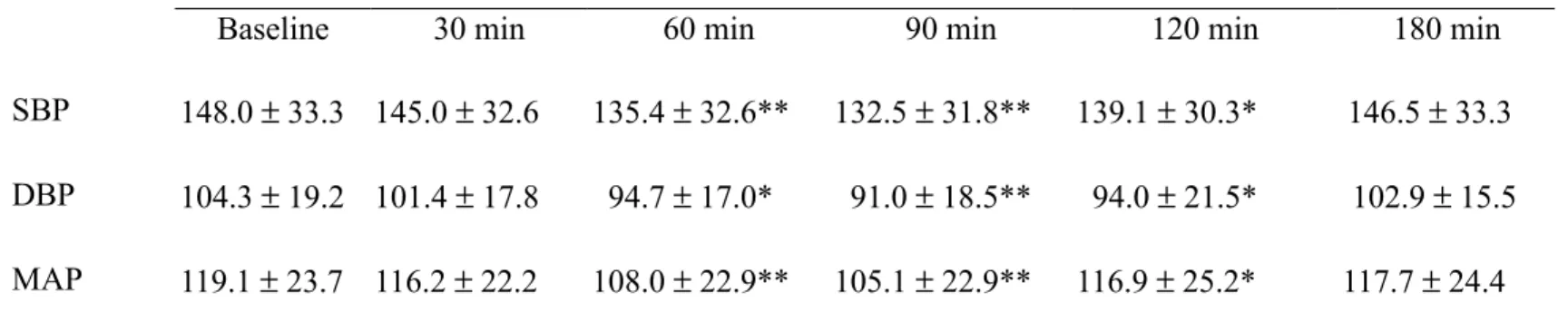
Effect of Stevioside on Blood Pressure by Nasogastric Feeding
After nasogastric feeding for 30 min, blood pressure was observed to lower effectively but did not reach statistical significance. Blood pressure began to decrease significantly at 60 min and to a maximun at 90 min, and returned to baseline level at 180 min (table1).
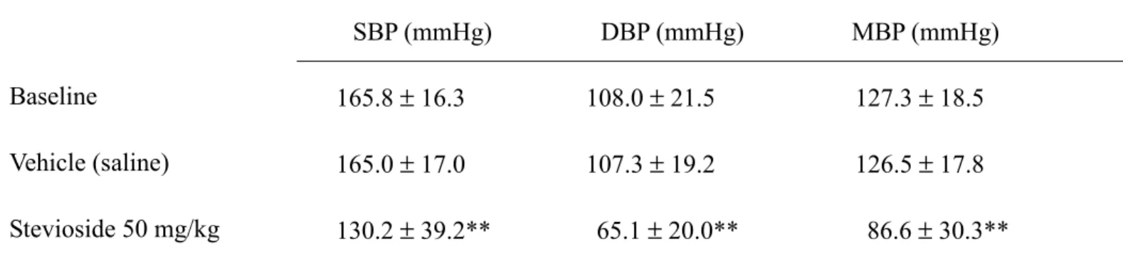
Effect of Stevioside on Blood Pressure by Intravenous Injection
The effect of stevioside on blood pressure was decreased more effective by intravenous administration than nasogastric feeding. Blood pressure decreased reaching a maximum at 5-10 min. The reduction of diastolic pressure was greater than that of systolic pressure (table 2).
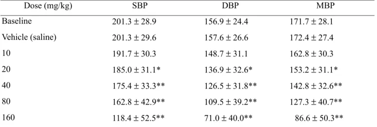
Effect of Stevioside on Renal Hypertension
Initially, intravenous administration of 10 mg/kg stevioside did not lower blood pressure significantly. From the dosage of 20 mg/kg, significant hypotension effect on renal hypertensive dogs was noticed in a dose-dependent manner. (table 3).
Effect of Stevioside on Blood Pressure via Vertebral Artery Injection
For evaluation of the direct effect on central nervous system (CNS), stevioside was injected through left vertebral artery to diminish the metabolic effect of liver on this compound. Blood pressure did not change significantly after stevioside was administered via vertebral artery (table 4). The hypotensive effect of stevioside was not related to the central nervous system.
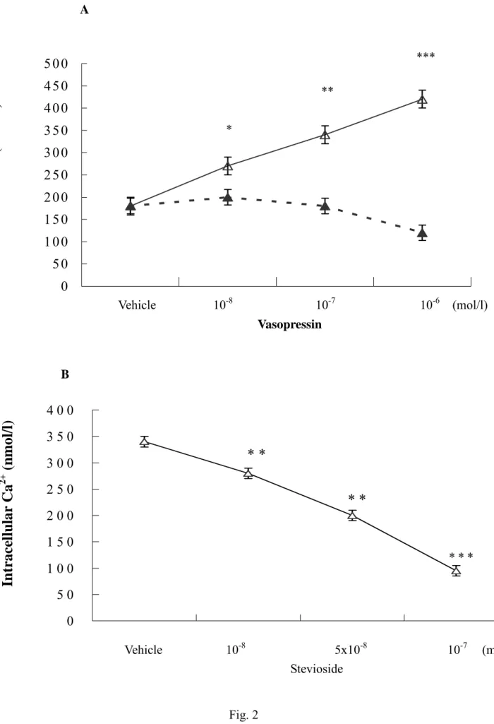
Effect of Stevioside on Ca2+ Influx in A7r5
Figure 2A shows that in Ca2+-containing medium, vasopressin increased the intracellular Ca2+ concentration dose-dependently in A7r5 cell suspension from 192.8 ± 20.2 nmol/l (vasopressin:0 mol/l) to 442.8 ± 32.8 nmol/l (vasopressin: 10-6 mol/l). But, vasopressin had no effect in Ca2+-free cell suspension,. This finding suggests that intracellular Ca2+ increase induced by vasopressin in A7r5 cells was mediated through Ca2+ influx from extracellular space.
In Ca2+-containing cell suspension, stevioside could dose-dependently inhibit the stimulatory effect of 10-6 mol/l vasopressin on intracellular Ca2+ from 346.8 ± 24.6 nmol/l (stevioside: 0 mol/l) to 112.4 ± 12.4 nmol/l (stevioside: 10-7 mol/l) (Fig.2B). Figure 2C shows that phenylephrine increased the intracellular Ca2+ in A7r5 whether the medium contained Ca2+ or not. Phenylephrine caused the intracellular Ca2+ concentration to increase from 186.8
± 18.8 nmol/l (phenylephrine: 0 mol/l) to 826.6 ± 28.8 nmol/l (phenylephrine: 10-7 mol/l) in Ca2+-containing medium, and to 410.4 ± 20.8 nmol/l (phenylephrine: 10-7 mol/l) in Ca2+-free medium. Stevioside caused intracellular Ca2+ concentration decrease by a dose-dependent manner in A7r5 pretreated with phenylephrine (10-8 mol/l) under Ca2+-containing medium (Fig. 2D). The intracellular Ca2+ was decreased from 464.8 ± 40.8 nmol/l (stevioside 0 mol/l) to 220.6 ± 19.8 nmol/l (stevioside 10-6 mol/l) in Ca2+-containing medium, whereas no significant change was observed in Ca2+-free medium, the intracellular Ca2+ concentration remained unchanged at 438.6 ± 38.4 nmol/l (stevioside 10-6 mol/l).
A23187 can increase intracellular Ca2+ concentration without depolarization.ref Data show that stevioside had no inhibitory effect on A23187 induced Ca2+ influx, the intracellular Ca2+
was still at the concentration of 1046.4 ± 110.8 nmol/l.
Discussion
The present data confirm that intravenous stevioside is an effective antihypertensive agent in anesthetized dogs. Previous studies have also shown that the Stevia extract is an effective hypotensive agent [14], and Melis et al. have shown that intravenous adminstration of pure stevioside ( 8,12, and 16mg/kg/h ) resulted in a significant dose-dependent decrease in mean arterial pressure from 121 to 72 mm Hg in anesthetized Wistar rats [15]. Our previous report also showed dose-dependnet hypotensive effect of stevioside on SHR [6]. In this study, our data showed that intravenous administration of stevioside to renal hypertensive dogs resulted in significantly and dose-dependently decrease of blood pressure. SHR is a common animal model for studying genetic hypertension. The mechanism of blood pressure elevation may be related to the abnormality of neurohormonal system (ACTH-corticoid and TSH-thyroxine) [16]. The plasma catecholamine value of immature SHR is higher than normotensive rats and increase of serum sodium concentration does not induce renin secretion. Animal model in this study is acquired hypertension by using renal hypertensive dogs through surgical ligation of one side renal artery. The development of hypertension is related to renin-angiotensin system.
So, stevioside not only reduces blood pressure in genetic hypertensive rats but also in acquired hypertensive dogs in which the mechanism of hypertension is different.
In our previous studies, we found that the effective dose of hypotension of stevioside when administering intravenously was around 50 mg/kg in hypertensive rats [6,17], so we chose to use this dose in the experiment using dogs as animal model. We also reported a double-blind placebo-controlled study of oral stevioside in human hypertension [18]. We found the dose of a 250 mg/capsule by oral administration trice a day is effective and safe in human, so we chose the dose of 200 mg/kg by nasogastric feeding to dogs in the present study.
Nasogastric administration was ineffective in lowering blood pressure, implicating that the
efficacy of stevioside was not good in blood pressure control by oral administration. The absorption of stevioside from gastrointerstinal tract may be low. Injection via vertebral artery did not result in significant blood pressure lowering implicates that the hypotensive action of stevioside was probably not through central effect.
The mechanism of the hypotensive action of stevioside has also been investigated previously, but proved to be inconclusive. Earlier studies have shown that hypotensive response to stevioside appears to occur through a Ca2+ antagonist mechanism similar to that with verapamil [8]. These investigators also showed that hypotension induced by stevioside in normotensive rat is inhibited by indomethacin, which is a potent inhibitor of prostaglandin (PG) synthesis [19]. Purdy et al have shown that PGI2 attenuated angiotensin II-mediated increases in cytosolic calcium in preglomerular vascular smooth muscle cells. This effect may be related to cAMP activated protein kinase C (PKC) pathway [20]. Thus, the inhibitory effect of calcium influx of stevioside might be also related to prostaglandin activities.
Our data also showed that stevioside could inhibit vasopressin or phenylephrine-induced intracellular calcium increase on cultured smooth muscle cells under calcium-contained medium. Szmigielski et al. have reported that phenylephrine-induced translocation of PKC from the cytosol to the membrane and this translocation was blocked by prazosin and verapamil [21]. It seems that the influx of calcium ions through calcium channel is probably necessary for alpha1-adrenoceptor mediated activation and translocation of PKC. We need further study to evaluate whether stevioside has blocking actions against vasopressin or alpha-1 adrenergic receptors.
Fura-2/AM Ca2+ concentration measurement is a standard method in performing experiment concerning Ca2+ influx. Our data also revealed that stevioside was effective in inhibiting Ca2+ influx in aortic smooth muscle cells. Calcium ionophore (A23187) can induce
Ca2+ channels.
In conclusion, the present study confirmed that stevioside is an effective antihypertensive agent, and its mechanism of antihypertension is probably through Ca2+ antagonism.
References
1. Frohlich ED: Hypertension, Evaluation and Treatment. pp. 1-21, Williams and Wilkins, Ltd. Baltimore. 1998
2. Joint National Committee on Detection, Evaluation, and Treatment of High Blood Pressure. The 1996 Report of the Joint National Committee on Detection, Evaluation, and Treatment of High Blood Pressure (JNC-VI). Arch Intern Med 1997; 157:
2413-2446.
3. Jachuck SJ, Brierley H, Jachuck S, Willzox PM: The effect of hypotensive drugs on the quality of life. J R Coll Gen Pract 1982; 32:103-105.
4. Williams GH, Croog SH, Levine S, Testa MA, Sudilovsky A: Impact of antihypertensive therapy on quality of life. Effect of hydrochlorothiazide. J Hypertens 1987; 5: 29-35.
5. Soejarto DD, Kinghorn AD, Farnsworth NR: Potential sweetening agents of plant origin.
III. Organoleptic evaluation of Stevia leaf herbarium samples for sweetness. J Nat Prod 1982; 45: 590-599.
6. Chan P, Xu DY, Liu JC, Chen YJ, Tomlinson B, Huang WP, Cheng JT: The effect of stevioside on blood pressure and plasma catecholamines in spontaneously hypertensive rats. Life Science 1998; 63: 1679-1684
7. Lee CN, Wong KL, Liu JC, Chen YJ, Cheng JT, Chan P: Inhibitory effect of stevioside on calcium influx to produce antihypertension. Planta Medica 2001; 67: 796-799.
8. Melis MS, Sainati AR: Effect of calcium and verapamil on renal function of rats during treatment with stevioside. J Ethnophar 1991; 33: 257-262
9. Conway BJ: Changes in sodium balance and hemodynamics during development of experimental renal hypertension in dogs. Cir Res 1968; 23 :763-767.
10. Henning M, Van Zwieten PA: Central hypotensive effect of α-myethyldopa. J Pharm
rat thoracic aorta. Exp Cell Res 1976; 98: 349-366.
12. Goldman WF, Bova S, Blaustein MP: Measurement of intracellular Ca2+ in cultured arterial smooth muscle cells using fura-2 and digital imaging microscopy. Cell Calcium 1990; 11: 221-231.
13. Grynkiewicz G, Poenie GM, Tsien RY: A new generation of Ca2+ indicators with greatly improved fluorescence properties. J Biol Chem 1985; 260: 3440-3450.
14. Boeckh EMA, Humboldt G: Efeitos cardiocirculatorios do extrato aquoso total em individuos normais e do esteviosideo em ratos. Ciência e Cultura. 1981; 32: 208-210.
15. Melis MS, Macial RE, Sainati AR: Effects of indomethacin on the action of stevioside on mean arterial pressure and on renal function in rats. IRCS Medical Science 1985; 13:
1230-1231
16. Persson AEG, Boberg U: Renal abnormalities in experimental models of hypertension:
the SHR versus the Milan HR. J Cardiovac Pharmacol 1988; 12:S27-S35.
17. Hsu YH, Liu JC, Kao PF, Lee CN, Chen YJ, Hsieh MH, Chan P: Antihypertensive effect of stevioside in different strains of hypertensive rats. Chin Med J (Taipei) 2002; 65:1-6.
18. Chan P, Tomlinson B, Chen YJ, Liu JC, Huang WP, Cheng JT: A double-blind placebo-controlled study of the effectiveness and safety of oral stevioside in human hypertension. Br J Clin Pharmacol 2000; 50:215-220.
19. Melis MS, Sainati AR: Participation of prostaglandins in the effect of stevioside on renal function and arterial pressure in rats. Braz J Med Bio & Res 1991; 24: 1269-1276.
20. Purdy KE, Arendshorst WJ: Prostaglandins buffer angiotensin II-mediated increases in cytosolic calcium in preglomerular vascular smooth muscle cells. Am J Physiol 1999;
277:F850-858.
21. Szmigielski A, Szmigielska H, Zalewska-Kaszubska J, Marczak G: The cooperation between the influx of extracellular calcium and alpha1-adrenceptor-induced translocation of protein kinase C. Pharm Res 1997; 36: 211-219.
22. Sanchez-Fernandez M, Katz GM, Suarez-Kurtz G, Kaczorowshi GJ, Reuben JP:
Mobilization of intracellular calcium in cultured vascular smooth muscle cells by uridine triphosphate and calcium ionophore. J Membr Bio 1993; 135: 273-28
Table 1. Time course of blood pressure changes in anesthetized dogs after nasogastric feeding of stevioside
Stevioside (200 mg/kg)
Baseline 30 min 60 min 90 min 120 min 180 min
SBP 148.0 ± 33.3 145.0 ± 32.6 135.4 ± 32.6** 132.5 ± 31.8** 139.1 ± 30.3* 146.5 ± 33.3 DBP 104.3 ± 19.2 101.4 ± 17.8 94.7 ± 17.0* 91.0 ± 18.5** 94.0 ± 21.5* 102.9 ± 15.5 MAP 119.1 ± 23.7 116.2 ± 22.2 108.0 ± 22.9** 105.1 ± 22.9** 116.9 ± 25.2* 117.7 ± 24.4
Values are mean ± SD. SBP=systolic blood pressure, DBP=diastolic blood pressure, MBP=mean arterial blood pressure.
* p <0.05; ** p <0.01. (vs baseline); the number of experimental animals = 8.
Table 2. Hypotensive effect of stevioside on anesthetized dogs by administering intravenously
SBP (mmHg) DBP (mmHg) MBP (mmHg)
Baseline 165.8 ± 16.3 108.0 ± 21.5 127.3 ± 18.5
Vehicle (saline) 165.0 ± 17.0 107.3 ± 19.2 126.5 ± 17.8
Stevioside 50 mg/kg 130.2 ± 39.2** 65.1 ± 20.0** 86.6 ± 30.3**
Abbreviations as in Table 1. ** p <0.01. (vs vehicle); the number of experimental animals = 8.
Table 3. Dose-dependentl hypotensive effect of stevioside administered intravenously on renal hypertensive dogs
Dose (mg/kg) SBP DBP MBP
Baseline 201.3 ± 28.9 156.9 ± 24.4 171.7 ± 28.1
Vehicle (saline) 201.3 ± 29.6 157.6 ± 26.6 172.4 ± 27.4
10 191.7 ± 30.3 148.7 ± 31.1 162.8 ± 30.3
20 185.0 ± 31.1* 136.9 ± 32.6* 153.2 ± 31.1*
40 175.4 ± 33.3** 126.5 ± 31.8** 142.8 ± 32.6**
80 162.8 ± 42.9** 109.5 ± 39.2** 127.3 ± 40.7**
160 118.4 ± 52.5** 71.0 ± 40.0** 86.6 ± 50.3**
Abbreviations as in Table 1. * p <0.05; ** p <0.01 (vs vehicle); the number of experimental animals = 8.
Table 4. Changes of blood pressure in anesthetized dogs after stevioside administering via left vertebral artery
Dose (mg/kg) SBP DBP MBP
Baseline 144.3 ± 29.6 102.9 ± 18.5 116.9 ± 23.4
Vehicle (saline) 145.0 ± 30.3 102.9 ± 18.5 116.9 ± 23.7 Stevioside (5 mg/kg) 143.6 ± 31.1 102.1 ± 17.8 116.2 ± 24.4 Abbreviations as in Table 1; the number of experimental animals = 8.
Legends of Figures
Fig. 1. The chemical structure of stevioside.
Fig. 2.Using Fura-2/AM to evaluate intracellular Ca2+ in A7r5 cells. (A) Vasopressin-induced dose-dependent increase of intracellular Ca2+ in A7r5 cultured with calcium-contained solution; (B) stevioside dose-dependently inhibited 10-6 mol/l vasopressin-induced intracellular Ca2+ increase; (C) phenylephrine-induced intracellular increase of Ca2+ on A7r5 cultured with calcium-contained or calcium-free solution dose-dependently; (D) stevioside dose-dependently inhibited 10-8 mol/l phenylephrine-induced intracellular Ca2+
increase in A7r5 cultured with calcium-contained solution. -∆- = Ca2+-containing solution; -▲- = Ca2+- free solution.
* p < 0.05, ** p <0.01,*** p <0.001.
O
CO 2
Glc Glc
Glc
Stevioside
Fig. 1.
0 5 0 1 0 0 1 5 0 2 0 0 2 5 0 3 0 0 3 5 0 4 0 0 4 5 0 5 0 0
0 5 0 1 0 0 1 5 0 2 0 0 2 5 0 3 0 0 3 5 0 4 0 0
* *
* *
* * *
Intr ace llu la r Ca
2+(n m o l/l)
*
**
***
Intr ace llu la r Ca
2+(n m o l/l)
Fig. 2 A
B
Vehicle 10-8 5x10⁻⁸ 10-7 (mol/l) Stevioside
Vehicle 10-8 10-7 10-6 (mol/l) Vasopressin
0 100 200 300 400 500 600 0 1 0 0 2 0 0 3 0 0 4 0 0 5 0 0 6 0 0 7 0 0 8 0 0 9 0 0 1 0 0 0
Vehicle 10-8 5x10⁻⁸ 10-7 (mol/l) Phenylephrine
C
Intr ace llu la r Ca
2+(n m o l/l)
**
**
*
**
Intr ace llu la r Ca
2+( nm o l/ l)
Vehicle 10-8 10-7 10-6 (mol/l) D
*
**
***
**
*