Open Access
Research article
Comparative analysis of differentially expressed genes in normal
and white spot syndrome virus infected Penaeus monodon
Jiann-Horng Leu
†1, Chih-Chin Chang
†2, Jin-Lu Wu
1,5, Chun-Wei Hsu
2,
Ikuo Hirono
3, Takashi Aoki
3, Hsueh-Fen Juan
4, Chu-Fang Lo
1,
Guang-Hsiung Kou*
1and Hsuan-Cheng Huang*
2Address: 1Institute of Zoology, National Taiwan University, Taipei 106, Taiwan, 2Institute of Bioinformatics, National Yang-Ming University,
Taipei 112, Taiwan, 3Graduate School of Marine Science and Technology, Tokyo University of Marine Science and Technology, 4-5-7 Konan,
Minato, Tokyo 108-8477, Japan, 4Department of Life Science, Institute of Molecular and Cellular Biology, National Taiwan University, Taipei 106,
Taiwan and 5Department of Biological Sciences, National University of Singapore, 117543, Singapore
Email: Jiann-Horng Leu - [email protected]; Chih-Chin Chang - [email protected]; Jin-Lu Wu - [email protected]; Chun-Wei Hsu - [email protected]; Ikuo Hirono - [email protected]; Takashi Aoki - [email protected]; Hsueh-Fen Juan - [email protected]; Chu-Fang Lo - [email protected]; Guang-Hsiung Kou* - [email protected]; Hsuan-Cheng Huang* - [email protected]
* Corresponding authors †Equal contributors
Abstract
Background: White spot syndrome (WSS) is a viral disease that affects most of the commercially
important shrimps and causes serious economic losses to the shrimp farming industry worldwide. However, little information is available in terms of the molecular mechanisms of the host-virus interaction. In this study, we used an expressed sequence tag (EST) approach to observe global gene expression changes in white spot syndrome virus (WSSV)-infected postlarvae of Penaeus
monodon.
Results: Sequencing of the complementary DNA clones of two libraries constructed from normal
and WSSV-infected postlarvae produced a total of 15,981 high-quality ESTs. Of these ESTs, 46% were successfully matched against annotated genes in National Center of Biotechnology Information (NCBI) non-redundant (nr) database and 44% were functionally classified using the Gene Ontology (GO) scheme. Comparative EST analyses suggested that, in postlarval shrimp, WSSV infection strongly modulates the gene expression patterns in several organs or tissues, including the hepatopancreas, muscle, eyestalk and cuticle. Our data suggest that several basic cellular metabolic processes are likely to be affected, including oxidative phosphorylation, protein synthesis, the glycolytic pathway, and calcium ion balance. A group of immune-related chitin-binding protein genes is also likely to be strongly up regulated after WSSV infection. A database containing all the sequence data and analysis results is accessible at http://xbio.lifescience.ntu.edu.tw/pm/.
Conclusion: This study suggests that WSSV infection modulates expression of various kinds of
genes. The predicted gene expression pattern changes not only reflect the possible responses of shrimp to the virus infection but also suggest how WSSV subverts cellular functions for virus multiplication. In addition, the ESTs reported in this study provide a rich source for identification of novel genes in shrimp.
Published: 16 May 2007
BMC Genomics 2007, 8:120 doi:10.1186/1471-2164-8-120
Received: 16 January 2007 Accepted: 16 May 2007 This article is available from: http://www.biomedcentral.com/1471-2164/8/120
© 2007 Leu et al; licensee BioMed Central Ltd.
This is an Open Access article distributed under the terms of the Creative Commons Attribution License (http://creativecommons.org/licenses/by/2.0), which permits unrestricted use, distribution, and reproduction in any medium, provided the original work is properly cited.
Background
White spot syndrome (WSS) is a highly contagious viral disease of penaeid shrimp. The cumulative mortality of diseased shrimp can reach 100% within 3–10 days. Since its first outbreak in 1993, WSS has caused serious eco-nomic losses to the shrimp farming industry worldwide. The causative agent, white spot syndrome virus (WSSV), is an enveloped, non-occluded, rod-shaped virus that con-tains a circular, double-stranded DNA of about 300 kb. This virus has an extremely wide range of potential hosts, infecting not only shrimps, but also other decapods [1,2]. WSSV infects most shrimp tissues and organs, and it rep-licates in the nuclei of infected cells. At the late stage of infection, either the nucleus or the whole cell disinte-grates, leading to loss of cellular architecture. Both genomic and proteomic approaches have revealed the unique characteristics of the virus, and the virus has been erected as the type species of the new family of
Nimaviri-dae [3-5].
Due to its serious impact on shrimp aquaculture, there is an urgent need to understand WSSV and to unveil the underlying mechanisms involved in WSSV pathogenesis in shrimp. Although considerable progress has been made in characterizing the virus, information on the host genes involved in WSSV pathogenesis is limited. To identify these host genes, one strategy is to isolate genes that are differentially expressed after WSSV infection. To that pur-pose, a variety of different approaches have been used, including an mRNA differential display technique [6], suppression subtractive hybridization [7], SSH and differ-ential hybridization [8], cDNA microarrays [9,10] and ESTs [11]. Both cDNA microarrays and EST libraries are particularly suitable for large-scale gene expression analy-sis, and both of these methods have been well developed in several model organisms. However, the application of these methods to shrimp is still in its infancy. Conse-quently, compared to other model organisms, only rela-tively few sequenced ESTs and microarray cDNA targets are available for shrimp. Furthermore, all of the studies cited above focused exclusively on the identification of immune-related genes with only immune-related organs, (ie, the hemocytes and the hepatopancreas [HP]) being analyzed. However, WSSV is a systemic virus that infects most shrimp tissues and organs, and it is logical to assume that in different cell types, the virus would be likely to modulate the expression of different host genes in order to promote its multiplication in the correspondingly dif-ferent cellular contexts. If so, then the gene expression changes induced by WSSV in immune-related cells should be different from those in non-immune cells. Therefore, in the present paper, rather than using a specific tissue or organ to investigate only the gene expression patterns of immune-related cells, we instead take a global view by using entire P. monodon postlarvae as our study subject.
Our large scale EST approach used two different cDNA libraries, one from normal and one from WSSV-infected
P. monodon postlarvae. The respective EST data were then
compared to predict the gene expression changes in host shrimp after WSSV infection.
As an additional benefit, this large scale EST study also increases our transcriptomic data for Crustacea and penaeid shrimp. This in turn will improve our under-standing of penaeid shrimp biology, which is important because the penaeid shrimp are economically valuable and yet they remain vulnerable to outbreaks of various viral diseases. Research into the genetics and genomics of shrimp has been gaining in importance over the past dec-ade, and a number of penaeid shrimp EST projects have already been undertaken. However, most of these projects and their associated EST libraries were small in scale [12-15]. Currently, the two largest penaeid shrimp EST studies have published 13,656 and 10,100 ESTs, respectively [16,17]. We hope that, together with the 15,981 addi-tional ESTs released with this report, this will provide a good foundation for further research into the genetics, genomics and even the proteomics of shrimp.
Results
Generation and analysis of EST libraries
Two cDNA libraries, PmTwN and PmTwI, were con-structed from normal and WSSV-infected postlarvae of P.
monodon, respectively. No normalization was applied to
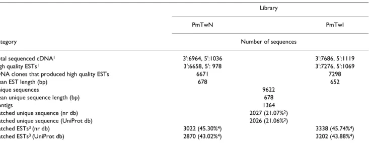
these two libraries. A total of 7,200 and 8,064 clones were randomly selected from the normal and infected libraries for DNA template preparation, respectively. After tem-plate quality screening, a total of 6,964 and 7,686 cDNA clones were sequenced from the 3' end from the normal and infected libraries, respectively. After base-calling, vec-tor sequence trimming and screening to eliminate low quality sequences and contamination from WSSV and other sources, a total of 6,658 and 7,276 high quality 3' ESTs were generated from the normal and infected librar-ies, respectively (Table 1).
After finishing the 3' end sequencing, as well as 3' end sequence assembly and annotation, we next performed DNA sequencing from the 5' end for a fraction of cDNA clones. We randomly chose the cDNA clones from the 3' EST contigs that showed no significant hits of the BlastX searches to the NCBI nr database. From the normal and infected libraries, 1,036 and 1,119 clones, respectively, were subjected to 5' end sequencing with SP6 primer. After base-calling, trimming the vector sequence and elim-inating low quality sequences, 978 and 1,069 high quality 5' ESTs were generated (Table 1). These were then com-bined with the high quality 3' ESTs for the final assembly and annotation.
Overall, these high quality ESTs were derived from 6,671 normal and 7,298 infected cDNA clones. The average lengths of the high quality sequences were 678 bp and 652 bp in the normal and infected libraries, respectively. The CAP3 assembly program produced 9,622 unique sequences. Of these unique sequences, 8,258 were sin-glets, consisting of only one EST, and the other 1,364 were contigs, consisting of at least two ESTs. Most contigs con-tained 2–4 ESTs, and the largest contig was formed by 854 ESTs. The average length of the unique sequences was 678 bp (Table 1).
We also noted that when both libraries were checked for contamination by WSSV sequence, 167 ESTs were found to be WSSV contaminants in the PmTwI library (E value < 10-25, score > 100), whereas there were no WSSV contam-inants in PmTwN. This result is consistent with the PCR screening results for the original, unchallenged postlarvae, and it reconfirmed that the shrimp used in the present study were WSSV-free.
Sequence similarity
BlastX found 2,027 (21.07%) unique sequences similar to known protein sequences in the NCBI nr protein database and 2,026 (21.06%) unique sequences that had GO anno-tations in the UniProt database. Working backwards from the unique sequences to the original ESTs in the normal library (PmTwN), this translates to 3,022 (45.30%) ESTs that match known protein sequences in the nr database,
and 2,870 (43.02%) ESTs with matches in the UniProt database. In the infected library (PmTwI), the correspond-ing number of matches are 3,338 (45.74%) and 3,202 (43.88%), respectively. These data are summarized in Table 1.
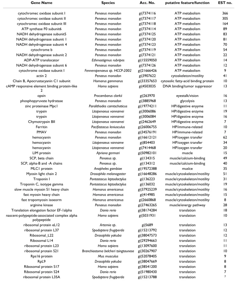
Identification of the most abundant genes in each library Table 2 and 3 list the 50 most abundant genes in each library. In the normal library (Table 2), most of these abundant genes can be classified into four major groups, including proteins involved in ATP metabolism, proteins involved in translation, proteins highly or specifically expressed in muscle, and proteins highly or specifically expressed in the HP. These results probably reflect the fact that the shrimp postlarvae were in an active growth stage. During active growth, both energy and cellular translation machinery would be needed to synthesize proteins and build muscle, while the HP would be actively engaged in synthesizing digestive enzymes, hemocyanin and other proteins. By contrast, in the infected library (Table 3), the most abundant genes no longer included the HP proteins, but instead included four other groups: immune-related proteins with chitin-binding or lectin domains, proteins involved in glycolysis, cuticle-related proteins and several different actin genes.
Identification of genes with differential abundance Among the known genes represented by EST matches, 360 genes were found exclusively in the normal library, 361 Table 1: Summary of cDNA libraries and EST data
Library
PmTwN PmTwI
Category Number of sequences
Total sequenced cDNA1 3':6964, 5':1036 3':7686, 5':1119
High quality ESTs1 3':6658, 5': 978 3':7276, 5':1069
cDNA clones that produced high quality ESTs 6671 7298
Mean EST length (bp) 678 652
Unique sequences 9622
Mean unique sequence length (bp) 678
Contigs 1364
Matched unique sequence (nr db) 2027 (21.07%2)
Matched unique sequence (UniProt db) 2026 (21.06%2)
Matched ESTs3 (nr db) 3022 (45.30%4) 3338 (45.74%4)
Matched ESTs3 (UniProt db) 2870 (43.02%4) 3202 (43.88%4)
1: The numbers of 3' and 5' end sequences are shown.
2: The percentage is calculated by dividing the number of unique sequences (9622) by the number of matched unique sequences (2027 and 2026, respectively).
3: Some cDNA clones have both 5' and 3' ESTs, and when both or either ESTs had matches in databases, the corresponding 5' and 3' ESTs from the same clone will be marked and only one was counted.
4: The percentage is calculated by dividing the number of cDNA clones that produced high quality ESTs (6671 or 7298) by the number of matched ESTs (3022 and 3338, respectively, or 2870 and 3202).
Table 2: The fifty most abundant genes in the normal library.
Gene Name Species Acc. No. putative feature/function EST no.
cytochromec oxidase subunit I Penaeus monodon gi|7374116 ATP metabolism 366
cytochromec oxidase subunit II Penaeus monodon gi|7374117 ATP metabolism 305 cytochromec oxidase subunit III Penaeus monodon gi|7374118 ATP metabolism 164
ATP synthase F0 subunit6 Penaeus monodon gi|7374114 ATP metabolism 89
NADH dehydrogenase subunit5 Penaeus monodon gi|7374125 ATP metabolism 83
NADH dehydrogenase subunit 1 Penaeus monodon gi|7374120 ATP metabolism 81
NADH dehydrogenase subunit 4 Penaeus monodon gi|7374123 ATP metabolism 70
cytochrome b Penaeus monodon gi|7374119 ATP metabolism 54
NADH dehydrogenase subunit 2 Penaeus monodon gi|7374121 ATP metabolism 22
ADP-ATP translocator Ethmostigmus rubripes gi|15559050 ATP metabolism 14
NADH dehydrogenase subunit 6 Penaeus monodon gi|7374126 ATP metabolism 12
cytochrome oxidase subunit I Fenneropenaeus sp. HCCP-2002 gi|21666426 ATP metabolism 9
actin 2 Penaeus monodon gi|3907622 cytoskeleton/motility 41
Chain B, Apocrustacyanin C1 subunit Homarus gammarus gi|33357653 cytosolic fatty-acid binding protein 10 cAMP responsive element binding protein-like
2
Homo sapiens gi|4503035 DNA binding/tumor suppressor 13
opsin Procambarus clarkii gi|263970 eyestalk/vision 16
phosphopyruvate hydratase Penaeus monodon gi|3885968 glycolysis 13
zinc proteinase Mpc1 Paralithodes camtschaticus gi|19774211 HP/digestive enyyme 11
trypsin Litopenaeus vannamei gi|3006086 HP/digestive enzyme 19
trypsin Litopenaeus vannamei gi|3006084 HP/digestive enzyme 16
Chymotrypsin BII Litopenaeus vannamei gi|2462649 HP/digestive enzyme 7
Ferritin Pacifastacus leniusculus gi|26006755 HP/immune-related 10
PMAV Penaeus monodon gi|34576191 HP/immune-related 7
hemocyanin Penaeus monodon gi|16612121 HP/oxygen transfer 62
hemocyanin Litopenaeus vanname gi|854403 HP/oxygen transfer 34
hemocyanin Litopenaeus vannamei gi|7414468 HP/oxygen transfer 20
LIM protein Apriona germari gi|50982101 muscle 15
SCP, beta chain Penaeus sp. gi|134315 muscle/calcium-binding 49
SCP, alpha-B and -A chains Penaeus sp. gi|134312 muscle/calcium-binding 40
MLC1 protein Anopheles gambiae gi|19572388 muslce 35
Myosin light chain 2 Drosophila melanogaster gi|16648286 muscle/cytoskeleton/motility 51 Troponin I Pontastacus leptodactylus gi|136223 muscle/cytoskeleton/motility 31 Troponin C, isotype gamma Pontastacus leptodactylus gi|136032 muscle/cytoskeleton/motility 19 slow muscle myosin S1 heavy chain Homarus americanus gi|37925239 muscle/cytoskeleton/motility 16 fast myosin heavy chain Homarus americanus gi|414985 muscle/cytoskeleton/motility 15 fast tropomyosin isoeorm Homarus americanus gi|2660868 muscle/cytoskeleton/motility 8
arginine kinase Penaeus monodon gi|27463265 muscle/energy pathway 28
Translation elongation factor EF-1alpha Danio rerio gi|38174284 translation 18 nascent-polypeptide-associated complex alpha
polypeptide
Homo sapiens gi|5031931 translation 10
ribosomal protein eL12 Artemia sp. gi|5689 translation 13
ribosomal protein L37 Spodoptera frugiperda gi|15213792 translation 12
Ribosomal_L22 Drosophila yakuba gi|38047573 translation 12
Ribosomal L14 Danio rerio gi|29294663 translation 11
ribosomal protein L23 Homo sapiens gi|13097600 translation 11
ribosomal protein S21 Branchiostoma belcheri tsingtaunese gi|30267907 translation 10
Rps16 protein Mus musculus gi|52078405 translation 9
RpL9 Drosophila yakuba gi|38047669 translation 8
Ribosomal protein S17 Homo sapiens gi|38541200 translation 8
Ribosomal protein S24 Danio rerio gi|51980430 translation 7
Table 3: The fifty most abundant genes in the WSSV-infected library.
GeneName Species Acc. No. putative feature/function EST no.
cytochromec oxidase subunit I Penaeus monodon gi|7374116 ATP metabolism 488 cytochromec oxidase subunit II Penaeus monodon gi|7374117 ATP metabolism 270 cytochromec oxidase subunit III Penaeus monodon gi|7374118 ATP metabolism 174
cytochrome b Penaeus monodon gi|7374119 ATP metabolism 112
ATP synthase F0 subunit 6 Penaeus monodon gi|7374114 ATP metabolism 86
NADH dehydrogenase subunit4 Penaeus monodon gi|7374123 ATP metabolism 60
NADH dehydrogenase subunit 5 Penaeus monodon gi|7374125 ATP metabolism 56
NADH dehydrogenase subunit 1 Penaeus monodon gi|7374120 ATP metabolism 43
NADH dehydrogenase subunit 2 Penaeus monodon gi|7374121 ATP metabolism 13
cytochrome oxidase subunit I Fenneropenaeus sp gi|21666426 ATP metabolism 13 ATP lipid-binding protein like protein Marsupenaeus japonicus gi|18700491 ATP metabolism 10
NADH dehydrogenase subunit 6 Penaeus monodon gi|7374126 ATP metabolism 9
Cytochrome c oxidase subunit IV Drosophila melanogaster gi|16197913 ATP metabolism 8
ADP-ATP translocator Ethmostigmus rubripes gi|15559050 ATP metabolism 7
CG14607-PA Drosophila melanogaster gi|24644788 Chitin binding Peritrophin-A domain/ immune
13 ENSANGP00000013986 Anopheles gambiae gi|31243037 Chitin binding Peritrophin-A domain/
immune
10 ENSANGP00000023091 Anopheles gambiae gi|31200471 Chitin binding Peritrophin-A domain/
immune
8 CG6055-PA Drosophila melanogaster gi|7297257 C-type lectin (CTL) and CTL-like
domains/immune
18
BCS-1 Balanus amphitrite gi|9186884 cuticle protein 324
Endocuticle structural glycoprotein SgAbd-8 Schistocerca gregaria gi|47605412 cuticle protein 32 calcification-associated peptide-1 Procambarus clarkii gi|33468738 cuticle protein 17
Cuticle protein AMP1A Homarus americanus gi|3287772 cuticle protein 11
DD9B Marsupenaeus japonicus gi|7008007 cuticle protein 10
cuticle protein 20 Manduca sexta gi|19548965 cuticle protein 9
actin1 Penaeus monodon gi|3907620 cytoskeleton/motility 17
Actin Bombyx mori gi|113216 cytoskeleton/motility 14
beta actin Homarus gammarus gi|34576243 cytoskeleton/motility 9
fructose 1,6-bisphosphate aldolase Homalodisca coagulata gi|46561746 glycolysis 69
phosphopyruvate hydratase Penaeus monodon gi|3885968 glycolysis 46
glyceraldehyde-3-phosphate dehydrogenase Procambarus clarkii gi|31338868 glycolysis 10 triosephosphate isomerase Archaeopotamobius sibirien gi|19848023 glycolysis 9
trypsin Litopenaeus vannamei gi|3006086 HP/digestive enyyme 9
LIM protein Apriona germari gi|50982101 muscle 31
SCP, alpha-B and -A chains Penaeus sp. gi|134312 muscle/calcium-binding 18 MLC1 protein Anopheles gambiae gi|19572388 muscle/cytoskeleton/motility 34 slow muscle myosin S1 heavy chain Homarus americanus gi|37925239 muscle/cytoskeleton/motility 11 Troponin I Pontastacus leptodactylus gi|136223 muscle/cytoskeleton/motility 9
arginine kinase Penaeus monodon gi|27463265 muscle/enery pathway 40
thioredoxin-1 Mesobuthus cyprius gi|51869033 redox reaction 14
Translation elongation factor EF-1alpha (GTPase) Danio rerio gi|38174284 translation 14
Ribosomal L18p Xenopus laevis gi|27371036 translation 13
Ribosomal protein S24 Danio rerio gi|51980430 translation 12
ribosomal protein eL12 Artemia sp. gi|5689 translation 9
RpL9 Drosophila yakuba gi|38047669 translation 9
ribosomal protein L35A Spodoptera frugiperda gi|15213788 translation 8
40S ribosomal protein S10 Ictalurus punctatus gi|15294031 translation 8
ribosomal protein L7 Spodoptera frugiperda gi|18253049 translation 8
Ribosomal L22 Drosophila yakuba gi|38047573 translation 7
Rps16 protein Mus musculus gi|52078405 translation 7
porin Anopheles gambiae gi|19697919 voltage dependent anion-selective channel
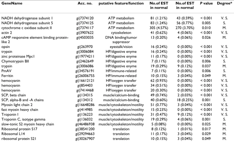
genes were found only in the infected library, and 264 genes were cross expressed in both libraries. Based on the number of homologous ESTs, Fisher's exact test found a significant increase in abundance for 23 genes (Table 4), and a significant decrease for 25 genes (Table 5).
Among the genes with increased abundance, several major groups can be identified, including four proteins with a chitin binding Peritrophin-A domain, seven cuti-cle-related proteins, four proteins involved in oxidative phosphorylation, two glycolytic enzymes and two ribos-omal proteins. Other increased-abundance genes include thioredoxin-1, actin and a protein homologous to CG6055-PA. The decreased-abundance genes can also be classified into several groups, including two SCP calcium-binding proteins, five cytoskeleton/motility-related pro-teins, three proteins involved in oxidative phosphoryla-tion, three ribosomal proteins and seven proteins that are produced mainly by the HP. These HP-produced proteins include four digestive enzymes, hemocyanin and two immune-related proteins, PmAV and ferritin. Other decreased-abundance genes include opsin and cAMP responsive element binding protein-like 2.
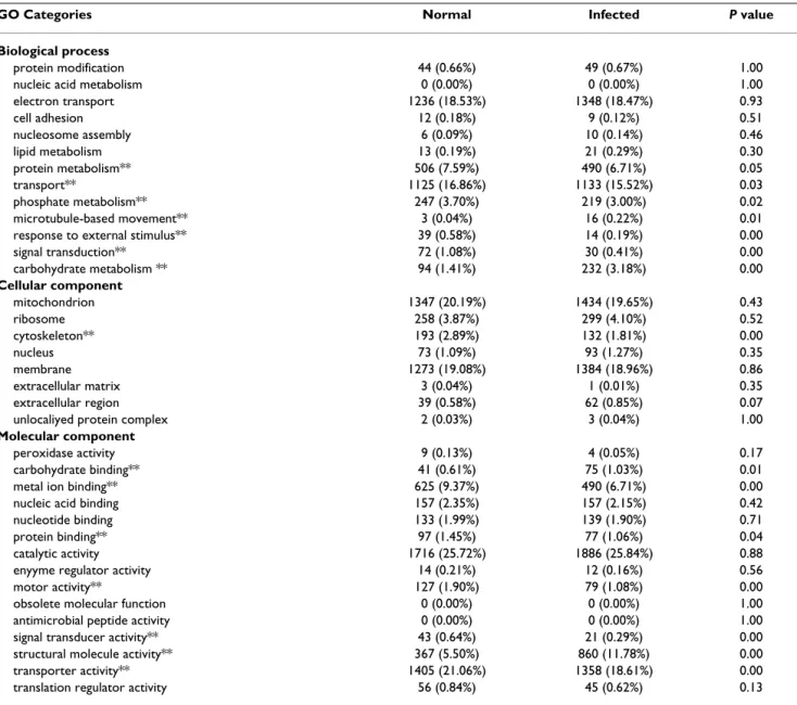
Functional classification based on Gene Ontology
The putative functions assigned to the unique sequences by the Gene Ontology (GO) classification scheme sug-gested that in the normal and infected libraries, respec-tively, 3,397 and 3,571 ESTs map to biological processes, 3,408 and 3,188 ESTs map to cellular components, and 2,012 and 2,375 ESTs map to molecular functions (Table 6). In both libraries, most of the corresponding biological process genes are involved in electron transport, transport, protein metabolism, phosphate metabolism and carbohy-drate metabolism (Table 6). Table 6 also shows that most of the cellular component genes encode proteins located in the mitochondria, membrane, ribosome, cytoskeleton, and nucleus, and that most of the molecular function genes are associated with catalytic activity, transporter activity, structural molecules, and metal ion binding. Analysis of GO categories showed a significant statistical difference (Fisher's exact test; P < 0.05) between the nor-mal and infected libraries for several biological processes, including carbohydrate metabolism, signal transduction, response to external stimulus, microtubule-based move-ment, phosphate metabolism, transport and protein metabolism (Table 6). Among the processes that appear Table 4: Unique genes with increased differential abundance in the normal and infected libraries.
GeneName Acc. no. putative feature/ function No. of EST in normal No. of EST in normal P value Degree*
cytochrome b gi|7374119 ATP metabolism 54 (0.81%) 112 (1.54%) < 0.001 V. S. cytochrome c oxidase subunit I gi|7374116 ATP metabolism 366 (5.49%) 488 (6.69%) 0.003 S. Cytochrome c oxidase subunit IV gi|16197913 ATP metabolism 0 (0.00%) 8 (0.11%) 0.008 S. ATPase subunit C gi|18700491 ATP metabolism 1 (0.02%) 10 (0.14%) 0.013 M. ENSANGP00000013986 gi|31243037 Chitin binding
Peritrophin-A domain
0 (0.00%) 10 (0.14%) 0.002 S. ENSANGP00000023091 gi|31200471 Chitin binding
Peritrophin-A domain
0 (0.00%) 8 (0.11%) 0.008 S. RE51076p gi|21064537 Chitin binding
Peritrophin-A domain
0 (0.00%) 7 (0.10%) 0.016 M.
CG14607-PA gi|24644788 Chitin binding Peritrophin-A domain
0 (0.00%) 13 (0.18%) < 0.001 V. S. CG6055-PA gi|7297257 C-type lectin (CTL)
and CTL-like domain
1 (0.02%) 18 (0.25%) < 0.001 V. S. BCS-1 gi|9186884 cuticle protein 4 (0.06%) 324 (4.44%) < 0.001 V. S. Endocuticle structural glycoprotein SgAbd-8 gi|47605412 cuticle protein 2 (0.03%) 32 (0.44%) < 0.001 V. S. calcification-associated peptide-1 gi|33468738 cuticle protein 0 (0.00%) 17 (0.23%) < 0.001 V. S. Cuticle protein AMP1A gi|3287772 cuticle protein 0 (0.00%) 11 (0.15%) 0.001 S.
DD9B gi|7008007 cuticle protein 1 (0.02%) 10 (0.14%) 0.013 M.
cuticle protein 20 gi|19548965 cuticle protein 1 (0.02%) 9 (0.12%) 0.023 M. Cuticle protein AM/CP1114 gi|5921935 cuticle protein 0 (0.00%) 6 (0.08%) 0.032 M. Actin gi|113216 cytoskeleton/motility 0 (0.00%) 14 (0.19%) < 0.001 V. S. actin 1 gi|3907620 cytoskeleton/motility 5 (0.08%) 17 (0.23%) 0.019 M. fructose 1,6-bisphosphate aldolase gi|46561746 glycolysis 5 (0.08%) 69 (0.95%) < 0.001 V. S.
phosphopyruvate hydratase gi|3885968 glycolysis 13 (0.20%) 46 (0.63%) < 0.001 V. S.
thioredoxin-1 gi|51869033 redox reaction 2 (0.03%) 14 (0.19%) 0.005 S.
ribosomal protein L10A gi|14994666 translation 0 (0.00%) 6 (0.08%) 0.032 M. ribosomal protein L7 gi|18253049 translation 1 (0.02%) 8 (0.11%) 0.041 M. *: The degree of significance is based on P value. V. S.: very strong (P < 0.001), S.: strong (0.001 ⬉ < 0.01), M.: moderate (0.01 ⬉ P < 0.05).
to be increased after WSSV infection, carbohydrate metab-olism showed the most significant change, mostly because this GO category includes the proteins involved in the glycolytic pathway as well as proteins with the chi-tin binding Peritrophin-A domain, and the abundance of both of these groups was highly increased (Table 3 and 4). Two categories, signal transduction and response to exter-nal stimulus, showed significantly decreased abundance in the infected library. We note that opsin is included in both of these two categories.
Two molecular functions, structural molecular activity and carbohydrate binding, appear to be elevated signifi-cantly in the infected library (Table 6). The structural molecular activity category consisted of cuticle-related proteins (including the highly abundant BCS-1 in the infected library), ribosomal proteins, and cytoskeletal proteins. The carbohydrate-binding category included proteins with C-type lectin (CTL) and CTL-like domains and proteins with the chitin binding Peritrophin-A domain, both of which showed significantly increased abundance in the infected library (Table 3 and 4). Signif-icantly decreased categories included metal ion binding, motor activity, transporter activity, signal transducer activ-ity and protein binding. The metal ion binding category consisted of a diverse array of proteins, including cyto-chrome c oxidase subunit II, various enzymes, the
cal-cium-binding proteins and cytoskeletal/muscle-related proteins.
In the cellular component group, only the cytoskeleton category was significantly different (Table 6). This cate-gory included the cytoskeleton/motility-related proteins, which had a decreased abundance in the infected library. We have constructed a database to host all the sequence data and the analysis results obtained from this study. The database can be accessed through a web interface [18].
Discussion
Shrimps are economically important cultured aquatic ani-mals. However, compared to other aquacultured animals, there have been relatively few studies on shrimp genom-ics. In the present study, a global analysis of 15,981 high-quality shrimp ESTs revealed 2,027 known genes and 7,595 unknown unique sequences. These ESTs will not only be a valuable addition to the current archived sequences from shrimps, but will also provide a major resource for the comparative analysis of gene expression profiles between normal and WSSV-infected shrimps. To the extend that changes in EST abundance (Table 4 and 5) are predictive of changes in gene expression, then the present data suggest that in P. monodon postlarvae, WSSV Table 5: Unique genes with decreased differential abundance in the normal and infected libraries.
GeneName Acc. no. putative feature/function No.of EST in normal
No.of EST in normal
P value Degree*
NADH dehydrogenase subunit 1 gi|7374120 ATP metabolism 81 (1.21%) 43 (0.59%) < 0.001 V. S. NADH dehydrogenase subunit 5 gi|7374125 ATP metabolism 83 (1.24%) 56 (0.77%) 0.005 S. cytochrome c oxidase subunit II gi|7374117 ATP metabolism 305 (4.57%) 270 (3.70%) 0.010 M.
actin 2 gi|3907622 cytoskeleton 41 (0.62%) 4 (0.06%) < 0.001 V. S.
cAMP responsive element binding protein-like 2
gi|4503035 DNA binding/tumor suppressor
13 (0.20%) 4 (0.06%) 0.026 M.
opsin gi|263970 eyestalk/vision 16 (0.24%) 0 (0.00%) < 0.001 V. S.
trypsin gi|3006084 HP/digestive enyme 16 (0.24%) 0 (0.00%) < 0.001 V. S.
zinc proteinase Mpc1 gi|19774211 HP/digestive enyme 11 (0.17%) 0 (0.00%) < 0.001 V. S.
Chymotrypsin BII gi|2462649 HP/digestive enyme 7 (0.11%) 0 (0.00%) 0.006 S.
trypsin gi|3006086 HP/digestive enyme 19 (0.29%) 9 (0.12%) 0.037 M.
PmAV gi|34576191 HP/immune-related 7 (0.11%) 0 (0.00%) 0.006 S.
Ferritin gi|26006755 HP/immune-related 10 (0.15%) 3 (0.04%) 0.049 M.
hemocyanin gi|16612121 HP/oxygen transfer 62 (0.93%) 0 (0.00%) < 0.001 V. S.
hemocyanin gi|854403 HP/oxygen transfer 34 (0.51%) 0 (0.00%) < 0.001 V. S.
hemocyanin gi|7414468 HP/oxygen transfer 20 (0.30%) 0 (0.00%) < 0.001 V. S.
SCP, beta chain gi|134315 muscle/calcium-binding 49 (0.74%) 2 (0.03%) < 0.001 V. S. SCP, alpha-B and -A chains gi|134312 muscle/calcium-binding 40 (0.60%) 18 (0.25%) 0.001 S. Myosin light chain 2 gi|16648286 muscle/cytoskeleton/motility 51 (0.77%) 3 (0.04%) < 0.001 V. S. fast myosin heavy chain gi|414985 muscle/cytoskeleton/motility 15 (0.23%) 0 (0.00%) < 0.001 V. S. Troponin I gi|136223 muscle/cytoskeleton/motility 31 (0.47%) 9 (0.12%) < 0.001 V. S. Troponin C, isotype gamma gi|136032 muscle/cytoskeleton/motility 19 (0.29%) 4 (0.06%) 0.001 S. slow-tonic S2 myosin heavy chain gi|46486938 muscle/cytoskeleton/motility 5 (0.08%) 0 (0.00%) 0.025 M.
Ribosomal protein S17 gi|38541200 translation 8 (0.12%) 1 (0.01%) 0.017 M.
Ribosomal L14 gi|29294663 translation 11 (0.17%) 3 (0.04%) 0.029 M.
infection modulates the expression of various kinds of genes. The predicted up-regulated genes include several proteins involved in oxidative phosphorylation, cuticular proteins, a protein with C-type lectin (CTL) and CTL-like domains, proteins with the chitin binding Peritrophin-A domain, two glycolytic enzymes, and thioredoxin-1. The predicted down-regulated genes include several proteins that are synthesized in the HP (digestive enzymes, two immune-related proteins, and the hemocyanin), five cytoskeleton/motility-related proteins, four proteins involved in oxidative phosphorylation, and opsin. Several ribosomal protein genes and actin genes are also pre-dicted to be differentially modulated by WSSV.
Cuticular proteins
This is the first study to suggest that WSSV infection strongly up-regulates the expression of cuticular proteins. One prominent feature of arthropods is the cuticular cov-ering of the whole body. Cuticles are highly organized structures made of chitin filaments embedded in a pro-teinaceous matrix, and they are produced as a layered, extracellular secretion from the underlying epidermis [19]. Pathological studies reveal that the cuticular epider-mis is one of the main target tissues of WSSV [20]. At the late stage of infection, this tissue is heavily infected, it loses its cellular architecture and becomes necrotic. Loos-ening of the cuticle and the appearance of white spots in Table 6: Gene Ontology of the sequences with significant Blastx hits in UniProt
GO Categories Normal Infected P value
Biological process
protein modification 44 (0.66%) 49 (0.67%) 1.00
nucleic acid metabolism 0 (0.00%) 0 (0.00%) 1.00
electron transport 1236 (18.53%) 1348 (18.47%) 0.93 cell adhesion 12 (0.18%) 9 (0.12%) 0.51 nucleosome assembly 6 (0.09%) 10 (0.14%) 0.46 lipid metabolism 13 (0.19%) 21 (0.29%) 0.30 protein metabolism** 506 (7.59%) 490 (6.71%) 0.05 transport** 1125 (16.86%) 1133 (15.52%) 0.03 phosphate metabolism** 247 (3.70%) 219 (3.00%) 0.02 microtubule-based movement** 3 (0.04%) 16 (0.22%) 0.01
response to external stimulus** 39 (0.58%) 14 (0.19%) 0.00
signal transduction** 72 (1.08%) 30 (0.41%) 0.00 carbohydrate metabolism ** 94 (1.41%) 232 (3.18%) 0.00 Cellular component mitochondrion 1347 (20.19%) 1434 (19.65%) 0.43 ribosome 258 (3.87%) 299 (4.10%) 0.52 cytoskeleton** 193 (2.89%) 132 (1.81%) 0.00 nucleus 73 (1.09%) 93 (1.27%) 0.35 membrane 1273 (19.08%) 1384 (18.96%) 0.86 extracellular matrix 3 (0.04%) 1 (0.01%) 0.35 extracellular region 39 (0.58%) 62 (0.85%) 0.07
unlocaliyed protein complex 2 (0.03%) 3 (0.04%) 1.00
Molecular component
peroxidase activity 9 (0.13%) 4 (0.05%) 0.17
carbohydrate binding** 41 (0.61%) 75 (1.03%) 0.01
metal ion binding** 625 (9.37%) 490 (6.71%) 0.00
nucleic acid binding 157 (2.35%) 157 (2.15%) 0.42
nucleotide binding 133 (1.99%) 139 (1.90%) 0.71
protein binding** 97 (1.45%) 77 (1.06%) 0.04
catalytic activity 1716 (25.72%) 1886 (25.84%) 0.88
enyyme regulator activity 14 (0.21%) 12 (0.16%) 0.56
motor activity** 127 (1.90%) 79 (1.08%) 0.00
obsolete molecular function 0 (0.00%) 0 (0.00%) 1.00
antimicrobial peptide activity 0 (0.00%) 0 (0.00%) 1.00
signal transducer activity** 43 (0.64%) 21 (0.29%) 0.00
structural molecule activity** 367 (5.50%) 860 (11.78%) 0.00
transporter activity** 1405 (21.06%) 1358 (18.61%) 0.00
translation regulator activity 56 (0.84%) 45 (0.62%) 0.13
the cuticular epidermis are two of the pathological charac-ters caused by WSSV infection.
The white spots in the cuticle of WSSV-infected shrimp represent abnormal deposits of calcium salts by the cutic-ular epidermis [21]. Table 4 suggests that after WSSV infection, a gene corresponding to crayfish calcification-associated peptide-1 (CAP-1) is very strongly up-regu-lated. Crayfish CAP-1 is isolated from the exoskeleton, has chitin-binding ability and, most importantly, anti-calcifi-cation activity [22]. CAP-1 mRNA is strongly expressed in the epidermal tissue during the postmolt stage [23]. Inoue et al. [23] proposed that CAP-1 might play an important role in calcification and cuticle formation in the exoskele-ton, and if so, then, the abnormal production of CAP-1 in WSSV-infected shrimp may cause the abnormal deposits of calcium salt, leading to the formation of white spots in the cuticle.
Table 4 includes a protein gene that is a homolog to BCS-1, a gene that was cloned from a subtracted barnacle cypris larval cDNA library by differential screening [24]. BCS-1 mRNA is specifically expressed in barnacle cypris larvae, and during the process of larval attachment and metamor-phosis, the amount of BCS-1 mRNA is decreased [24]. The function of BCS-1 remains unknown. However, Scan-Prosite analysis of the BCS-1 protein sequence revealed that BCS-1 contains the chitin-binding type R&R domain profile, which is a structural feature of insect cuticular teins. This suggests that BCS-1 is a barnacle cuticular pro-tein, and the homolog to BCS-1 in Table 4 is therefore described as a cuticle protein.
Pathological studies have shown that WSSV infection seri-ously damages the cuticular epidermis, and this study now suggests that WSSV infection strongly up-regulates the gene expressions of various cuticular proteins. It remains unclear how and why WSSV infection induces the expression of cuticular protein genes, and whether this benefits WSSV. These are questions that deserve further study.
Gene expression in non-primary WSSV-target organs: HP, muscle and compound eye
Compared to the cuticular epidermis, the HP, muscle and compound eye are only lightly infected by WSSV, and these organs remain intact at the late stage of infection [20]. In HP, WSSV mainly infects the myoepithelial cells of the hepatopancreatic sheath and the fibroblast of the connective tissue, whereas the epithelium of the tubules, which synthesize the hemocyanin and digestive enzymes [25], are rarely infected. However, our EST analysis sug-gests that the RNAs of hemocyanin and several digestive enzymes are strongly reduced after WSSV infection. This suggests that although the epithelium of the tubules are
refractory to WSSV infection, at least some of their physi-ological functions (such as gene transcription) are dra-matically affected by the infection of other cell types in HP. If expression of digestive enzymes is down-regulated in HP, then this, together with the fact that WSSV infec-tion targets the stomach, might well explain why WSSV-infected shrimp reduce their food consumption.
The compound eye and muscle are also only lightly infected by WSSV. Even so, it seems that infection severely decreases the transcription of several genes that are prima-rily and/or highly expressed in these two organs, suggest-ing that WSSV infection would definitely affect the functions of both organs.
Proteins with the chitin binding peritrophin-A domain The chitin binding Peritrophin-A domain is found in chi-tin binding proteins, particularly the peritrophic matrix proteins of insects and animal chitinases [26,27]. The per-itrophic matrix (PM) lines the midgut of insects and it is believed that the PM facilitates digestion and forms a pro-tective barrier to prevent invasion by bacteria, viruses and parasites [28]. There are several classes of PM proteins, and one of these protein classes, the peritrophins, is stud-ied extensively and has been found in several insects [26,27,29]. Recent studies have shown that peritrophin proteins also exist in crustaceans [30-32]. Khayat et al. [30] were the first to identify two peritrophin-like cDNAs that are highly expressed during oogenesis in Penaeus
sem-isulcatus and the two proteins are components of the
cor-tical rods, forming a jelly layer after fertilization. A similiar protein was also isolated from the mature ovary of
Marsu-penaeus japonicus [31] Another peritrophin-like protein
has also been identified in Fenneropenaeus chinensis. This peritrophin mRNA is constitutively expressed in the ova-ries, and can only be induced by E. coli to express in hemo-cytes, heart, stomach, gut, and gills [32]. In addition, the recombinant protein can bind to Gram-negative bacteria and chitin, suggesting that it may play a role in immune defense and other physiological responses. Interestingly, as shown in Table 4, the four proteins with a chitin bind-ing Peritrophin-A domain can only be identified in the infected library, which suggests that their expressions are induced after WSSV infection and that they may therefore play some roles in the shrimp's antiviral immune response.
C-type lectins
In P. monodon postlarvae, the protein homologous to Dro-sophila CG6055-PA showed greatly increased abundance after WSSV infection (Table 4). The Drosophila CG6055-PA protein has a C-type lectin (CTL) and CTL-like domain, which is a structural module that has Ca2+ -dependent carbohydrate-binding activity. Proteins that have this module are generally known as C-type (Ca2+
-dependent) lectins to distinguish them from the other (Ca2+-independent) types of animal lectins. Animal C-type lectins play important roles in innate and adaptive immunity through pathogen recognition and cellular interactions [33]. In invertebrates, C-type lectins are involved in various immune responses, including the acti-vation of the proPO system [34], antibacterial activity [35], and the promotion of phagocytosis [36]. In shrimps, several C-type lectins have been identified. PmLec is a P.
monodon C-type lectin that is able to bind to bacterial
lipopolysaccharide (LPS) to enhance hemocyte phagocy-tosis [36]. Fclectin is a C-type lectin gene cloned from the hemocytes of Chinese shrimp Fenneropenaeus chinensis [37]. Fclectin mRNA is mainly expressed in hemocytes and its expression is greatly affected after challenge by bac-teria, LPS or WSSV [37]. Neither PmLec nor Fclectin was found in our ESTs, but another C-type lectin, PmAV, was represented. The PmAV gene has been identified in WSSV-resistant P. monodon, and the recombinant protein shows a strong antiviral activity toward a fish virus in vitro [38]. Unlike the CG-6055-PA homolog, however, Table 5 sug-gests that PmAV was strongly down-regulated. A third C-type lectin, the gene homologous to Drosophila RH18728p, was also identified in our EST database, but its expression does not appear to change significantly after WSSV infection.
Calcium-binding proteins
Table 5 suggests that expression of the sarcoplasmic cal-cium-binding protein (SCP) α and β subunits is strongly down-regulated after WSSV infection. Shrimp, lobster and crayfish SCPs exist as dimers of two different polypeptide chains, the α and β [39-41], and they function as cytosolic Ca2+ buffers. In crayfish, although the SCP is ubiquitously expressed in many tissues, it is most abundant in muscle [42]. If the P. monodon SCP α and β subunits are also both highly expressed in muscle, then this would be consistent with our observation that, just like several other muscle-specific transcripts, these two genes showed decreased abundance after WSSV infection (Table 5). In this connec-tion, we also note that WSSV itself encodes several pro-teins with EF-hand calcium-binding motifs [4]. This means that WSSV can potentially modulate the calcium ion concentration of infected cells by decreasing the expression of shrimp cell SCP and by simultaneously expressing the viral-encoded calcium-binding proteins. Glycolytic pathway
Until recently, glycolytic enzymes were considered as "straightforward" enzymes with no sophisticated regula-tory properties. However, these enzymes are now known to perform various functions in addition to their innate glycolytic function, and they play an important role in several biological and pathophysiological processes [43-45]. Two glycolytic enzyme genes, phosphopyruvate
hydratase and fructose 1,6-bisphosphate aldolase, showed increased abundance after WSSV infection. Phos-phopyruvate hydratase is a key protein in the glycolytic pathway, catalyzing the conversion of 2-phosphoglycerate to phosphoenopyruvate, but it can also be a receptor for plasminogen [46,47] or a transcriptional repressor [45]. Together with phosphoglycerate kinase and tubulin, phosphopyruvate hydratase forms an active transcription initiation complex that enhances transcriptional elonga-tion of the Sendai virus genome [48]. In addielonga-tion, this enzyme has also been described as a stress protein induced by hypoxia [49]. The non-glycolytic functions of the other glycolytic enzyme, aldolase, presently remain unknown. Nevertheless, taken together, all these studies suggest that it would be worthwhile to further investigate whether either or both of these enzymes play an essential role during WSSV infection.
Oxidative phosphorylation
Several proteins involved in oxidative phosphorylation and encoded by mitochondrial DNA showed differential abundance at the RNA level after WSSV infection (Table 4 and 5). WSSV infection decreases the abundance of NADH dehydrogenase subunits 1 and 5 and cytochrome c oxidase subunit II, but simultaneously increased the abundance of cytochrome b, cytochrome c oxidase subu-nit I and IV and ATPase subusubu-nit C. Numerous studies have shown that viruses affect mitochondria in different ways. Morphological changes in mitochondria are induced by human immunodeficiency virus (HIV) [50], human T-cell leukemia virus type 1 [51] and Rubella virus [52]. Changes in location are observed after infection by Human herpesvirus 1 (HHV-1) and Hepatitis B virus [53,54]. The mitochondrial respiratory chain is affected by simian virus 40 [55], Poliovirus [56], HHV-1 and influ-enza virus [57]. The present study now suggests for the first time that virus infection might also modulate the amounts of mitochondrial mRNAs. However, it remains to be determined whether such changes could affect the mitochondrial oxidative phosphorylation and hence the generation of ATP.
Actin genes
WSSV infection also differentially affected the abundance of actin genes (Table 4 and 5). Actins are conserved pro-teins that participate in muscle contraction, cell motility, cell division, and cytoskeletal structure [58]. In almost all eukaryotes, actins are encoded by members of multigene families, and these different actin genes are expressed dif-ferently across different cell types, tissues, and develop-mental stages [59]. In vertebrates, three main groups of actin isoforms, α, β and γ, have been identified. The α-actins are found in muscle tissues and the β and γ-α-actins coexist in most cell types as components of the cytoskele-ton. The actin and the microtubule cytoskeleton play
important roles in the life cycle of every virus, from the beginning of virus attachment to the host cell to the final assembly and egress of virus [60]. In spite of this, how-ever, some viruses actively degrade some specific host mRNAs, including β-actin, to shut off host cell protein synthesis [61,62]. Two recent studies have revealed the interplay between WSSV and the shrimp actin gene. One of the major WSSV structural proteins, VP26, interacts with actin [63], and the actin mRNA becomes unstable after WSSV infection [10]. The present study now suggests that the expression of these P. monodon actins is differen-tially modulated after WSSV infection. Two isoforms (gi|113216 and gi|3907620) showed increased abun-dance (Table 4), whereas one (gi|3907622) showed decreased abundance (Table 5). We also note that rela-tively speaking, the P. monodon actins are not very well documented. For instance, while Artemia is shown to have 8–10 actin genes and 4 isoforms, one of which is muscle specific, and crab (Gecarcinus latefalis) has seven or eight documented actin genes [64], only two P. monodon actin genes have so far been released to a public database. The previously undocumented actin genes listed in our EST libraries (see Table 2) now add several more actins to that list.
The predicted modulation of actin isoforms by WSSV also means that actin is not a good choice of reference gene in RNA level studies of WSSV virus/host interactions. This is in addition to the already known difficulty that while the vertebrate muscular actins are recognizable from their amino acid sequence, the invertebrate actins cannot be distinguished in the same way because they all resemble the vertebrate cytoplasmic actins [65,66]. Clearly, further studies will be needed to characterize and classify these P.
monodon actin genes by their tissue distribution patterns,
with a particular focus on the isoforms that have their expressions modulated by WSSV.
Conclusion
In conclusion, the 15,981 high-quality ESTs generated in this study provide a rich source for identification of novel genes in shrimp and for comparative analysis of gene expression patterns in normal and WSSV-infected shrimp. An EST-based strategy not only greatly facilitates in silico expression profiling, it also provides an experimental approach to elucidate WSSV pathogenesis and to investi-gate the shrimp's response to virus infection. Our data suggest that in postlarval shrimp, WSSV infection strongly affects the physiological functions of several organs/tis-sues, including the HP, muscle, eyestalk and cuticle, and that the expressions of several genes in these organs/tis-sues are strongly modulated. In addition, WSSV is pre-dicted to affect several basic cellular metabolic processes, including oxidative phosphorylation, protein synthesis, glycolysis, and calcium ion balance.
Methods
Shrimp, virus and challenge
The postlarvae used to construct the cDNA libraries were at the PL20 stage, with an average length and weight of 9 mm and 0.02 g, respectively. These postlarvae were derived from WSSV-free Penaeus monodon spawners. They were cultivated in the Tungkang marine laboratory of Tai-wan's Fisheries Research Institute. The WSSV suspension used to challenge the postlarvae was prepared from frozen (-80°C), WSSV-infected black tiger shrimp that had been collected during a natural outbreak of WSS in 1994 [67]. The postlarvae were challenged by immersion. At 66 hours after infection, they were collected in cryotubes and stored in liquid nitrogen for later RNA extraction. They were further confirmed by PCR to be WSSV-infected. Nor-mal, WSSV-free postlarvae were also collected and stored in liquid nitrogen and their WSSV-free status was con-firmed by PCR.
RNA extraction and cDNA library construction
For each library, RNA was extracted from a pool of about 50 entire animals weighing approximately 1 g in total. Total RNAs were extracted from normal and WSSV-infected postlarvae using RNAzol B reagent (Teltest Ltd. Friendswood, TX), and the mRNAs were purified from the total RNAs using the QuickPrep™ Micro mRNA Purifica-tion Kit (GE healthcare) following the protocol provided by the manufacturer. The respective cDNA libraries were constructed using the λZAP-cDNA library construction kit (Stratagene) according to the manufacturer's instructions. In brief, the first-strand cDNA was synthesized from 5 µg of mRNA with the oligo-d(T) linker-primer, 5'-(GA)10ACTAGTCTCGAG(T)18-3', and the MMLV (Molony murine leukemia virus) reverse transcriptase supplied in the kit. The second strand of cDNA was synthesized with DNA polymerase I in the presence of RNase H, and then the cDNA was blunt ended with pfu DNA polymerase. Both strands of the cDNA were ligated with the EcoR I adapter, and digested by Xho I. cDNA fractionation was performed using Sepharose CL-4B gel filtration, and the cDNA was then inserted into the EcoR I-Xho I site of the Uni-Zap phage vector. The resulting phage libraries were converted to pBluescript phagemid libraries by massive in
vivo excision using ExAssist helper phage. About 8,000
white colonies were randomly isolated from both normal and infected libraries on LB-ampicillin plates containing IPTG and X-gal, and plasmid DNA was extracted from each library.
Sequencing
Randomly selected clones were inoculated into individual wells of 96-well plates with 170 µl LB media containing 8 µg ampicillin, and the plates were incubated at 37°C for 18 hours. The DNA templates were prepared using Mon-tage Plasmid Miniprep96 kits, and checked by
electro-phoresis in 1% agarose gel. Sequencing was carried out using the BigDye version 3.0 sequencing reaction kit using the optimal protocol provided by the manufacturer. DNA sequencing from the 3' end and 5' end of the cDNA was conducted with T7 or SP6 primers, respectively, on a high-throughput automated sequencer (MJ Research BaseSta-tion and ABI3730, USA) using standard protocols. Sequence analysis
The raw traces for ESTs were subjected to base-calling by running Phred (Q > 13). pBluescript vector sequences were trimmed using Cross_match with default parameters (minimatch 12, penalty -2, minscore 20). Those ESTs hav-ing a length of more than 100 bp after vector trimmhav-ing were subject to further analysis. BlastN [68] was used to find matching sequences with E value < 10-5 in the whole WSSV genome (GenBank accession no. AF440570), and also to screen out possible contaminants from bacterial chromosomal DNA, RNA, and lambda phage DNA. The interspersed repeats and low complexity sequences in the ESTs were then masked by RepeatMasker using the Dro-sophila repeat sequence as reference. Low quality sequences, including short sequences (less than 100 bp) and those with a high percentage of nucleotide A (adeno-sine) or N (uncertain read) were considered uninforma-tive and were eliminated from further analysis. The ESTs that passed through the above quality check procedures were considered high quality ESTs. The high quality ESTs in both the normal and infected libraries were combined and assembled to form contigs using CAP3 [69] with the overlapping percentage parameter set to > 95% in order to obtain highly reliable contig sequences. ESTs that did not form contigs designated singlets. Collectively, the result-ant contigs and singlets are referred to as unique sequences.
Functional annotation
Putative functions of the unique sequences were discov-ered by using BlastX to translate each nucleotide query sequence into all reading frames and then searching for matches in the NCBI non-redundant database. Significant hits (with E value < 10-10) in the NCBI nr database were followed up with protein function searches in the UniProt database [70], which provides value-added information reports for protein functions. The UniProt reports consist of Gene Ontology (GO) annotations that classify proteins by biological process, cellular component, and molecular function. Each unique sequence was tentatively assigned GO classification based on annotation of the single "best hit" match in UniProt. These data were then used to clas-sify the corresponding genes according to their GO func-tions.
Expression analysis and statistical evaluation of EST occurrence
The unique sequences were considered to have increased abundance if they had a significantly greater number of hits (ie. more ESTs) in the WSSV-infected library com-pared to the normal library. Conversely, unique sequences were considered to have decreased abundance if they had significantly more hits in the normal library. The statistical significance of homologous ESTs with dif-ferential abundance was determined using Fisher's exact test [71-73], which is widely used to evaluate 2 × 2 contin-gency tables. Fisher's exact test produces a significance value P ranging between 0 and 1, where a value close to 0 implies that there is a significant differential abundance of the gene or the annotated function between the normal and infected libraries. The significance of differential abundance genes with a P value smaller than 0.001 was considered "very strong". P values between 0.001 and 0.01, and between 0.01 and 0.05 were considered "strong" and "moderate", respectively.
Availability and requirements
Project name: Penaeus monodon Functional Genomics Database
Project home page: http://xbio.lifescience.ntu.edu.tw/ pm/
Operating system: Platform independent Programming language:PHP
Abbreviations
WSSV: white spot syndrome virus HP: hepatopancreas
Authors' contributions
JHL extracted RNAs from shrimps, and constructed the cDNA library. IH and TA carried out the sequence sis. CCC, CWH and HCH carried out the sequence analy-sis, functional annotation, and expression analysis. CFL and GHK conceived and directed the project. CFL and HCH designed the study. JHL, JLW, CCC, HFJ, and HCH drafted the manuscript. All authors read and approved the final manuscript.
Acknowledgements
This investigation was supported financially by the National Science Council grants (NSC94-2317-B-002-022, 2317-B-002-009 and NSC95-2317-B-002-010) and the National Science and Technology Program for Agricultural Biotechnology from Council of Agriculture (95AS-6.2.1-ST-a1-23). We are indebted to Paul Barlow for his helpful criticism.
References
1. Lo CF, Ho CH, Peng SE, Chen CH, Hsu HC, Chiu YL, Chang CF, Liu KF, Su MS, Wang CH, Kou GH: White spot syndrome
baculovi-rus (WSBV) detected in cultured and captured shrimp, crabs and other arthropods. Dis Aquat Org 1996, 27:215-226.
2. Lo CF, Leu JH, Ho CH, Chen CH, Peng SE, Chen YT, Chou CM, Yeh PY, Huang CJ, Chou HY, Wang CH, Kou GH: Detection of
bacu-lovirus associated with white spot syndrome (WSBV) in penaeid shrimps using polymerase chain reaction. Dis Aquat
Org 1996, 5:133-141.
3. van Hulten MC, Witteveldt J, Peters S, Kloosterboer N, Tarchini R, Fiers M, Sandbrink H, Lankhorst RK, Vlak JM: The white spot
syn-drome virus DNA genome sequence. Virology 2001, 286:7-22.
4. Yang F, He J, Lin X, Li Q, Pan D, Zhang X, Xu X: Complete genome
sequence of the shrimp white spot bacilliform virus. J Virol
2001, 75:11811-11820.
5. Huang C, Zhang X, Lin Q, Xu X, Hu Z, Hew CL: Proteomic
analy-sis of shrimp white spot syndrome viral proteins and charac-terization of a novel envelope protein VP466. Mol Cell
Proteomics 2002, 1:223-231.
6. Astrofsky KM, Roux MM, Klimple KR, Fox JG, Dhar AK: Isolation of
differentially expressed genes from white spot syndrome virus (WSV) infected Pacific blue shrimp (Penaeus
styliros-tris). Arch Virol 2002, 147:1799-1812.
7. Pan D, He N, Yang Z, Liu H, Xu X: Differential gene expression
profile in hepatopancreas of WSSV-resistant shrimp (Penaeus japonicus) by suppression subtractive hybridization.
Dev Comp Immunol 2005, 29:103-112.
8. He N, Qin Q, Xu X: Differential profile of genes expressed in
hemocytes of White Spot Syndrome Virus-resistant shrimp (Penaeus japonicus) by combining suppression subtractive hybridization and differential hybridization. Antivir Res 2005, 66:39-45.
9. Dhar AK, Dettroi A, Roux MM, Klimpel KR, Read B: Identification
of differential expressed genes in shrimp (Penaeus stylirostris) infected with white spot syndrome virus by cDNA microar-rays. Arch Virol 2003, 148:2381-2396.
10. Wang B, Li F, Dong B, Zhang X, Zhang C, Xiang J: Discovery of the
genes in response to white spot syndrome virus (WSSV) infection in Fenneropenaeus chinensis through cDNA micro-array. Mar Biotechnol (NY) 2006, 8:491-500.
11. Rojtinnakorn J, Hirono I, Itami T, Takahashi Y, Aoki T: Gene
expres-sion in heamocytes of kuruma prawn, Penaeus japonicus, in response to infection with WSSV by EST approach. Fish Shell
Immunol 2002, 13:69-83.
12. Lehnert SA, Wilson KJ, Byrne K, Moore SS: Tissue-specific
expressed sequence tags from the black tiger shrimp
Penaeus monodon. Mar Biotechnol 1999, 1:465-76.
13. Gross PS, Bartlett TC, Browdy CL, Chapman RW, Warr GW:
Immune gene discovery by expressed sequence tag analysis of hemocytes and hepatopancreas in the pacific white shrimp, Litopenaeus vannamei, and the Atlantic white shrimp, L setiferus. Dev Comp Immunol 2001, 25:565-77.
14. Supungul P, Klinbunga S, Pichyangkura R, Jitrapakdee S, Hirono I, Aoki T, Tassanakajon A: Identification of immune-related genes in
hemocytes of black tiger shrimp (Penaeus monodon). Mar
Bio-technol 2002, 4:487-94.
15. Yamano K, Unuma T: Expressed sequence tags from eyestalk of
kuruma prawn, Marsupenaeus japonicus. Comp Biochem Physiol
A Mol Integr Physiol 2006, 143:155-61.
16. O'Leary NA, Trent HF III, Robalino J, Peck MET, Mckillen DJ, Gross PS: Analysis of multiple tissue-specific cDNA libraries from
the Pacific whiteleg shrimp, Litopenaeus vannamei. Integrative
and Comparative Biology 2006, 46:931-939.
17. Tassanakajon A, Klinbunga S, Paunglarp N, Rimphanitchayakit V, Udomkit A, Jitrapakdee S, Sritunyalucksana K, Phongdara A, Pong-somboon S, Supungul P, Tang S, Kuphanumart K, Pichyangkura R, Lursinsap C: Penaeus monodon gene discovery project: The
generation of an EST collection and establishment of a data-base. Gene 2006, 384:104-12.
18. Penaeus monodon Functional Genomics Database [http://
xbio.lifescience.ntu.edu.tw/pm/]
19. Anderson SO, Hojrup P, Roepstoref P: Insect cuticular proteins. Insect Biochem Molec Biol 1995, 25:153-176.
20. Chang PS, Lo CF, Wang YC, Kou GH: Identification of white spot
syndrome associated baculovirus (WSBV) target organs in
shrimp, Penaeus monodon, by in situ hybridization. Dis Aquat
Org 1996, 27:131-139.
21. Wang CS, Tang KFJ, Kou GH, Chen SN: Light and electron
micro-scopic evidence of white spot disease in the giant tiger shrimp, Penaeus monodon (Fabricius), and the kuruma shrimp, Penaeus japonicus (Bate), cultured in Taiwan. Journal
of fish Diseases 1997, 20:323-331.
22. Inoue H, Ozaki N, Nagasawa H: Purification and Structural
Determination of a Phosphorylated Peptide with Anti-calci-fication and Chitin-binding Activities in the Exoskeleton of the Crayfish, Procambarus clarkii. Biosci Biotechnol Biochem 2001, 65:1840-1848.
23. Inoue H, Ohira T, Ozaki N, Nagasawa H: Cloning and expression
of a cDNA encoding a matrix peptide associated with calcifi-cation in the exoskeleton of the crayfish. Comp Biochem Physiol
B Biochem Mol Biol 2003, 136:755-765.
24. Okazaki Y, Shizuri Y: Structures of six cDNAs expressed
specif-ically at cypris larvae of barnacles, Balanus amphitrite. Gene
2000, 250:127-135.
25. Lehnert SA, Johnson SE: Expression of hemocyanin and
diges-tive enzyme messenger RNAs in the hepatopancreas of the Black Tiger Shrimp Penaeus monodon. Comp Biochem Physiol B
Biochem Mol Biol 2002, 133:163-171.
26. Elvin CM, Vuocolo T, Pearson RD, East IJ, Riding GA, Eisemann CH, Tellam RL: Characterization of a major peritrophic
mem-brane protein, peritrophin-44, from the larvae of Lucilia
cup-rina. cDNA and deduced amino acid sequences. J Biol Chem
1996, 271:8925-8935.
27. Shen Z, Jacobs-Lorena M: A type I peritrophic matrix protein
from the malaria vector Anopheles gambiae binds to chitin. Cloning, expression, and characterization. J Biol Chem 1998, 273:17665-17670.
28. Lehane MJ: Peritrophic matrix structure and function. Annu Rev Entomol 1997, 42:525-505.
29. Gaines PJ, Walmsley SJ, Wisnewski N: Cloning and
characteriza-tion of five cDNAs encoding peritrophin A domains from the cat flea, Ctenocephalides felis. Insect Biochem Mol Biol 2003, 33:1061-1073.
30. Khayat M, Babin PJ, Funkenstein B, Sammar M, Nagasawa H, Tietz A, Lubzens E: Molecular characterization and high expression
during oocyte development of a shrimp ovarian cortical rod protein homologous to insect intestinal peritrophins. Biol
Reprod 2001, 64:1090-9.
31. Kim YK, Kawazoe I, Tsutsui N, Jasmani S, Wilder MN, Aida K:
Isola-tion and cDNA cloning of ovarian cortical rod protein in kuruma prawn Marsupenaeus japonicus (Crustacea: Decap-oda: Penaeidae). Zoolog Sci 2004, 21:1109-19.
32. Du XJ, Wang JX, Liu N, Zhao XF, Li FH, Xiang JH: Identification
and molecular characterization of a peritrophin-like protein from fleshy prawn (Fenneropenaeus chinensis). Mol Immunol
2006, 43:1633-44.
33. Weis WI, Taylor ME, Drickamer K: The C-type lectin
super-family in the immune system. Immunol Rev 1998, 163:19-34.
34. Yu XQ, Gan H, Kanost MR: Immulectin, an inducible C-type
lec-tin from an insect, Manduca sexta, stimulates activation of plasma prophenol oxidase. Insect Biochem Mol Biol 1999, 29:585-597.
35. Suzuki T, Takagi T, Furukohri T, Kawamura K, Nakauchi M: A
cal-cium-dependent galactose-binding lectin from the tunicate
Polyandrocarpa misakiensis. Isolation, characterization, and
amino acid sequence. J Biol Chem 1990, 265:1274-1281.
36. Luo T, Yang H, Li F, Zhang X, Xu X: Purification,
characteriza-tion and cDNA cloning of a novel lipopolysaccharide-binding lectin from the shrimp Penaeus monodon. Dev Comp Immunol
2006, 30:607-17.
37. Liu YC, Li FH, Dong B, Wang B, Luan W, Zhang XJ, Zhang LS, Xiang JH: Molecular cloning, characterization and expression
anal-ysis of a putative C-type lectin (Fclectin) gene in Chinese shrimp Fenneropenaeus chinensis. Mol Immunol 2007, 44:598-607.
38. Luo T, Zhang XB, Shao ZZ, Xu X: PmAV, a novel gene involved
in virus resistance of shrimp Penaeus monodon. FEBS Lett 2003, 551:53-57.
39. Wnuk W, Jauegai-Adell J: Polymorphism in high affinity calcium
binding proteins from crustacean sarcoplasm. Eur J Biochem